Saposin lipoprotein particles and libraries from crude membranes

a technology of lipoproteins and libraries, applied in the field of life sciences, can solve the problems of delayed research on membrane lipidomes and membrane proteomes, incompatible with membrane protein or lipid functional analysis in their natural environment, and inability to soluble detergent-free systems, etc., to achieve flexibility and/or variation, maintain protein structure and/or function, and the effect of flexible siz

Active Publication Date: 2022-05-31
SALIPRO BIOTECH AB
View PDF7 Cites 0 Cited by
  • Summary
  • Abstract
  • Description
  • Claims
  • Application Information

AI Technical Summary

Benefits of technology

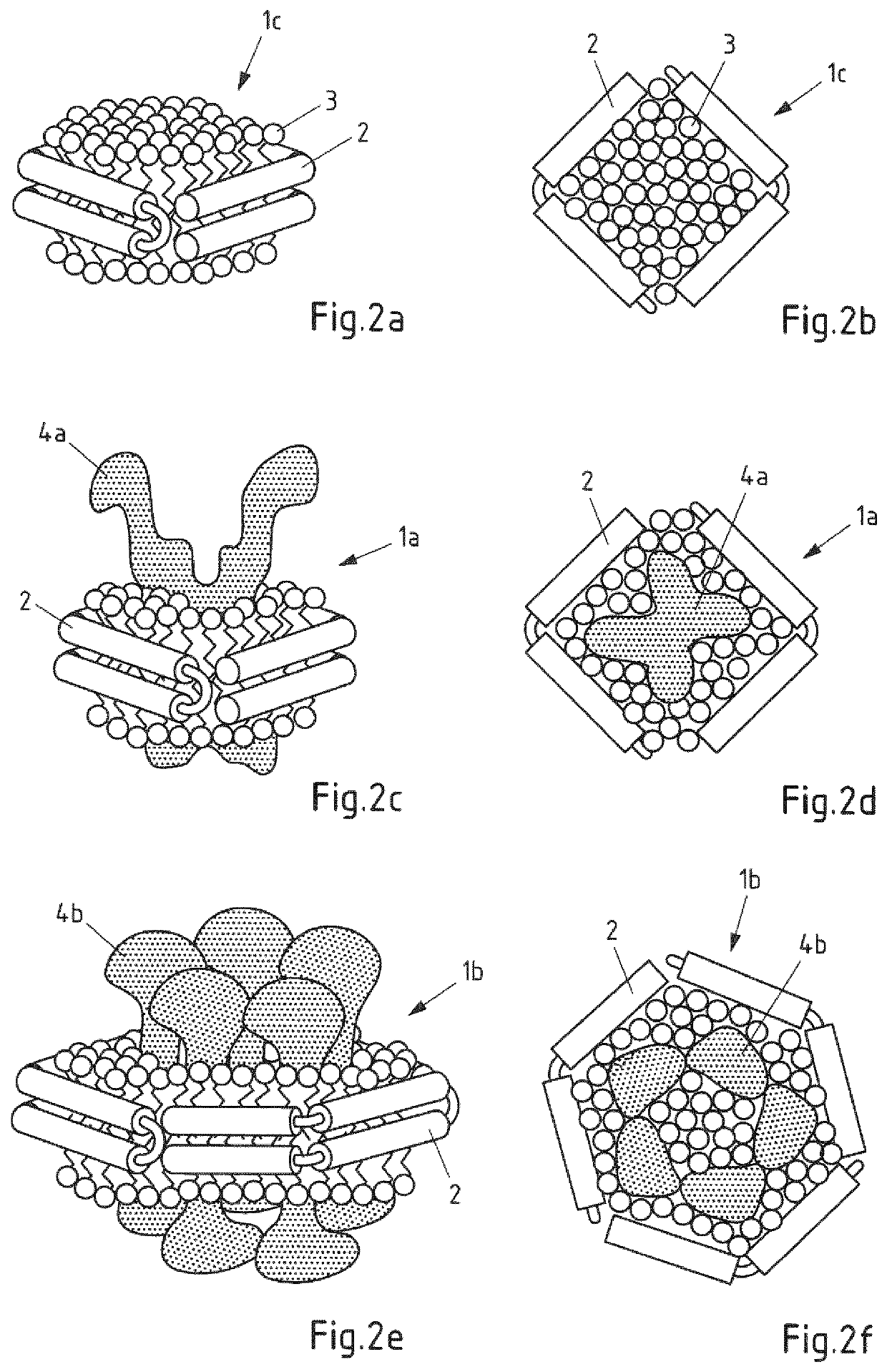
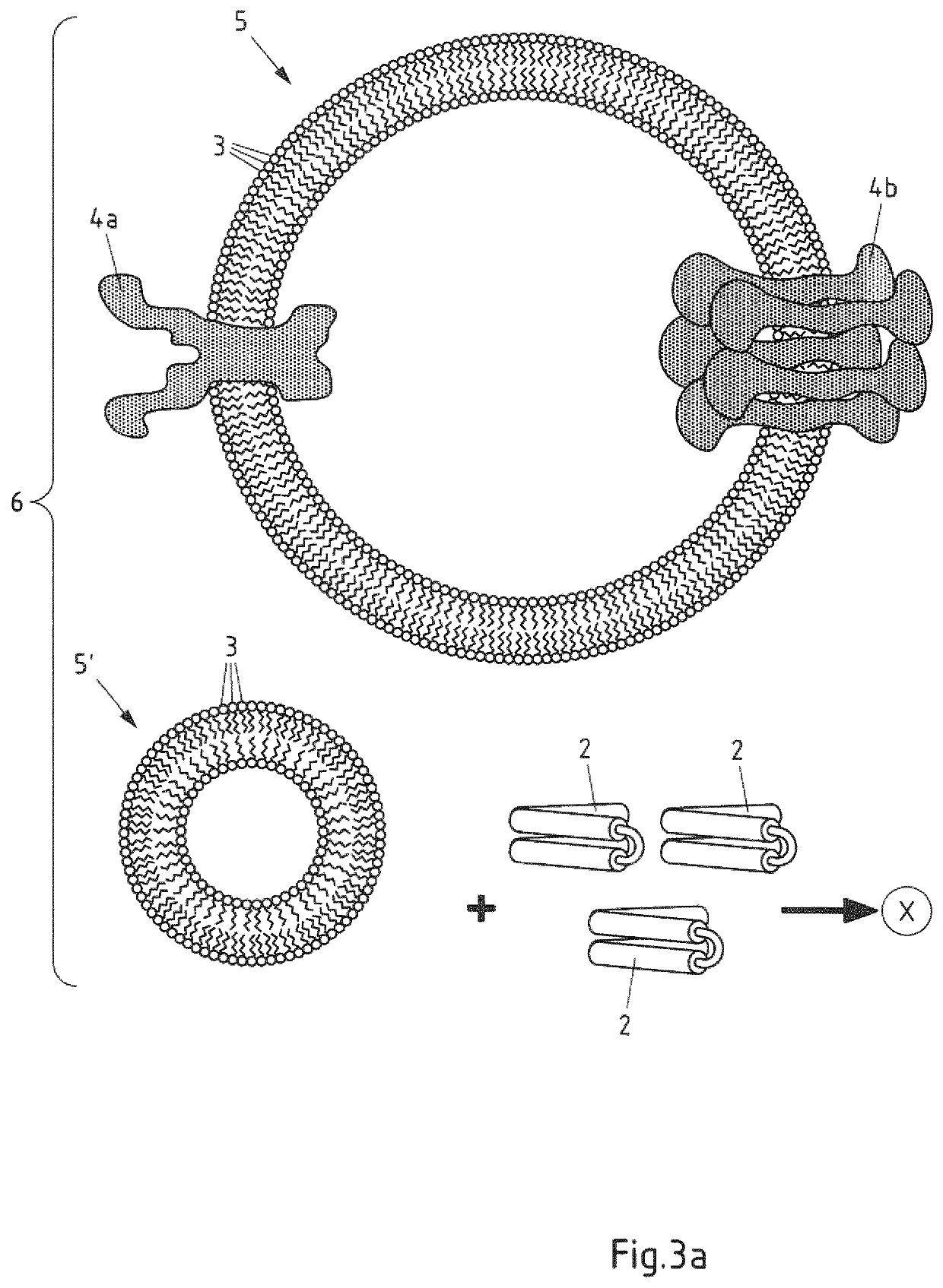
[0045]The process according to the invention has the advantage that it is relatively easy to conduct and surprisingly yields libraries of Salipro particles that reflect the diversity and complexity of the crude membrane used as starting material and incorporate and preserve the lipid and membrane proteins components contained therein in their natural membrane environment. A wide variety of membranes can be used in the process according to the invention. The crude membrane vesicles used in step a) are prepared from crude membranes of cell or organelles. Crude membrane vesicles obtained from cell and / or organelle membranes of many organisms can be used. It was surprising that Salipro particles could be obtained also when complex membranes of eukaryotic, prokaryotic or archaeal cells were employed as starting material instead of the highly purified, detergent-solubilized starting materials required in the prior art preparation methods.
[0240]In another aspect, the particles of the invention are useful as vaccination formulation, as carrier thereof or as drug delivery vehicle. Many pathogenic antigens that could be particularly potent in vaccinations are exposed on the surface and / or comprised in the outer cell membrane of eukaryotic or prokaryotic pathogens or disease cells (e.g. cancer cells) in a patient. These antigens can, e.g., be derived from pathogenic lipids, other hydrophobic biomolecules or membrane proteins. The term pathogen as used herein does not comprise viruses. With the particles of the invention, such antigens can effectively be incorporated into the particles which then can be used as antigen-presenting delivery vehicles in vaccination formulations. Along these lines, the particles of the invention are also useful to serve as antigen-presenting delivery vehicles for generating antibodies against lipids or membrane proteins in suitable host animals, preferably in mammals such as e.g. rabbits, goats, lamas, mice and primates.

Problems solved by technology

Unfortunately, membranes (and the membrane proteins and lipids contained therein) are not soluble in the detergent-free systems that are usually used in drug screening procedures and most functional assays employed in life science research.
The latter may be useful for protein or lipid identification techniques, such as mass spectrometry, but is incompatible with functional analysis of membrane proteins or lipids in their natural environment, an underlying requirement for successful research and drug development in this area.
These difficulties in purification, isolation and in functional analysis have delayed research on membrane lipidomes and membrane proteomes.
As described above, the preparation and subsequent use of membrane proteome libraries has thus far been hampered by the insolubility of membranes and membrane components in detergent free systems.
Interactions between membrane protein targets and soluble ligands are difficult to study in vitro due to the insolubility of membrane proteins in non-detergent systems.
Hydrophobic compounds such as membrane proteins and lipids are notoriously difficult to handle and represent two of the major challenges for pharmaceutical or life-science research, and applications: (i) rendering insoluble hydrophobic compounds such as lipids or membrane proteins soluble in aqueous solutions and (ii) the handling and administration of such hydrophobic matter as therapeutic, research or diagnostic agents.
Yet, membrane proteins are difficult to study due to their insolubility and tendency to aggregate when extracted from their natural lipid bilayer environment.
Here, detergent micelles are most commonly employed which may, however, negatively impact on biocompatibility, can have adverse affects on membrane protein activity and may interfere with experimental conditions for assays.
Another major pharmacological challenge is represented by the administration and delivery of hydrophobic proteins and / or lipids as therapeutic or diagnostic agents.
Due to the limited solubility of these hydrophobic agents, they are prone to aggregation, leading to locally highly concentrated drug particles that may cause high toxicity, unwanted immune responses and render the drug inactive (Allen and Cullis, SCIENCE, 303 (5665): 1818-1822, Mar. 19, 2004).
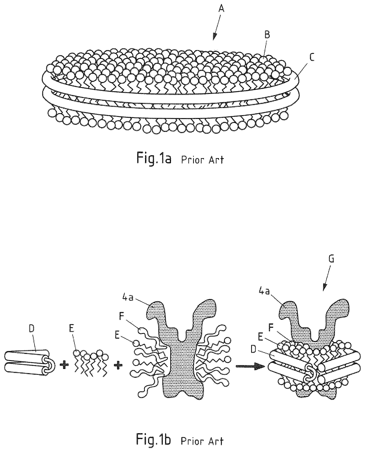
However, there are several drawbacks with this currently available nanodisc technology in that, for example, a removal of detergent is required during assembly of the particles.
Moreover, the size homogeneity provided by the tight double-belt like fit of the apolipoprotein-derived MSP seems to go at the expense of a fixed minimum particle size and a limitation as to the maximum diameters obtainable with the methods of the prior art.
Hence, the technology of this publication is limited to a pH value of 4.75, to the aforementioned size of the particles, and includes a laborious upstream liposome preparation step.
However, due to the 3.2 nm size limitation of the saposin A derived particles reported in Popovic et al., it seems that—if at all—only small molecules could be incorporated into such particles at the acidic pH disclosed therein.
In addition, the 10 nm Apolipoprotein A derived nanodiscs may be too large for certain applications.
Furthermore, the methods described in all of the above apolipoprotein- or Saposin-related prior art are sophisticated in their level of experimental detail and rely on well-defined systems of purified, detergent-solubilized components.
Thus, they are not expected to work when directly employing crude membranes as starting material, which are characterized by a highly complex structure and composition.
If one first were to purify and solubilize the membrane lipidome and proteome from a cell or organelle membrane, one would, however, remove the membrane proteins and lipids from their natural environment and context which can lead to a loss of function, loss of content, complexity and corresponding bias in the library's composition.
In addition, such process would be complicated and elaborate because not all membrane proteins and membrane lipids have the same requirements regarding handling, detergent solubilization and stability.
This makes investigation of the natural occurring membrane environment impossible.
In addition, these known procedures carry the risk of denaturation of the protein or destabilization of lipids and lipid domains during purification and extraction.
Moreover, it cannot be excluded that the proteins and / or lipids loose their native structure and function when divorced from its natural environment.
The handling and incorporation of such complex membranes without the detergent solubilization and purification steps taught in the prior art methods appears nearly impossible to conduct as the complex natural membrane structures are not expected to be as easy to handle as purified components.
Taken together, the membranes of prokaryotic, archaeal and eukaryotic organisms show a high degree of complexity, that arises from the intricate interplay between membrane proteins and a variety of lipids surrounding them in their native context.
The prior art does not contemplate, and certainly does not teach or suggest, a feasible proposal for preparing native state membrane proteome and lipidome libraries in the form of Saposin-derived particles.

Method used


Image

  • Saposin lipoprotein particles and libraries from crude membranes
  • Saposin lipoprotein particles and libraries from crude membranes
  • Saposin lipoprotein particles and libraries from crude membranes

Examples


example 1a

[0292]Crude yeast cell membrane fractions were obtained from GFP-GLUT5 expressing yeast cells. The membrane fraction, which contained spontaneously formed crude membrane vesicles, was incubated with detergent and Saposin A, followed by removal of detergent-micelles using gel-filtration chromatography / size-exclusion chromatography (SEC) in detergent-free buffer. This lead to the self-assembly of the membrane components present in the initial mixture into a library of nanoscale Salipro particles. In particular, monodisperse Salipro particles comprising membrane lipids and GFP-GLUT5 could be identified within this library.

[0293]1. Membrane Preparation

[0294]Crude yeast membranes were obtained from yeast cells expressing rat GLUT5 from a GAL1 inducible TEV cleavable GFP-His8 2μ vector pDDGFP2 known in the prior art. The vector was transformed into the S. cerevisiae strain FGY217 (MATa, ura3-52, lys2Δ201, and pep4Δ) which then overexpressed GFP-GLUT5 in its cell membrane.

[0295]To generate...

example 1b

[0303]1. Setup

[0304]Crude yeast membranes containing crude membrane vesicles and rat GLUT5 were prepared as described in example 1a.

[0305]2. Variation of the Process for Salipro Particle Library Formation.

[0306]Two different approaches to make Salipro-GFP-GLUT5 particles from crude membrane extract were evaluated.

[0307]For the first approach, 25 μl of saposin A (0.75 mg / ml) were added to 1 μl crude membrane extract containing crude membrane vesicles and GFP-GLUT5 and incubated for 5 min at 37° C. Thereafter 24 μl HN-D buffer was added to the mix and incubated for an additional 5 min at 37° C. The sample was then centrifuged for 10 min at 13 krpm and SEC analysis was performed as in example 1a.

[0308]For the second approach, 1 ul crude membrane extract containing crude membrane vesicles and GFP-GLUT5 were first supplemented by adding 24 ul HN-D buffer, followed by incubation at 37° C. for 5 min. The sample was then centrifuged for 10 min at 13 krpm. The lysate suspension was collected...

example 2

[0311]1. Setup

[0312]Crude yeast membranes containing rat GFP-GLUT5 and crude membrane vesicles were prepared as described in example 1a.

[0313]2. Salipro Formation, Titration

[0314]20 μl of yeast crude membranes were mixed and solubilized with 60 μl HN buffer and 20 μl HN buffer supplemented with 5% DDM at 4° C. for 1 h. The membrane lysate was then cleared from debris and protein aggregation by ultracentrifugation using a TLA-55 rotor at 47 krpm (100,000 g) for 30 min. 5 μl of the cleared membrane lysate, which contained crude membrane vesicles, was then mixed with different volumes (12, 20, 30 and 40 μl) of saposin A (4 mg / ml, HN buffer) and incubated 5 min at 37° C. Thereafter the sample volumes were adjusted to 50 μl with HN buffer and centrifuged 10 min at 13 krpm. SEC analysis was performed (using a Shimadzu HPLC system) and 35 μl sample was injected to a 5 / 150 Superdex 200 increase column (GE healthcare) with a flow rate at 0.3 ml / min and the presence of the fluorescent GFP tag...


PUM

PropertyMeasurementUnit
diameteraaaaaaaaaa
diameteraaaaaaaaaa
diameteraaaaaaaaaa

Abstract

The invention is directed to a process for preparing a library of saposin lipoprotein particles, wherein the particles comprise membrane components from a cell or an organelle membrane and a lipid binding polypeptide that is a saposin-like protein belonging to the SAPLIP family of lipid interacting proteins or a derivative form thereof, wherein the process comprises the steps of a) providing a mixture of crude membrane vesicles obtained from a cell or an organelle membrane; b) contacting the mixture of step a) with the lipid binding polypeptide in a liquid environment; and c) allowing for self-assembly of the particles. The invention also provides a process for preparing a purified saposin lipoprotein particle comprising the steps of preparing a library according to the process described above and the additional step of f) purifying the saposin lipoprotein particle from the library. In addition, the invention provides a library of saposin lipoprotein particles and saposin lipoprotein particles obtainable according to the processes of the invention. These can be used in medicine, in particular in preventing, treating or lessening the severity of a disease or for use in a diagnostic method, a cosmetic treatment or for use as vaccination formulation or as a tool for drug development, drug screening, drug discovery, antibody development, development of therapeutic biologies, for membrane or membrane protein purification, for membrane protein expression, for membrane and / or membrane protein research, in particular lipidomics and proteomics, preferably for the isolation, identification and / or study of membranes and / or membrane proteins or creation of a lipidome or proteome database.

Description

CROSS-REFERENCE TO RELATED APPLICATIONS[0001]This application claims the benefit of and priority under 35 U.S.C. § 371 to International Patent Cooperation Treaty Application No. PCT / EP2017 / 071043, filed Aug. 21, 2017, which claims the benefit of priority to European Patent Application No. 16184843.7, filed Aug. 19, 2016, the entire contents of both disclosures are incorporated herein by reference in their entireties.[0002]The invention relates to processes for preparing a library of lipoprotein particles from cell or organelle membranes and purifying lipoprotein particles therefrom as well as to a library of lipoprotein particles and purified lipoprotein particles themselves and their uses.BACKGROUND OF THE INVENTION[0003]In the last decade, research in the life sciences, and drug discovery in particular, has been rapidly changing. Advances in molecular biology, computing, genomics, proteomics and lipidomics, permitted to develop novel methods for research and drug discovery.[0004]I...

Claims


Application Information

Patent Timeline
Patent Type & Authority Patents(United States)
IPC IPC(8): A61K9/127G01N33/68A61K38/18A61K39/00A61K47/46A61K9/50A61K47/42
CPCG01N33/6842A61K9/127A61K38/18A61K39/0012A61K9/5068A61K9/5089A61K47/42A61K47/46G01N2570/00
Inventor FRAUENFELD, JENSLÖVING, ROBIN
Owner SALIPRO BIOTECH AB
Why Eureka
Try Eureka