Method of detecting single gene copies in-situ

a single gene and copy technology, applied in the field of nucleic acid detection and localization techniques, can solve the problems of radioactive toxicity and environmental concerns, other viral diseases are also frequently difficult to detect or distinguish clinically, and the signal generated by fluorescent markers is typically faded

Inactive Publication Date: 2002-02-14
VENTANA MEDICAL SYST INC
View PDF9 Cites 120 Cited by
  • Summary
  • Abstract
  • Description
  • Claims
  • Application Information

AI Technical Summary

Problems solved by technology

Other viral diseases are also frequently difficult to detect or distinguish clinically.
Although radioactive labels are effective, they are associated with radioactive toxicity and environmental concerns.
Unfortunately, the signal generated by fluorescent markers typically fades over time.
Additionally, the cost and availability of fluorescent microscopy equipment and trained personnel is greater than conventional brightfield microscopy.
However, the sensitivity required for detecting such multiple copy sequences is much lower than the sensitivity required for detection of unique copy sequences.
However, none of these were able to distinguish single gene copies in-situ.
While such has been d...

Method used


Image

  • Method of detecting single gene copies in-situ
  • Method of detecting single gene copies in-situ
  • Method of detecting single gene copies in-situ

Examples


example 2

Detection of Single Gene Copies

[0077] The slides produced in Example 1 were mounted with Permount (Fisher Scientific, NJ) and coverslipped.

[0078] Using brightfield microscopy, microphotographs were made on Kodak Ektachrome color slide film (EL 400), using a Zeiss Axiophot 20 epi-fluorescence microscope (Zeiss, West Germany) equipped with a 100X Plan-Neofluar oil immersion objective or a 100X Plan-APO oil immersion objection (Zeiss, West Germany), a Zeiss MC-100 camera (Zeiss, West Germany), and a blue or neutral density filter.

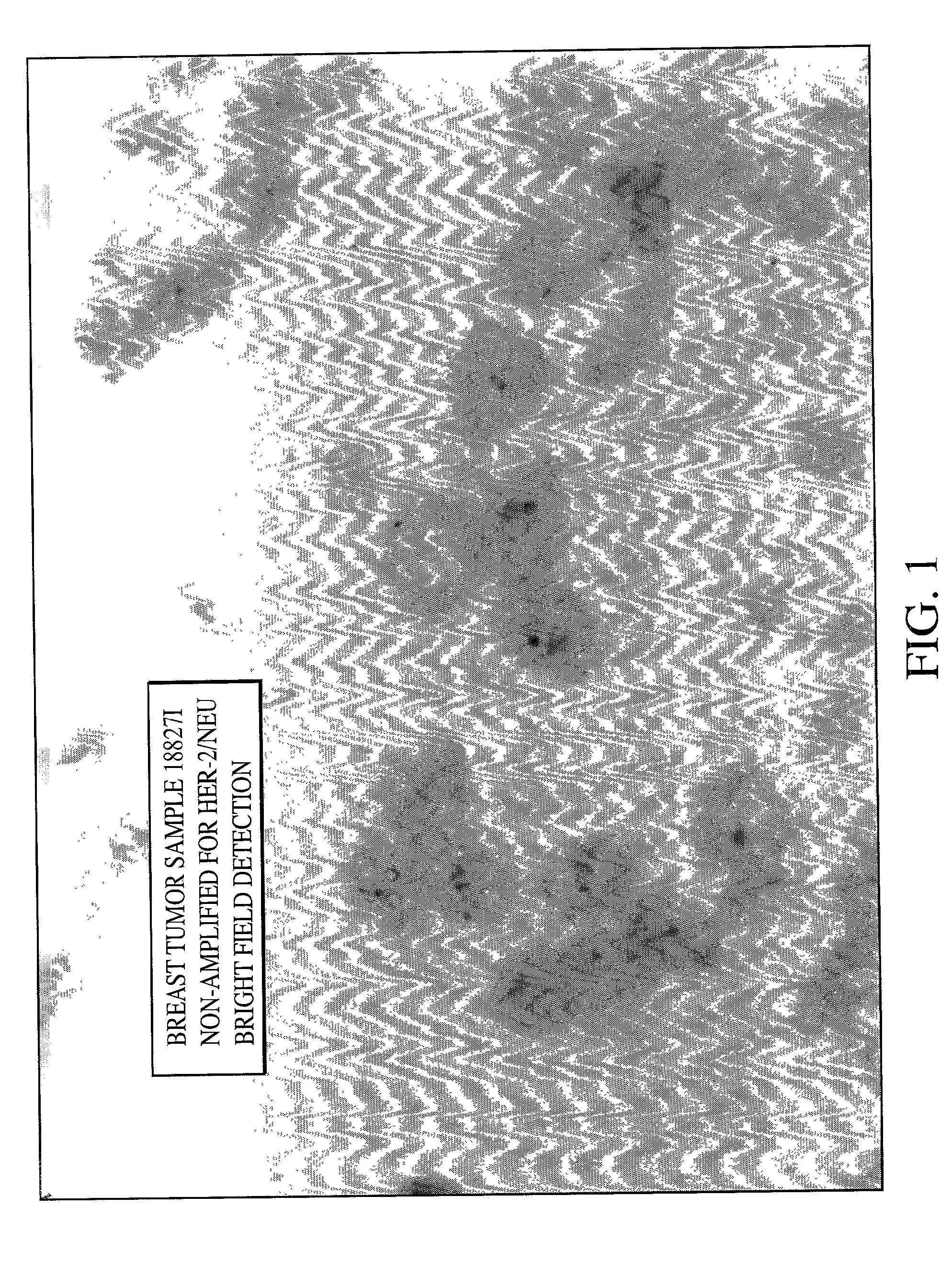
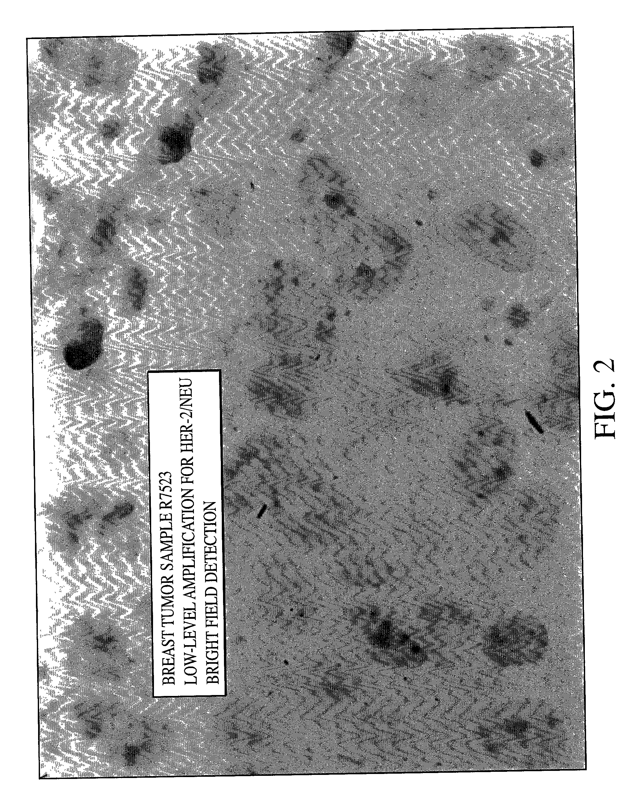
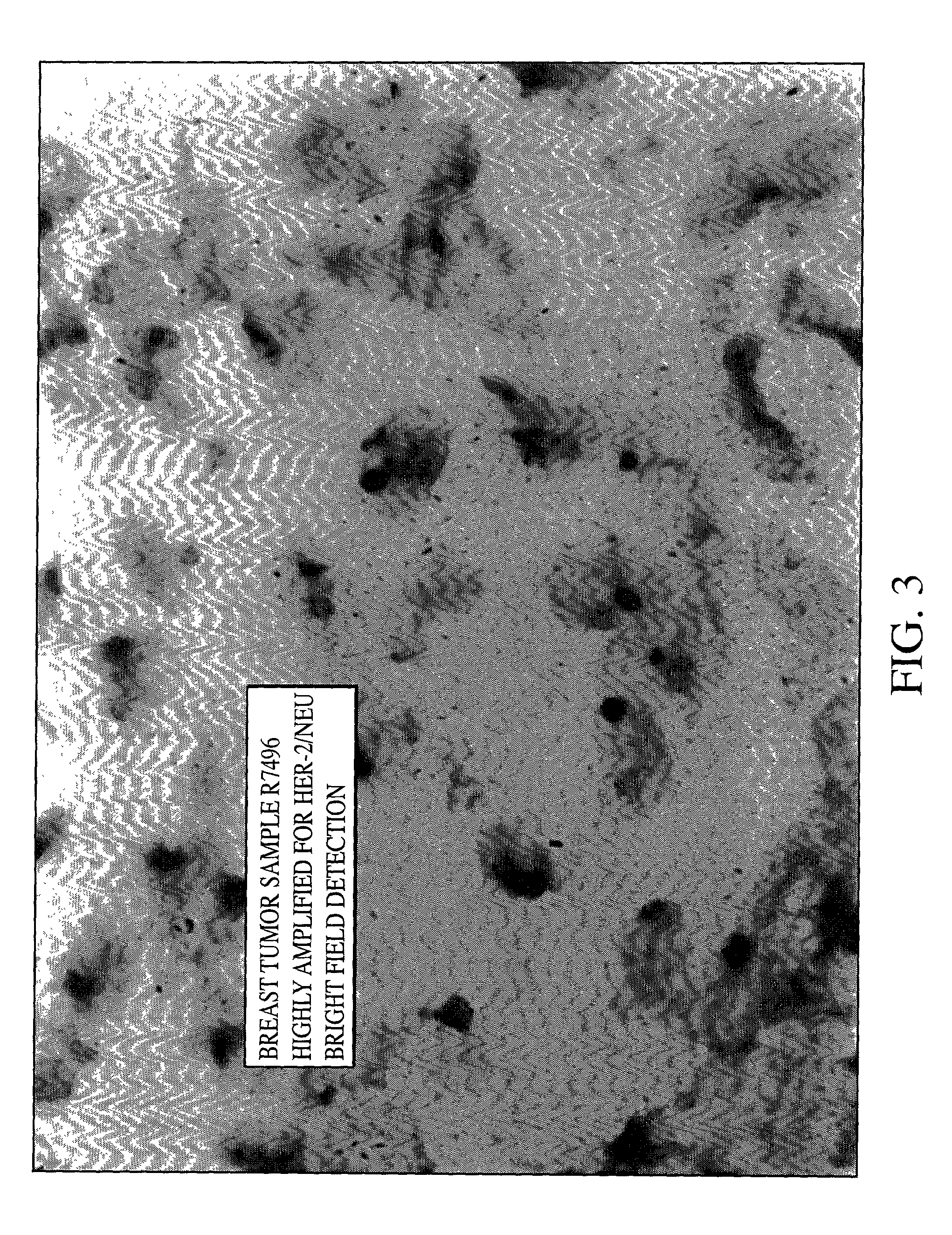
[0079] As can be seen from FIGS. 1, 2 and 3 single blue signal was visible at the location of each copy of the HER-2 / neu gene. The expected results for visualization of the HER-2 / neu gene in normal metaphase chromosomes from the MDA-MB468 breast tumor cell line is two; one on each chromatin, and the number of the blue signals in normal interphase nuclei is also two, or possibly two pairs if the cell is in G2 phase. In the cultured breast tumor cell lines MDA-M...

example 3

Detection of High Risk Human Papilloma Virus in Gynecologic Tissue Specimen

[0080] Six separate commercially available plasmids, i.e., pGem2, pUC13, pGem1, pLINK322, pGEM1 and pUCI3 containing entire genomes of HPV types 16 (DNA sequence available from GenBank, Accession No. K02718), 18 (DNA sequence available from GenBank, Accession No. X05015), 31 (DNA sequence available from GenBank, Accession No. J04353), 33 (DNA sequence available from GenBank, Accession No. M12732), 35 (DNA sequence available from GenBank, Accession No. M74117) and 51 (DNA sequence available from GenBank, Accession No. M62877) respectively, were labeled by nick translation with digoxigenin dCTP. Alternatively, one may clone the HPV into a plasmid by standard molecular biology techniques within the skill of the art. The labeled plasmids were then mixed together to form a single reagent. Incorporation of the digoxigenin nucleotide into the labeled DNA was verified by dot-blot procedure. DNA fragment size was dete...

example 4

Automated ISH Staining and Single Gene Copy Detection

[0093] The paraffin-embedd method of Example 1 was repeated on a DISCOVERY.TM. automated slide stainer discribed in PCT / US99 / 04181 with all steps being performed automatically. Using the same reagents and conditions of Example 1, individual spots representing single gene copies were observed using the detection methods of Example 2.


PUM


Abstract

A method for detecting single copies of a gene in-situ using brightfield microscopy is used in detection of nucleic acid sequences. Probes are directly or indirectly labeled with alkaline phosphatase with NBT/BCIP used as the chromogen.

Description

BACKGROUND TO THE INVENTION[0001] 1. Field of the Invention[0002] The present invention relates to techniques for detection and localization of nucleic acid and, more particularly, to the use of enzyme catalyzed chromogenic compositions in such detection and localization.[0003] 2. Description of Related Art[0004] In-situ hybridization (ISH) techniques are an important tool for detection of nucleic acid sequences, i.e., both DNA and RNA. Unique nucleic acid sequences occupy precise positions in chromosomes, cells and tissues and in-situ hybridization allows the presence, absence and / or amplification status of such sequences to be determined without major disruption of the sequences.[0005] It is known that certain nucleic acid sequences are associated pathologic conditions in living organisms. For example, the presence of certain genes and viral nucleic acids have been implicated in precancerous and cancerous pathology. Genetic diseases are also diagnosed by determining the presence, ...

Claims


Application Information

Patent Timeline
IPC IPC(8): C12Q1/68
CPCC12Q1/6804C12Q1/6816C12Q1/6841C12Q1/6886Y10T436/143333C12Q2563/125C12Q2563/131C12Q2521/525C12Q2600/156
Inventor LIGHT, ELIZABETH S.
Owner VENTANA MEDICAL SYST INC
Why Eureka
Try Eureka