
Cytoskeletal active agents for glaucoma therapy

a cytoskeletal and active agent technology, applied in the direction of biocide, drug composition, peptide/protein ingredients, etc., can solve the problems of abnormal resistance, loss of vision, disabling vision reduction, etc., and achieve the effect of reducing intraocular pressure and facilitating fluid flow

Inactive Publication Date: 2002-04-18
YEDA RES & DEV CO LTD +1
View PDF0 Cites 36 Cited by
  • Summary
  • Abstract
  • Description
  • Claims
  • Application Information

AI Technical Summary

Benefits of technology

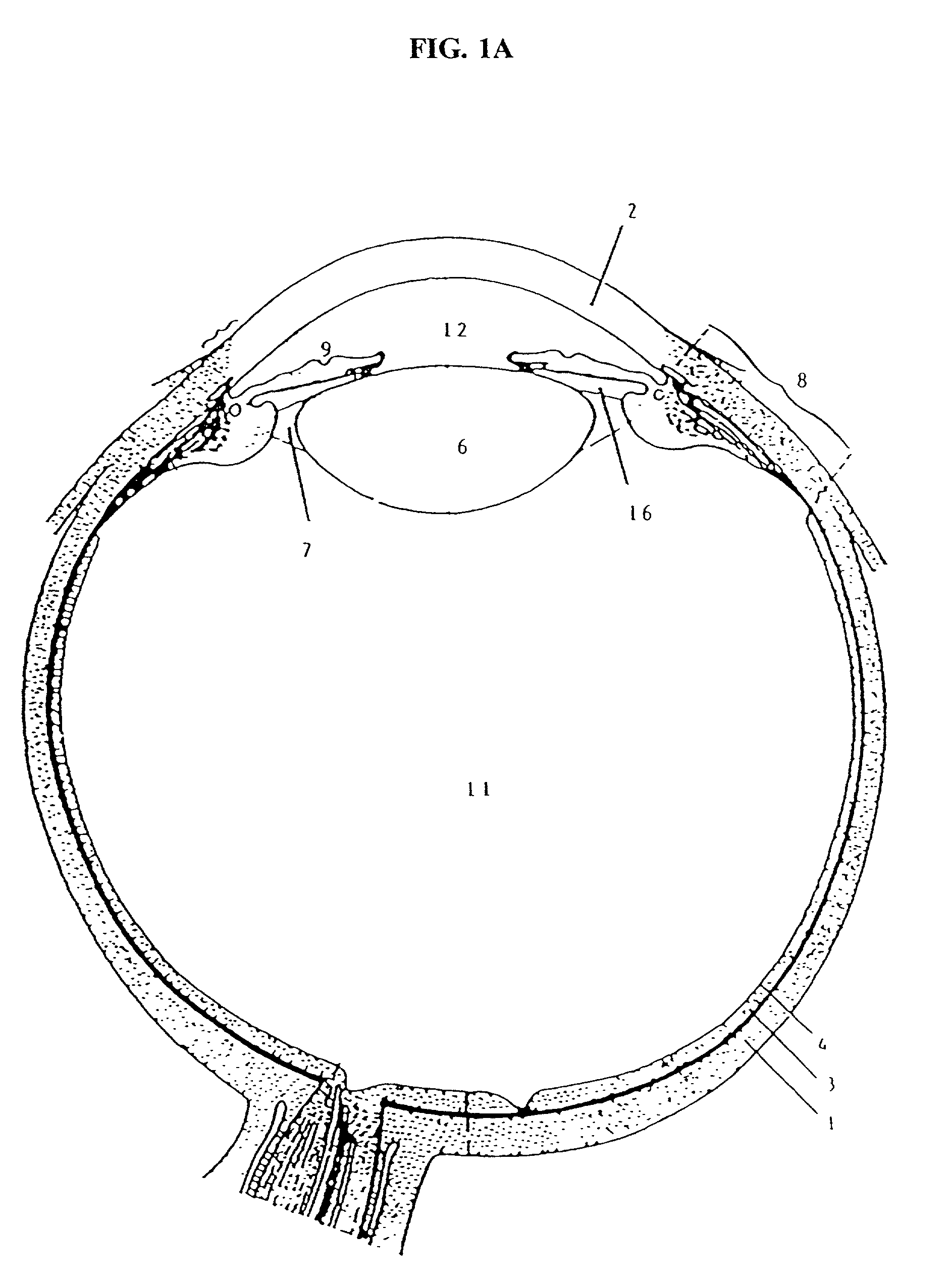
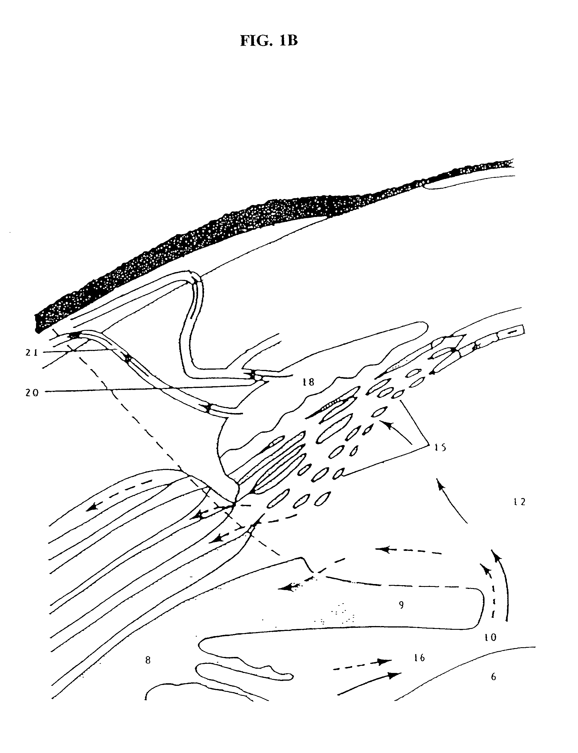
[0076] The present invention relates to the treatment of glaucoma. While the present invention does not depend on an understanding of the mechanism by which successful treatment is accomplished, it is believed that the compounds of the present invention cause transient, drug-induced relaxation of cells and / or disssociation of C-C junctions and C-ECM junctions, which facilitates fluid flow around the cells, through the trabecular meshwork, and across the Schlemm's5 canal inner wall monolayer, leading to reduction in intraocular pressure.

Problems solved by technology

Importantly, the loss of vision can result from intraocular pressures only slightly above or even within the statistically normal range, in eyes which are unusually pressure-sensitive, over a period of years.
Moreover, extremely high pressures (e.g., 70 mm Hg.) may cause blindness within only a few days (See e.g., Kaufman and Mitta, "Medical Therapy Of Glaucoma," in Kaufman and Mittag (eds
Open angle glaucoma is characterized by abnormally high resistance to fluid drainage from the eye.
The adverse effects associated with the miotic agents include painful ciliary or accommodative spasm and consequent induced fluctuating myopia and blurred vision (in patients under the age of approximately 45 years), ciliary or conjunctival congestion, ocular pain, headache, and the miosis itself, which can cause a disabling reduction of vision in elderly patients with early cataract formation (the combination of the small pupil and the cloudy lens critically reduce the amount of light reaching the retina).
The adverse ocular effects of both agents include ocular congestion or hyperemia, ocular pain and headache, and blurred vision.
In addition, epinephrine occasionally causes untoward systemic effects, such as palpitation, tachycardia, and hypertension.
Systemic cardiovascular and pulmonary adverse effects are of greater concern, are not uncommon especially in predisposed individuals (e.g., the elderly, patients with underlying cardiovascular or respiratory system disease, or those taking .beta.- adrenergic blocking agents systemically for other conditions), and may occasionally be life-threatening.
Many adverse effects have been associated with the oral carbonic anhydrase inhibitors, including gastrointestinal, central nervous system, hematologic, renal, electrolyte (especially dangerous in patients with heart disease taking digitalis and other salt-wasting diuretics concurrently), and metabolic disturbances.
Of course, the potential incidence of adverse effects increases when more than one agent is being used in a treatment regimen (See generally, McEvoy (ed.
As set forth above, untreated glaucoma can result in severe consequences, including blindness.
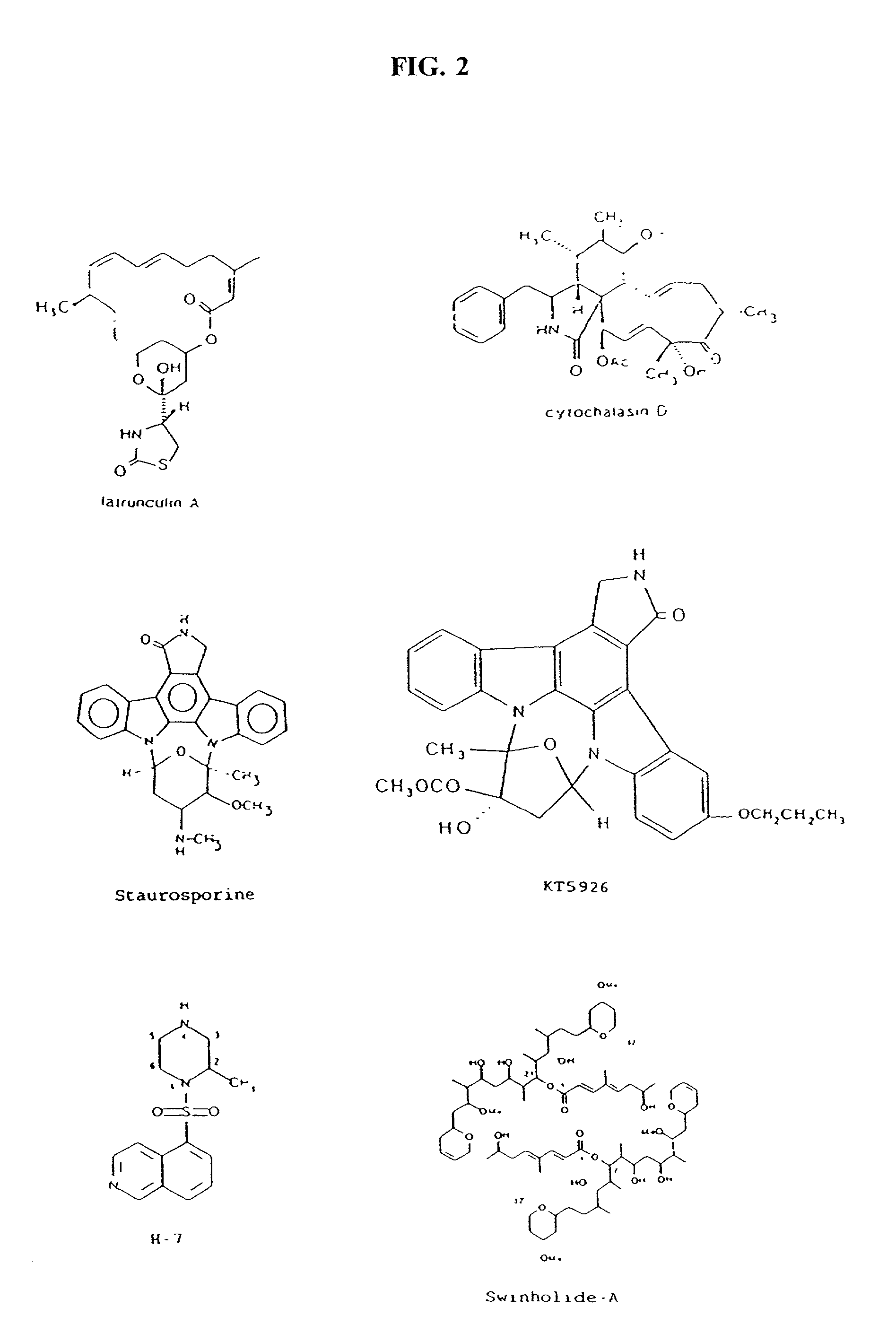
In some embodiments, the administration of latrunculin(s) (including but not limited to latrunculin-A) results in the binding of monomeric (G-) actin, which inhibits polymerization and leads to disruption of the actin networks.
These compounds primarily inhibit myosin light- chain kinase, thus inhibiting actomyosin-driven contractility; upon long exposure (>30 minutes), they cause deterioration of the entire microfilament system.
More specifically, compounds which interfere with the formation of the actin microfilaments, promote disruption of the filaments, interfere with actin assembly into bundles, or disturb the relationship between actin and myosin or the plaque will disrupt cell junctions and cause cells to pull apart from each other or from the extracellular matrix.

Method used


Image

  • Cytoskeletal active agents for glaucoma therapy
  • Cytoskeletal active agents for glaucoma therapy
  • Cytoskeletal active agents for glaucoma therapy

Examples


example 1

Dose-Dependent Affect Of H-7 On Total Outflow Facility Occurs Through A Direct Effect On The Trabecular Meshwork

[0246] This Example describes experiments to examine the dose-dependent effect of H-7 on total outflow facility, as measured by 2-level constant pressure perfusion of the anterior chamber (described above) of both eyes simultaneously with Brny's solution.

[0247] Initially, it should be noted that the protocols for the testing of H-7 (and some of the other compounds, described below) were as follows: (i) after determination of vehicle-only baselines, one eye received drug and the other eye received vehicle without drug; (ii) analogous to the first protocol, with the exception that after the post-drug determination, both eyes received vehicle without drug to determine reversibility of the drug effect; (iii) in monkeys with unilateral disinsertion of the ciliary muscle, both eyes received drug after determination of vehicle-only baseline; (iv) drug-free baseline facility was a...

example 2

H-7 Does Not Significantly Effect Corneal Endothelial Cell Counts At A Concentration Effective At Increasing Total Outflow Facility

[0254] In this Example, the effect of 300 &82 m H-7 on corneal endothelium was determined in cynomolgus monkeys via transcorneal intracameral injection.

[0255] The basic experimental protocol was as follows. Following administration of ketamine (10 mg / kg) anesthesia, slit lamp examination was performed to provide a baseline evaluation of the cornea, lens, and anterior chamber. Thereafter, the monkeys were anesthetized with IM pentobarbital-sodium (35 mg / kg). A 3 mM H-7 solution (2.1858 mg H-7 dissolved in 2 ml Brny's solution, pH adjusted to 6-7) was manufactured, and 10 .mu.l of that solution was administered to one eye and 10 til of Brny's solution to the opposite eye (keeping the needle away from the area to be photographed during the transcorneal intracameral injection). Specular micrographs were taken at 1 and 3 hours after H-7 administration to dete...

example 3

The Effect Of Topical Administration Of H-7 On Outflow Facility In Cynomolgus Monkeys

[0259] Generally speaking, agents used in the treatment of glaucoma are administered topically as an eyedrop, rather than being injected into the eye. This Example describes the results on outflow facility following the topical administration (i.e., via an eyedrop) of four different concentrations of H-7.

[0260] Following administration of ketamine (10 mg / kg) anesthesia, slit lamp examination was performed on the monkeys. Thereafter, the monkeys were anesthetized with IM pentobarbital-sodium (35 mg / kg). Baseline facility was measured (7 periods) by 2-level constant pressure perfusion of the anterior chamber; thereafter, the perfusion system was closed.

[0261] Four H-7 solutions of differing concentrations were manufactured: (i) a 90 mM solution (0.3 mg H7 dissolved in 10 .mu.l of 10% DMSO with Brny's solution, pH of the resulting solution adjusted to approximately 7); (ii) a 150 mM solution (1.1 mg H7...


PUM

PropertyMeasurementUnit
intraocular pressuresaaaaaaaaaa

Abstract

Methods for the treatment of glaucoma are provided by the present invention. The compounds described cause a perturbation of the actin cytoskeleton in the trabecular meshwork or the modulation of its interactions with the underlying membrane. Perturbation of the cytoskeleton and the associated adhesions reduces the resistance of the trabecular meshwork to fluid flow and thereby reduces intraocular pressure.

Description

[0001] The present application is a Continuation-in-Part of U.S. patent application Ser. No. 09 / 621,124, filed Jul. 21, 2000, which is a Continuation of U.S. patent application Ser. No. 09 / 022,228, filed on Feb. 11, 1998, which is a Continuation-In-Part of U.S. patent application Ser. No. 08 / 604,568, filed 02 / 21 / 96, which is now U.S. Pat. No. 5,798,380, which issued on Aug. 25, 1998.[0003] The present invention relates to the treatment of ocular disorders of the eye and more particularly to the treatment of glaucoma.[0004] I. Description And Characterization Of Glaucoma[0005] Glaucoma is an ophthalmologic disorder responsible for visual impairment. It is the fourth most common cause of blindness and the second most common cause of visual loss in the United States, and the most common cause of irreversible visual loss among African-Americans. Generally speaking, the disease is characterized by a progressive neuropathy caused at least in part by deleterious effects resulting from incr...

Claims


Application Information

Patent Timeline
Patent Type & Authority Applications(United States)
IPC IPC(8): A61K31/00A61K31/365A61K31/403A61K31/407A61K31/427A61K31/496A61K31/551A61K45/06
CPCA61K31/00A61K31/365A61K31/403A61K45/06A61K31/427A61K31/496A61K31/551A61K31/407
Inventor KAUFMAN, PAUL L.GEIGER, BENJAMIN
Owner YEDA RES & DEV CO LTD
Learn More