Magnetic resonance imaging methods and compositions

a magnetic resonance imaging and composition technology, applied in the field of magnetic resonance imaging (mri), can solve the problems of coarse spatial resolution, sensitivity reduction, and low spatial resolution achieve the effect of improving the accuracy of .sup.23na mri images, reducing the cost of mri imaging, and improving the accuracy of mri imaging

Inactive Publication Date: 2003-06-26
THE JOHN HOPKINS UNIV SCHOOL OF MEDICINE
View PDF8 Cites 28 Cited by
  • Summary
  • Abstract
  • Description
  • Claims
  • Application Information

AI Technical Summary

Problems solved by technology

Clinical applications of .sup.23Na MRI, however, are limited by the low in vivo concentrations and NMR sensitivity of endogenous sodium, which translates to coarse spatial resolution and lower sensitivity compared to the .sup.1H MRI.
In the heart, the problem of low spatial resolution of .sup.23Na MRI images is further compromised by the high sodium content of ventricular blood which hinders differentiation of the ventricular wall from the ventricular cavity, and hence the differentiation of elevated sodium in myocardial infarction (MI).
While these methods can attenuate ventricular sodium blood signals, these methods are associated with increased echo times (TE) or the need for cardiac gating, leading to detrimental signal-to-noise ratio (SNR) losses and significant prolongation of the total image acquisition times. Longer TE times lead to increased susceptibility induced artifacts and decreased SNR values in .sup.23Na images.
An additional disadvantage with these existing .sup.1H MRI imaging methods is that the close proximity of the longitudinal (T.sub.1), fast (T.sub.2,) and slow (T.sub.2f) sodium transverse relaxation times of blood and myocardium preclude the use of conventional inversion recovery or spin-echo techniques to attenuate or null ventricular blood signals.

Method used


Image

  • Magnetic resonance imaging methods and compositions
  • Magnetic resonance imaging methods and compositions
  • Magnetic resonance imaging methods and compositions

Examples


Embodiment Construction

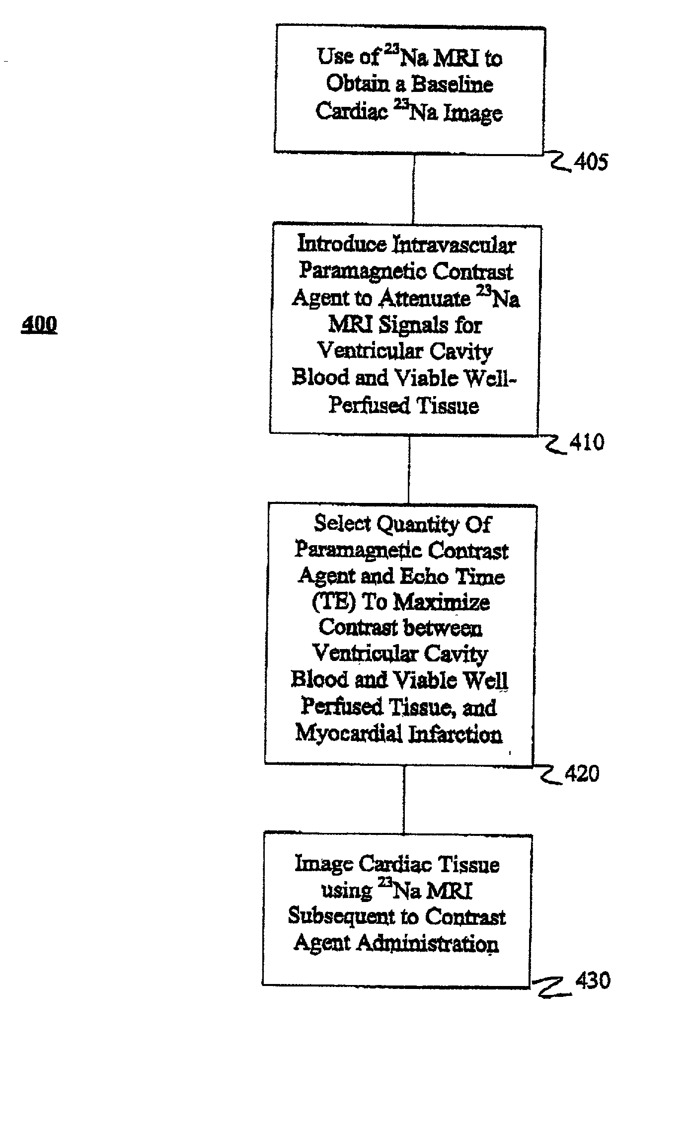
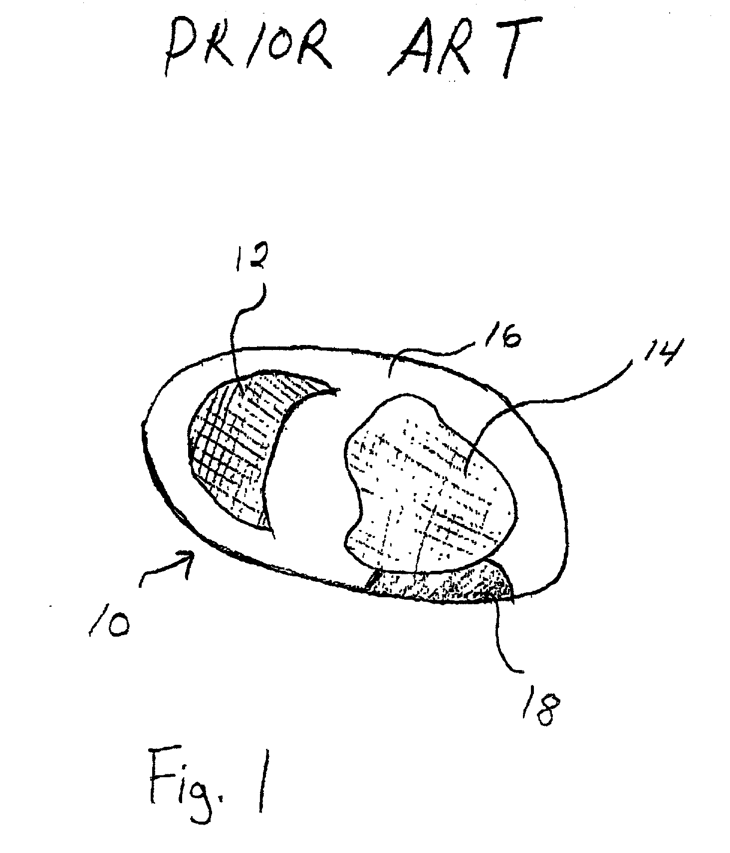
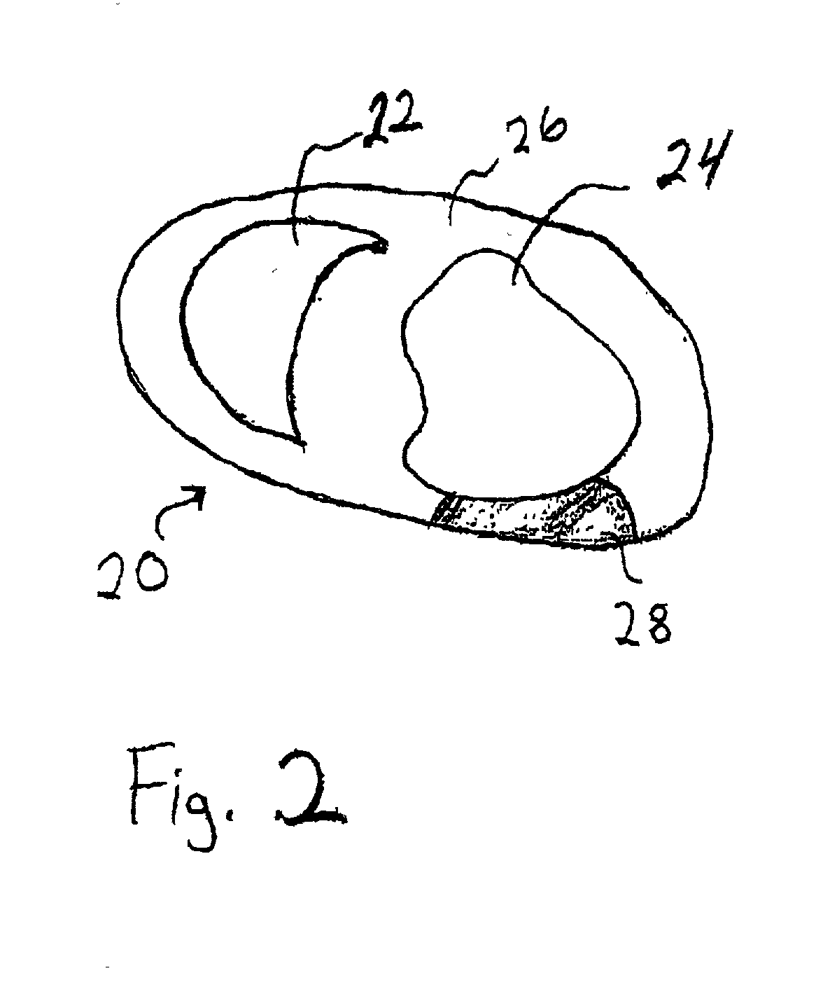
[0062] Experiments were performed and data were collected employing the present method in a canine model of myocardial infarction. Specifically, the experiments were directed to evaluating variation in the blood T.sub.1 and T.sub.2 relaxation times with increasing MION-46 amounts and determining the optimal dose for contrast-enhanced .sup.23Na cardiac MRI in normal canine hearts in vivo.

[0063] All MRI studies were performed with a commercially available 1.5T MRI system (Signa, Horizon, Echo Speed, 5.7 Epic platform, GE Medical Systems, Milwaukee, Wis.) equipped with broadband spectroscopy capabilities, using a 16-pole quadrature .sup.23Na birdcage coil tuned to 16.89 MHz and interfaced with a quadrature hybrid splitter. .sup.23Na MRI was performed with a three-dimensional (3D) twisted projection imaging (TPI) sequence using conventional scanner hardware with gradient having a maximum amplitude of 2.2 mT / m, a maximum slew rate of 12 mT / cm / s, and with a slew rate duty cycle limit of 2...


PUM

PropertyMeasurementUnit
echo timeaaaaaaaaaa
core sizeaaaaaaaaaa
diameteraaaaaaaaaa

Abstract

Methods and compositions are provided for magnetic resonance imaging of biological tissue, particularly methods and compositions for 23Na and 39K magnetic resonance imaging of cardiac tissue. In particular, methods of 23Na magnetic resonance imaging (MRI) cardiac imaging is presented for attenuating 23Na signals corresponding to ventricular cavity blood and viable well-perfused tissue and for visualizing myocardial infarction. Cardiac tissue is imaged using 23Na MRI after the introduction of an intravascular paramagnetic contrast agent. Optimally, the intravascular paramagnetic contrast agent is MION-46. The MION-46 suppresses the blood signal intensity in sodium images of ventricular cavities and signal for viable cardiac tissue. The quantity of MION-46 and the echo time (TE) for 23Na MRI of cardiac tissue may be selected to minimize signal intensity differences between ventricular cavity blood and well-perfused viable myocardium; maximize signal intensity differences between non-viable myocardium and ventricular cavity blood in myocardial infarction; and maximize signal intensity differences between non-viable myocardium and well-perfused viable myocardium in myocardial infarction.

Description

[0001] The present application claims the benefit of U.S. provisional application No. 60 / 260,524 filed Jan. 10, 2001, which is incorporated by referenced in its entirety.[0003] 1. Field of the Invention.[0004] The present invention relates to methods of magnetic resonance imaging (MRI), including use of new contrast agents. Methods and compositions of the invention are especially useful for imaging of cardiac tissue and, in particular, to the use of a paramagnetic contrast agent to decrease contrast and thereby allowing delineation between blood cavities (i.e., atrial and ventricular) and viable cardiac tissue, and non-viable, infarcted cardiac tissue.[0005] 2. Background of the Invention.[0006] Certain intravascular contrast agents have been used in proton (.sup.1H) magnetic resonance imaging (MRI) of the cardiovascular system. In addition, certain iron oxide particles have been reported for use as MRI contrast agents for imaging the liver and spleen.[0007] Sodium (.sup.23Na) magne...

Claims


Application Information

Patent Timeline
Patent Type & Authority Applications(United States)
IPC IPC(8): A61K49/10G01R33/28
CPCA61K49/126G01R33/5601B82Y5/00A61K49/1863
Inventor CONSTANTINIDES, CHRIS D.
Owner THE JOHN HOPKINS UNIV SCHOOL OF MEDICINE
Why Eureka
Try Eureka