
Treatment of substrates for immobilizing biomolecules

a biomolecule and substrate technology, applied in biomass after-treatment, instruments, coatings, etc., can solve the problems of poor spot morphology and fluorescent background that is often both relatively high and non-uniform, and the fluorescence detection sensitivity is severely compromised, and achieves good stability, reproducibility, and low background noise.

Inactive Publication Date: 2004-06-24
CORNING INC
View PDF3 Cites 0 Cited by
  • Summary
  • Abstract
  • Description
  • Claims
  • Application Information

AI Technical Summary

Benefits of technology

[0020] The present invention provides a simplified and reproducible method of providing substrates for immobilizing biomolecules. Experimentation has indicated that the substrates produced according to the invention have good stability, reproducibility and exhibit low background noise. Additional features and advantages of the invention will be set forth in the following description. It is to be understood that both the foregoing general description and the following detailed description are exemplary and are intended to provide further explanation of the invention as claimed.

Problems solved by technology

Poor quality slides result in low nucleic acid binding efficiency, poor spot morphology and fluorescent background that is often both relatively high and non-uniform.
Fluorescence detection sensitivity is severely compromised by background signals, which may originate from endogenous sample constituents / surface to which the target is immobilized or from nonspecific hybridization of probes to the target.
The intrinsic auto-fluorescence of the arrays obscures the sensitivity of gene expression analysis to a large extent by hindering the detectability of the low-level specific fluorescent signals.
Attempts to diminish or eliminate auto-fluorescence by selecting filters that reduce the transmission of emission relative to excitation wavelength or by selecting filters that absorb and emit at longer wavelengths are partially successful, but still have limitations.
Although narrowing the fluorescence detection bandwidth increases the resolution, it also compromises the overall fluorescence intensity detected.
In addition, during storage or printing, adsorption and oxidation of some biological or chemical contaminants, could result in the emission of fluorescence.
However, this is very difficult to achieve.
A drawback of this approach is that this method may take long time and may end up either losing DNA targets partially or incomplete washing off the contaminants.
Furthermore, methods used in the elimination of autofluorescence problems in bulk tissue samples would not be expected to be useful in the elimination of autofluorescense on substrates used for microarrays.
However, applicants experiments have found that curing of the slides in an oven was largely ineffective in reducing auto-fluorescence.

Method used


Image

  • Treatment of substrates for immobilizing biomolecules
  • Treatment of substrates for immobilizing biomolecules
  • Treatment of substrates for immobilizing biomolecules

Examples


example 1

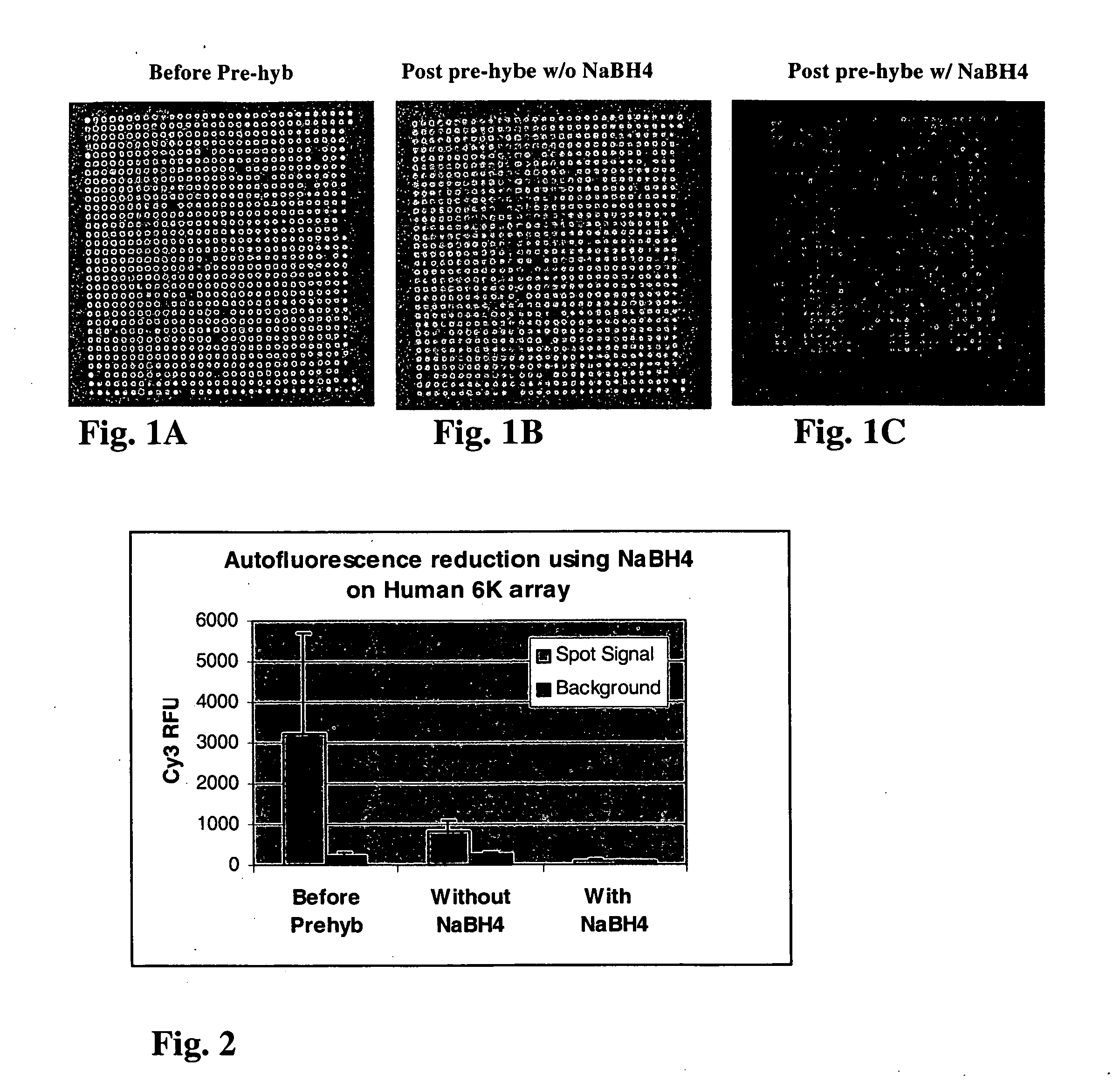
[0083] FIGS. 1A-1C show representative Cy3 images of a 1000 gene sub-grid of a microarrays of 2000 cDNA human DNA clones printed on CMT GAPS.TM. slides before and after treatment with 0.25% sodium borohydride, under a PMT setting of 950 volts. Referring to FIG. 1A which is an image of a slide before washing with any solution, both positive and negative spots are observed, indicating that the target quality is inconsistent since the spot fluorescent intensity either higher or lower than the background intensity on the GAPS surface. Analysis of the images showed that the normal prewash couldn't get rid of autofluorescence on both spots and the surface. FIG. 1B shows an image of a slide washed according to the procedures described above without sodium borohydride. Analysis of the images in FIG. 2B showed that the prewash without reducing agent could not reduce autofluorescence on both spots and the surface. FIG. 1C shows an image of a slide treated according to the procedures above wit...

example 2

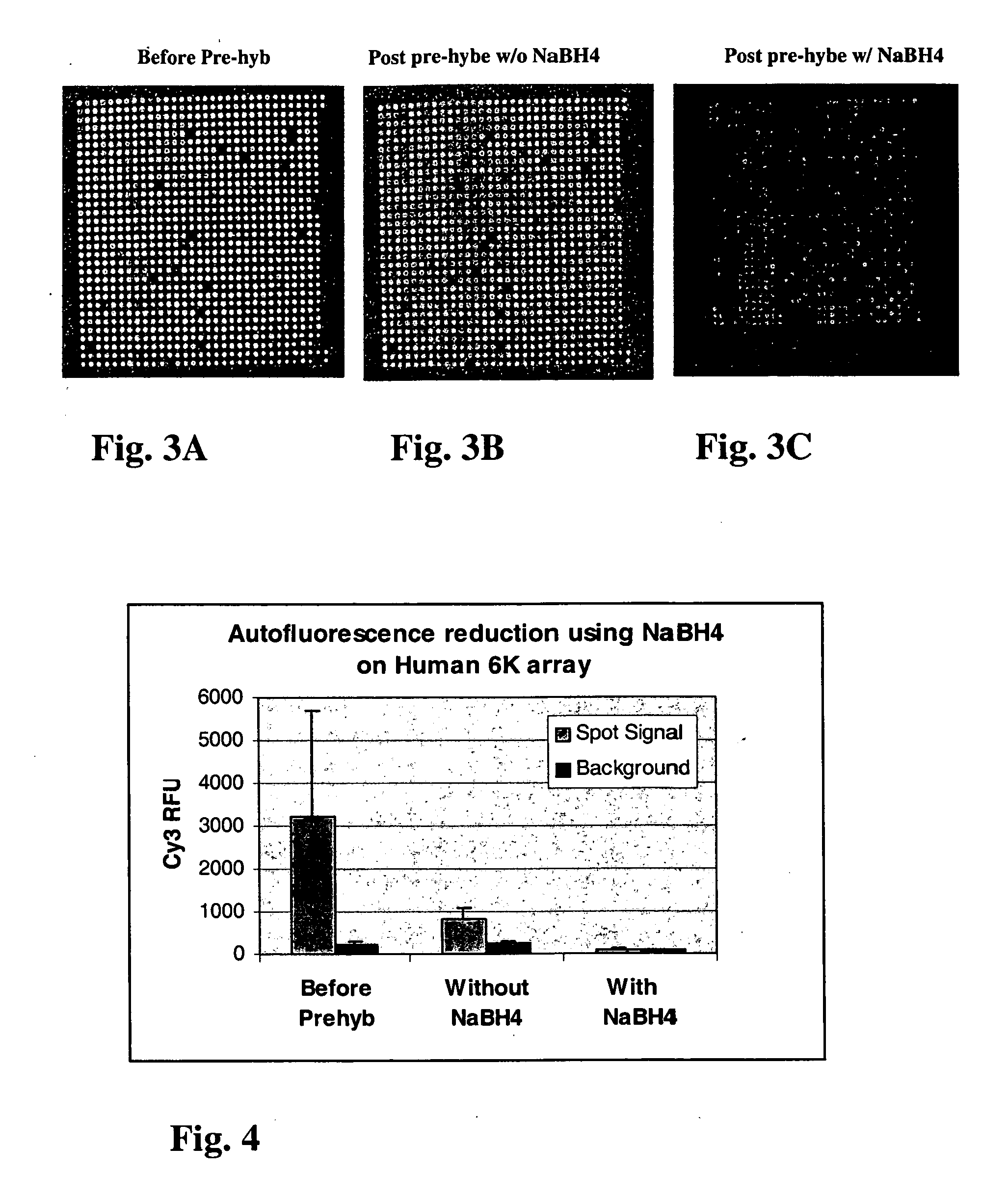
[0084] FIGS. 3A-C show representative Cy3 images of a 1000 gene sub-grid of an array of microarrays of 6000 cDNA human DNA clones printed on a CMT GAPS.TM. slides before and after treatment with 0.25% sodium borohydride, under a PMT setting of 800 volts. FIG. 3A is an image of an untreated slide. FIG. 3B is an image of a slide washed without treatment with reducing agent. FIG. 3C is an image of a slide washed and treated with sodium borohydride. FIG. 4 is a bar graph comparing the Cy3 RFU readings for the untreated slide, the slide washed without reducing agent and the slide treated with sodium borohydride. As shown in FIGS. 3A-C and FIG. 4, treatment significantly reduces auto-fluorescence on the microarray. Analysis of the image in FIG. 2B shows that even though a conventional pre-wash can remove auto-fluorescence on target spots partially, it can not remove autofluorescence on the surface at all. However, as can be seen from FIG. 2C, when treated with reducing agent, the autofluo...

example 3

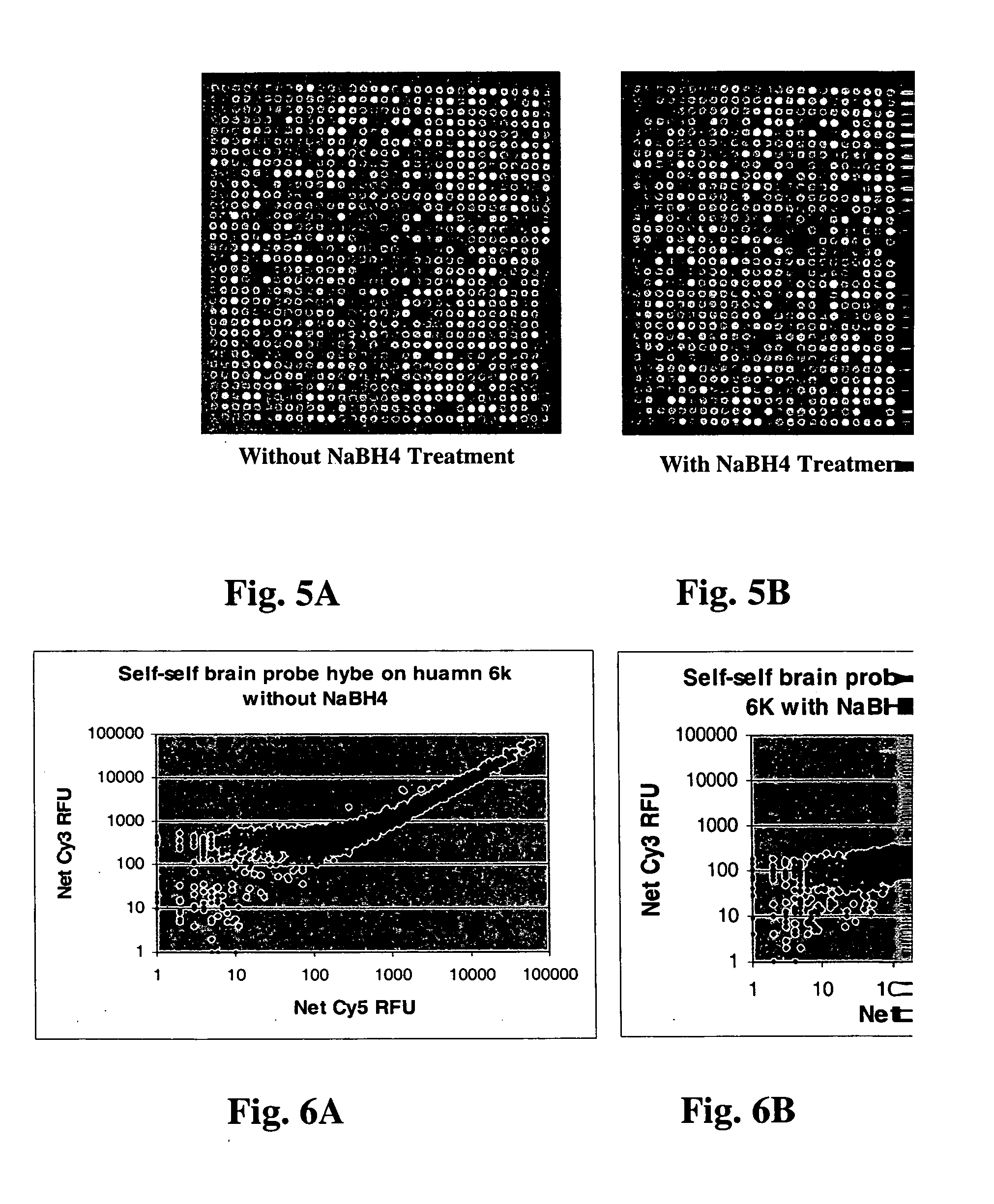
[0085] To test if reducing agent treatment affects the target hybridization, self-self hybridization of microarrays of 6000 cDNA human DNA cancer clones printed on a CMT GAPS.TM. slides (treated and untreated with sodium borohydride) with 0.25 .mu.g of total human brain RNA through linear amplification and reverse transcription labeling was also performed. FIG. 5A is an image a slide washed without reducing agent, and FIG. 5B is an image of a slide washed and treated with sodium borohydride according to the procedures described above. FIG. 6A is a graph of net Cy3 RFU versus net Cy5 RFU for the slide without sodium borohydride treatment and FIG. 6B is a graph of net Cy3 RFU versus net Cy5 RFU for the slide with sodium borohydride treatment. FIG. 6A shows more genes have higher Cy3 / Cy5 between 100-1000 on the untreated slides due to the Cy3 autofluorescence, when compared with the graph in FIG. 6B. This suggests that without sodium borohydride treatment, the Cy3 auto-fluorescence sig...


PUM

PropertyMeasurementUnit
concentrationaaaaaaaaaa
fluorescentaaaaaaaaaa
volumeaaaaaaaaaa

Abstract

A method of treating a substrate for immobilizing a biomolecule and substrates produced by the method are disclosed. The method includes contacting at least a portion of a substrate with a reducing agent such as a hydride. Treatment with an appropriate reducing agent substantially eliminates autofluorescence on substrates.

Description

[0001] This invention relates to substrates for use in immobilizing biomolecules and methods of making such substrates. More particularly, the present invention relates to treating substrates with a reducing agent and substrates produced by such treatment.[0002] Analysis of the structure, organization and sequence of nucleic acid molecules is important in the prediction, diagnosis and treatment of human disease and in the study of gene discovery, expression and development. One laboratory tool used in the analysis of nucleic acid molecules is the microarray or high density array (HDA), which is a microarray containing a large number of targets per square centimeter of array surface. The microarray provides the framework for immobilization of nucleic acid molecules for analysis on a rapid, large-scale basis. Microarrays generally include a substrate having a large number of positionally distinct nucleic acid targets attached to a surface of the substrate for subsequent hybridization ...

Claims


Application Information

Patent Timeline
Patent Type & Authority Applications(United States)
IPC IPC(8): G01N33/543
CPCG01N33/54393
Inventor BAO, YIJIALI, GUANGSHANRAGHAVACHARI, NALINIHE, MINGQIANXIE, XINYINGXIE, YUMING
Owner CORNING INC
Learn More