
Method and apparatus for characterization of chromophore content and distribution in skin using cross-polarized diffuse reflectance imaging

a technology of diffuse reflectance and chromophore, applied in the field of optical imaging of skin, can solve the problems of adverse affecting development, clinical significance of pws, and potentially devastating psychological and physical complications

Active Publication Date: 2005-02-10
RGT UNIV OF CALIFORNIA
View PDF6 Cites 64 Cited by
  • Summary
  • Abstract
  • Description
  • Claims
  • Application Information

AI Technical Summary

Benefits of technology

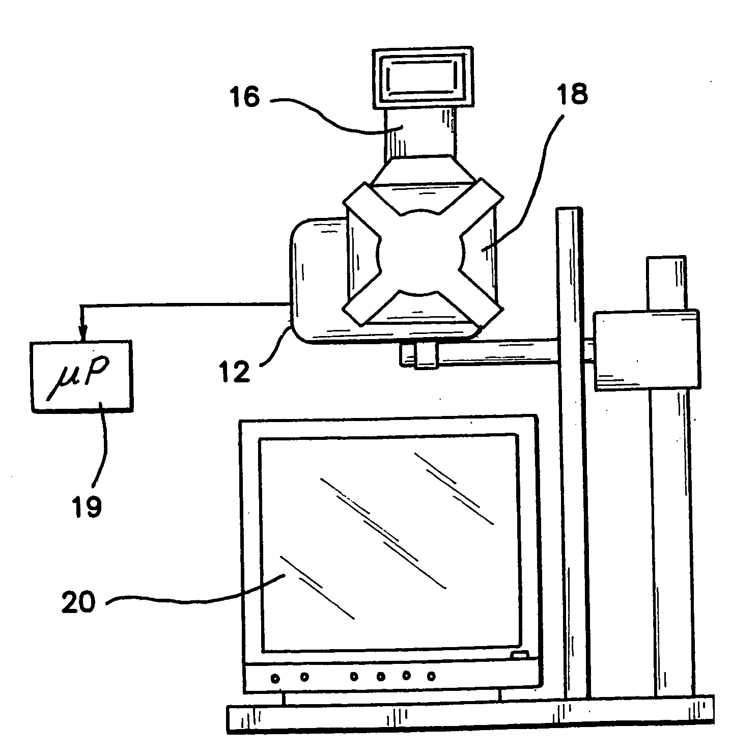
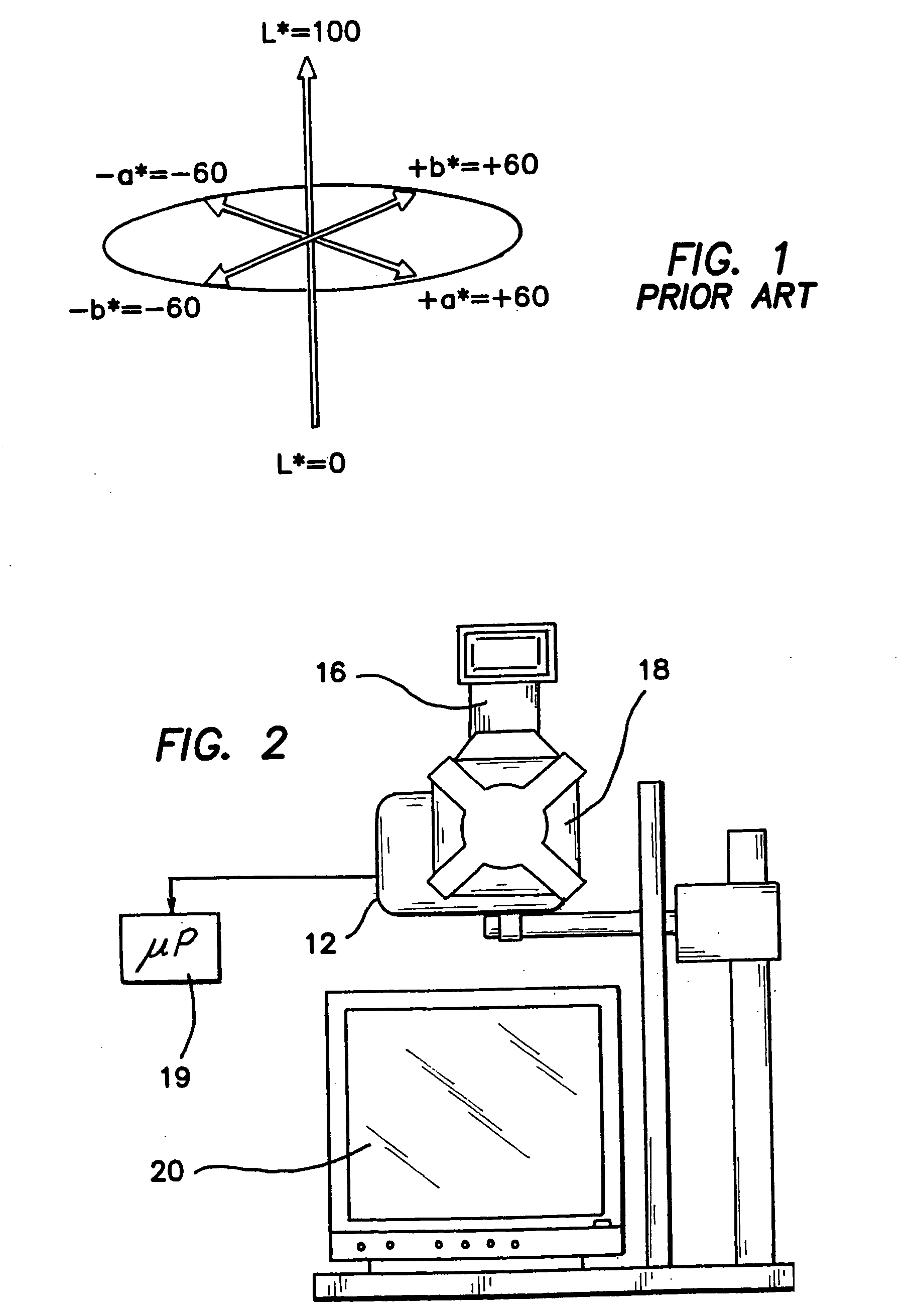
The invention is a noncontact, low-cost imaging system which rapidly and quantitatively characterizes skin. A digital camera is modified to incorporate crossed-polarizers, and combined with a novel image analysis method. The unique analysis technique provides quantitative color information with a Commission Internationale de I'Eclairage (CIE) L*a*b* image that can be used to deduce spatially resolved melanin and erythema indices. Color images and processed CIE color space images will enable pixel-by-pixel tissue analysis.
The digital imaging system of the invention provides color information of an entire port wine stain or other skin chromophore content with a single image. Polarization optics produce marked reduction in specularly reflected light in the images. A patient positioning device allows for repeatable positioning of the patient's head or body portion. The digital nature of the system provides a near real-time mapping of melanin and erythema or other skin chromophore metrics. The cross-polarized diffuse reflectance color imaging system obtains subsurface skin color information and acquisition of facial images in a reproducible fashion at a fixed distance from an illumination source.

Problems solved by technology

Since most of the malformations occur on the face, PWS is a clinically significant problem in the majority of patients.
PWS should not be considered a cosmetic problem but a disease with potentially devastating psychological and physical complications.
Personality development is adversely influenced in virtually all patients by the negative reaction of others to a “marked” person.
Clinical results using these methods were unsatisfactory due to cosmetically unacceptable scarring post treatment.
To date, none of these has been accepted as a reliable predictor of therapeutic outcome.
Unfortunately, if the ultimate standard required is complete blanching of the lesion, even after many repeat treatments, complete blanching is rarely achieved where an average success rate of below 10% is experienced.
We believe that this occurs primarily because the attending physician is unable to select the optimal treatment parameters for a specific PWS lesion.
However, due to a dearth of clinically accepted devices for determining these parameters, clinicians must still rely on subjective qualities such as PWS skin appearance and personal experience to determine treatment parameters (e.g., wavelength, pulse duration, spot size, light dose, and cryogen spray cooling factors) to use on each patient.
Furthermore, if the erythema metric changes minimally between two successive visits, the clinician could alter treatment parameters or deem the patient as a nonresponder and stop treatment.
Although reflectance measurement techniques can provide valuable information on PWS skin, they are generally limited in usefulness by practical considerations.
They provide information on only a small area (˜10-15 mm in diameter) per measurement; thus, it may be time consuming to measure an entire PWS.
Furthermore, since these devices are required to be in contact with skin, variations in contact pressure can induce artifacts in the measured reflectance values.
Application of excessive pressure can result in transient blanching of the PWS due to blood vessel collapse, resulting in potentially large error in measured values.
Furthermore, image quality may be affected by shadowing, glare, nonuniform illumination, changes in spectral qualities of the illumination source with time and artifacts resulting from environmental lighting.
Medical imaging is critical for quality health care, yet remains unavailable to many patients in small hospitals, rural communities and underdeveloped nations.

Method used


Image

  • Method and apparatus for characterization of chromophore content and distribution in skin using cross-polarized diffuse reflectance imaging
  • Method and apparatus for characterization of chromophore content and distribution in skin using cross-polarized diffuse reflectance imaging
  • Method and apparatus for characterization of chromophore content and distribution in skin using cross-polarized diffuse reflectance imaging

Examples


Embodiment Construction

Numerous factors affect the quality of information contained in each digital image. As an optical system is used to capture data from patients over a period of months to years, it is critical to characterize the sources of variation in the system and determine the sensitivity of the data to expected changes that may occur in the system components over time. The effects of curved surfaces on recovered indices, quality of crossed-polarizer extinction, drift in “color temperature” of the illumination device, errors in white balance, errors in repositioning target tissue and effects of small angular displacements in illumination and collection will impact the data analysis. We have quantified the variance associated with each of these factors and subsequently take steps to control or minimize the impact of those factors. Statistical error propagation analysis is used to determine the overall magnitude of multiple sources of error on CIE L*a*b* values computed from each raw image.

Ther...


PUM


Abstract

A digital imaging system provides color information of an entire port wine stain or other skin condition with a single image in CIE L*a*b* color space (L*, a*) derived from RGB pixel data (R, G, B). Cross-polarization optics produce marked reduction in specularly reflected light in the images. A patient positioning device allows for repeatable positioning of the patient's head or body portion. The digital nature of the system provides a near real-time mapping of melanin and erythema or other skin chromophore metrics. The cross-polarized diffuse reflectance color digital imaging system obtains subsurface skin color information and acquisition of facial images in a reproducible fashion at a fixed distance from an illumination source at optimized angles of view depending on the region of interest being imaged.

Description

BACKGROUND OF THE INVENTION 1. Field of the Invention The invention is directed to the field of optical imaging of skin and in particular to a noncontact imaging system for rapidly and quantitatively characterizing skin using a digital camera incorporating crossed-polarizers and an image analysis method. 2. Description of the Prior Art Port wine stain (PWS) birthmark is a congenital, progressive vascular malformation of the skin that occurs in an estimated 7 children per 1,000 live births and typically occur on the face and neck. Approximately 1,500,000 individuals in the United States and thirty-two million people worldwide have PWS birthmarks. Histopathological studies of PWS show a normal epidermis overlying an abnormal plexus of dilated blood vessels located in the dermis. Epidermal thickness (50-150 μm) and melanin absorption, as well as PWS blood vessel diameter (30-300 μm) and depth distribution (150-1000 μm) vary on an individual patient basis and even between different ...

Claims


Application Information

Patent Timeline
Patent Type & Authority Applications(United States)
IPC IPC(8): A61B5/00A61B5/103
CPCA61B5/0059A61B2560/0233A61B5/445A61B5/444
Inventor JUNG, BYUNGJOCHOI, BERNARDDURKIN, ANTHONY J.NELSON, J. STUART
Owner RGT UNIV OF CALIFORNIA
Learn More