

Novel fluorescent proteins
a fluorescent protein and protein technology, applied in the field of new fluorescent proteins, can solve the problems of loss of the fluorescent properties of gfp, not being useful in biological systems, etc., and achieve the effect of improving the usefulness of fluorescent proteins
- Summary
- Abstract
- Description
- Claims
- Application Information
AI Technical Summary
Benefits of technology
Problems solved by technology
Method used
Image

Examples
example 1
Cloning of cDNA Encoding GFP
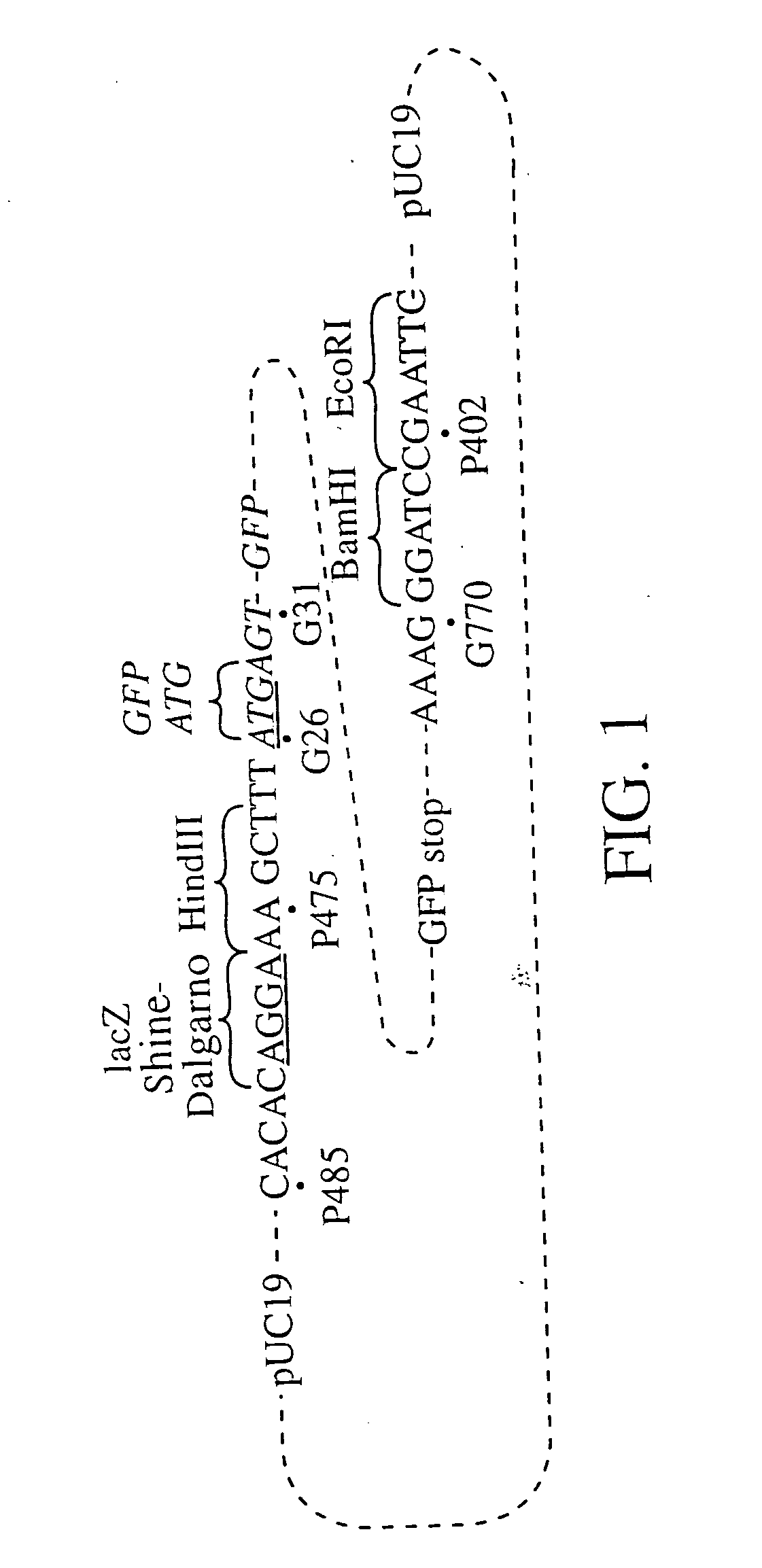
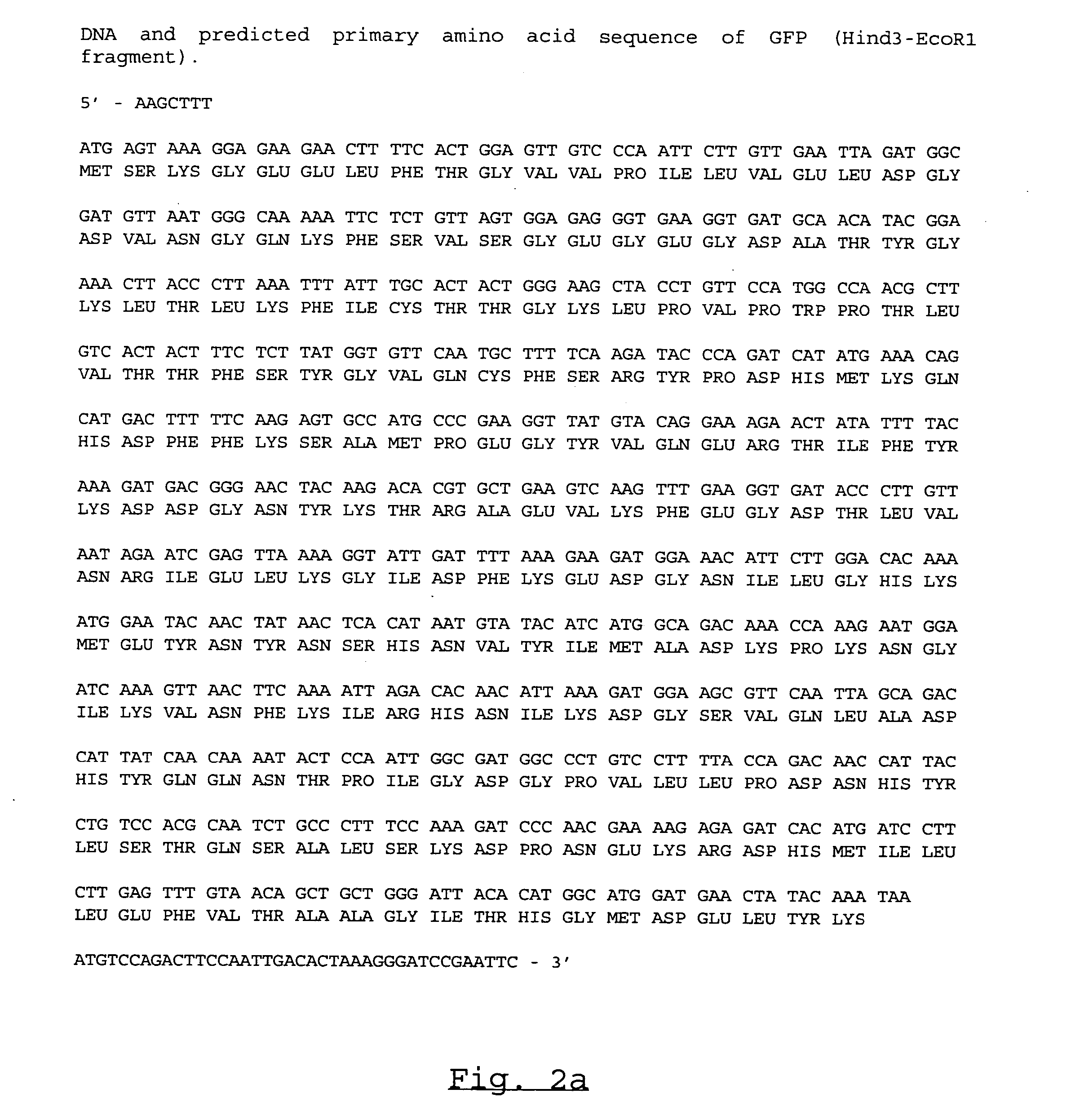
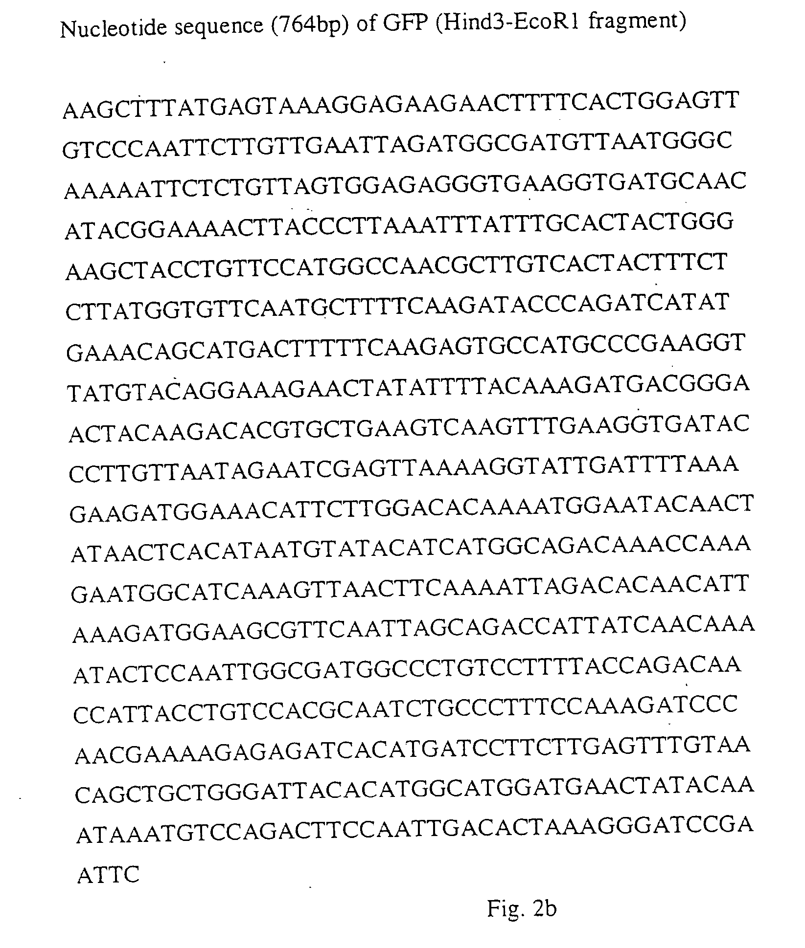
[0069] Briefly, total RNA, isolated from A. victoria by a standard procedure (Sambrook et al., Molecular Cloning. 2., eds. (1989) (Cold Spring Harbor Laboratory Press: Cold Spring Harbor, N.Y.), 7.19-7.22) was converted into cDNA by using the AMV reverse transcriptase (Promega, Madison, Wis., USA) as recommended by the manufacturer. The cDNA was then PCR amplified, using PCR primers designed on the basis of a previously published GFP sequence (Prasher et al., Gene 111 (1992), 229-223; GenBank accession No. M62653) together with the UlTma™ polymerase (Perkin Elmer, Foster City, Calif., USA). The sequences of the primers were:
GFP2:TGGAATAAGCTTTATGAGTAAAGGAGAAGAACTTTTandGFP-1:AAGAATTCGGATCCCTTTAGTGTCAATTGGAAGTCT
[0070] Restriction endonuclease sites inserted in the 5′ (a HindIII site) and 3′ (EcoRI and BamHI sites) primers facilitated the cloning of the PCR amplified GFP cDNA into a slightly modified pUC19 vector. The details of the construction are as fol...
example 2
[0075] F64L-GFP was constructed as follows: An E.coli expression vector containing Y66H-GFP was digested with restriction enzymes Nco1 and Xba1. The recognition sequence of Nco1 is located at position 173 and the recognition sequence of Xba1 is located at position 221 in the F64L-Y66H-GFP sequence listed below. The large Nco1-Xba1 vector fragment was isolated and ligated with a synthetic Nco1-Xba1 DNA linker of the following sequence:
[0076] One DNA strand has the sequence:
5′-CATGGCCAACGCTTGTCACTACTCTCTCTTATGGTGTTCAATGCTTTT-3′(SEQ ID NO:7)
[0077] The other DNA strand has the sequence:
5′-CTAGAAAAGCATTGAACACCATAAGAGAGAGTAGTGACAAGCGTTGGC-3′(SEQ ID NO:8)
[0078] Upon annealing, the two strands form a Nco1-Xba1 fragment that incorporates the sequence of the GFP chromophore SYG with the F64L substitution preceding SYG. The DNA sequence and predicted primary amino acid sequence of F64L-GFP is shown in FIG. 4 below.
[0079] The S65T-GFP mutation was described by Heim et al (Nature vol. 373 ...
example 3
Influence of The F64L Substitution On GFP And Its Serivatives When Expressed In Mammalian Cells
[0106] F64L-Y66H-GFP, F64L-GFP, and F64L-S65T-GFP were cloned into pcDNA3 (Invitrogen, Calif., USA) so that the expression was under control of the CMV promoter. Wild-type GFP was expressed from the pGFP-N1 plasmid (Clontech, Ca, USA) in which the CMV promoter controls the expression. Plasmid DNA to be used for transfection were purified using Jetstar Plasmid kit (Genomed Inc. NC, USA) and was dissolved in distilled water.
[0107] The precipitate used for the transfections were made by mixing the following components: 2 μg DNA in 44 μl of water were mixed with 50 μl 2xHBS buffer (280 mM NaCl, 1.5 mM Na2HPO4, 12 mM dextrose, 50 mM HEPES) and 6.2 μl 2M CaCl2. The transfection mix was incubated at room temperature for 25 minutes before it was added to the cells. HEK 293 cells (ATCC CRL 1573) were grown in 2 cm by 2 cm coverglass chambers (Nunc, Denmark) with approximately 1.5 ml medium (Dulbe...
PUM
| Property | Measurement | Unit |
|---|---|---|
| Temperature | aaaaa | aaaaa |
| Fluorescence | aaaaa | aaaaa |
Abstract
Description
Claims
Application Information