Rapid and non-invasive optical detection of internal bleeding

Inactive Publication Date: 2005-03-24
HO WINSTON ZONH +2
7 Cites 95 Cited by

AI-Extracted Technical Summary

Problems solved by technology

Internal bleeding caused by injuries, such as blunt force, sharp objects (i.e., knife, gun, broken bone fragments), can damage internal organs and blood vessels.
Internal blood loss, like in intra-cranial space, can pool in surrounding tissues and may build up pressure upon vital organs that cause cardiac and respiratory arrests.
Those signs and symptoms are related to the loss of blood acutely or chronically: a rapid blood loss may result in sudden death, whereas a slow blood loss may be neglected by the healthcare professionals and contributes to the loss of life of patients.
Unfortunately, there is no any definitive method available at present time without having an exploratory surgery.
Although ultrasound is a radiation-free technique, it does not provide the nature of fluid character in internal cavities of human body, such as pus, ascites, or blood.
It is also an expensive procedure.
The MRI though, is a radiation-free technique; however, it has similar disadvantages.
Hormon...

Method used

Human tissues are highly scattering and absorptive media for ultraviolet and visible light. It is difficult for ultraviolet and visible light to penetrate the tissue more than 5 mm, while near infrared can easily reach 10 mm or more. The employment of NIR photons provides the opportunity to probe deeper tissue layers, excite the fluorophore more effectively, produce more fluorescent photons, and transmit more fluorescence signal for detection. Therefore, the employment of proper wavelength for optical probing and fluorescent compound are critical for this application. The total fluorescence intensity, F, is proportional to the integration of the total fluorescence over the excitation volume V, and is given by the spatial integral of
Many NIR fluorescence compounds are potential candidates for the present application. One of examples, indocyanine green (ICG), because of its low toxicity, has been used in many clinical applications. Indocyanine green, molecular weight 775, is a tricarbocyanine type of green dye. FIG. 2 and FIG. 3 show the NIR absorption, 650-850 nm, and NIR fluorescence spectra, 650-900 nm, of ICG, respectively. ICG has little absorption in the visible light. However, it is easily excited by an NIR light source with high quantum efficiency. Diode laser light sources with a wavelength between 630-645 nm is suitable for ICG excitation. The fluorescent peak has a large red shifted relatively to the excitation wavelength. The fluorescence peak at 810 nm is within the NIR window for tissue optics. Due to ICG has a very large fluorescent quantum yield and a distinct peak at 810 nm, a sensitivity of 0.5 μg/ml can be achieved easily. FIG. 3(a), (b), and (c) show the fluorescence spectra of ICG in blood samples with various concentration between 0.5-500 μg/ml
R(r,θ) is the point source response function, which is a measure of probability that an emitted fluorescence photon generated at position (r,θ) in the sampling volume, V, that will reach the detector at radial position, r, and at the acceptance angle, θ, of the fluorescence collection light guide. This response function can be treated as a conventional rigid rotation function and is dependent on the tissue's optical properties. By proper selecting of excitation light source, a wavelength between 400 nm and 800 nm, and fluorescent compound, a wavelength between 500 nm and 950 nm, it is possible to diagnose internal bleeding non-invasively.
The fluorescent compound in leaked blood is probed externally with a light beam 7 confined in an optical probe or a light guide 20. The concentration of the fluorescent compound in the blood is in the range of 1-500 μg/ml. Thin tissue with no or minimal capillary blood vessel is the preferred area for optical probing. The potential areas for optical probing are vaginal canal, posterior fornix of vaginal wall, cervical region, rectum, frontal fontanel, occipital fontanel, and other relativ...

Benefits of technology

The present non-invasive optics-based probe and medical device has the advantages of simple, real time, and easy operation. The internal bleeding diagnostic device provides rapid and accurate results to assist clinician's decision-making. It should be understood, however, that the detail description and specific ex...

Abstract

A rapid and non-invasive optical method and device for diagnosing internal bleeding or hemorrhage in a human body by detecting leaked blood comprising administering a fluorescent compound parenterally; providing a light beam containing a wavelength absorbable by the florescent compound, wherein the light beam is illuminated at and transmitted through a tissue region into the human body; and analyzing fluorescence signal produced from the fluorescent compound in the leakage of blood for diagnosing the presence or absence of internal bleeding. The invention provides an accurate, rapid, easy-to-use, and inexpensive method for diagnosing internal bleeding, particularly in the fields of gynecology, obstetrics, neonatology, surgery bleeding, post-surgery bleeding, emergency medicine, and veterinary medicine for cases suspected of internal hemorrhage.

Application Domain

Ultrasonic/sonic/infrasonic diagnosticsSurgery +8

Technology Topic

Internal bleedingNon invasive +12

Image

  • Rapid and non-invasive optical detection of internal bleeding
  • Rapid and non-invasive optical detection of internal bleeding
  • Rapid and non-invasive optical detection of internal bleeding

Examples

  • Experimental program(1)

Example

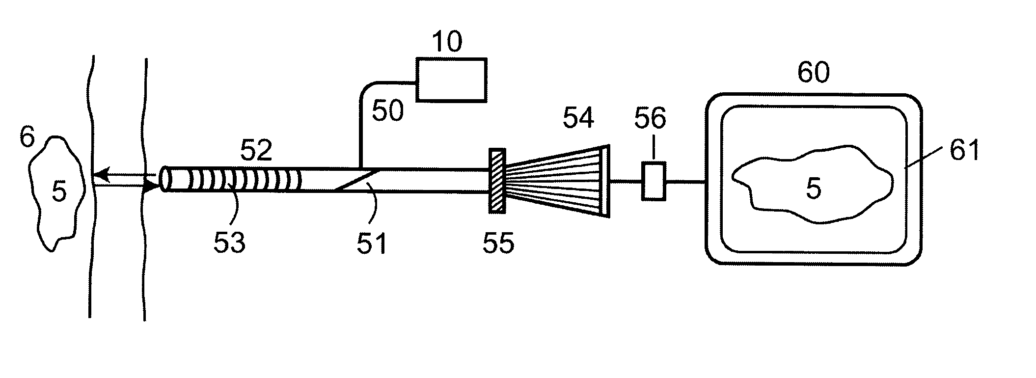
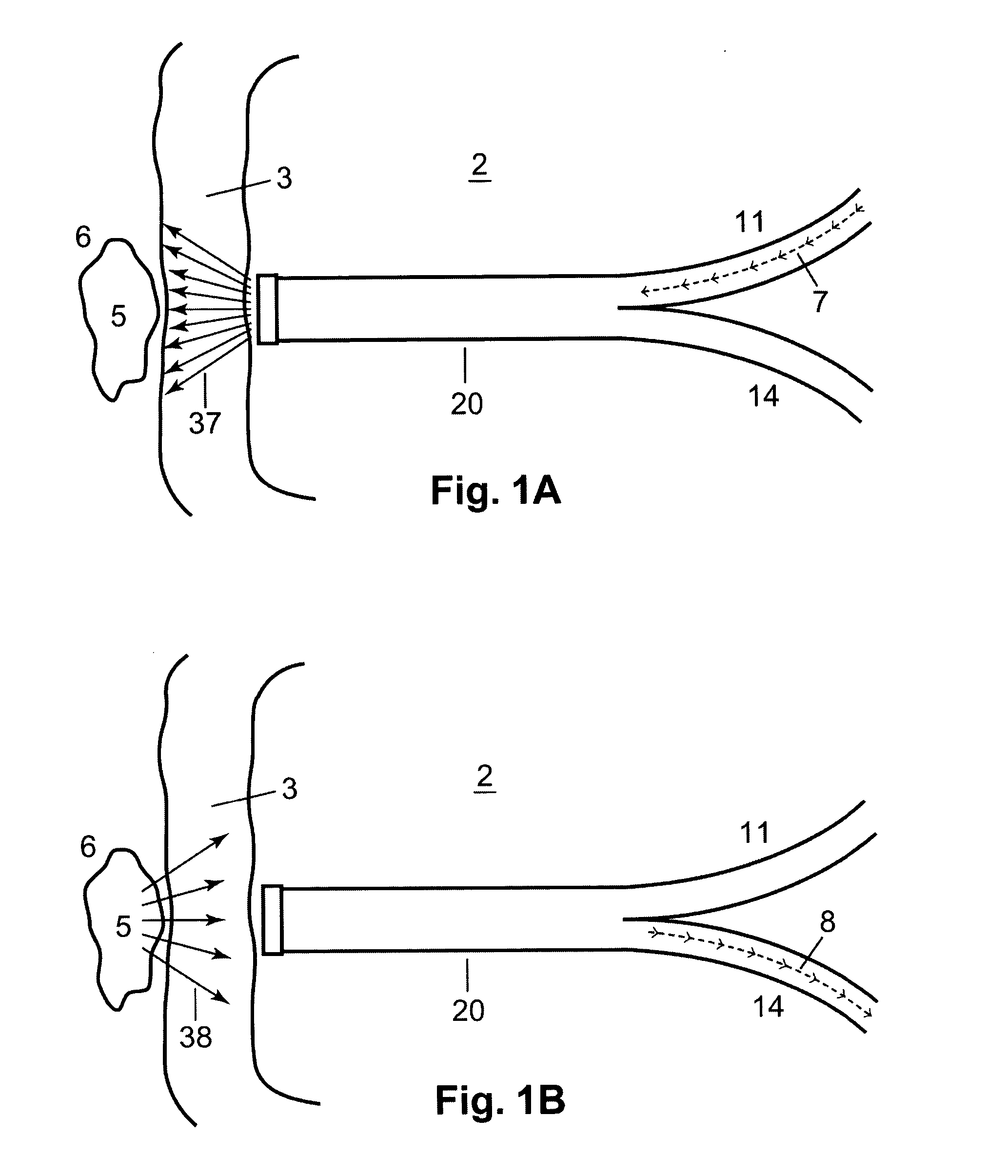
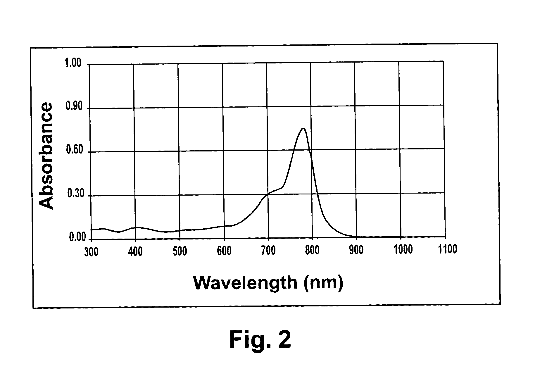
The preferred embodiments of the present invention described below relate particularly to a non-invasive optical method and device for diagnosing internal bleeding or hemorrhage in a human body by detecting leaked blood comprising: administering a fluorescent compound parenterally; providing a light source having a light beam, wherein said light beam contains a wavelength absorbable by said fluorescent compound, wherein said light beam is illuminated at and transmitted through a tissue region into said human body; and after administering said fluorescent compound for a few minutes, analyzing a fluorescence signal produced from said fluorescent compound in said leaked blood for diagnosing the presence or absence of internal bleeding in said human body. While the description sets forth various embodiment specific details, it will be appreciated that the description is illustrative only and should not be construed in any way as limiting the invention. Furthermore, various applications of the invention, and modifications thereto, which may occur to those who are skilled in the art, are also encompassed by the general concepts described below.
Once a clinician or doctor determines that a patient may have internal bleeding, patients will be administered with fluorescent compound parenterally either intravenously, or intramuscularly (if intravenous injection is not accessible or the case of illness is chronic). The dosage of the fluorescent compound should be effective for producing the fluorescence signal. The typical dosage is in the range of 0.1-10 mg/Kg body weight. Following intravenous or other parenteral administration, the fluorescent compound is quickly transported throughout the body and contained in the bloods vessels. The fluorescent compound can be circulated and distributed to any part of the body within about 3-5 minutes or in a short period of time. If internal bleeding occurs, the blood leaks out the circulation system, as shown in FIG. 1, and proliferates into nearby body cavity 6, such as abdomen. When the leakage of blood accumulated, it forms a pool 5 or a mass of blood. Fluorescent compound thus provides a marker for detecting leaked blood. Internal bleeding occurs frequently in the fields of gynecology, obstetrics, neonatology, surgery bleeding, post-surgery bleeding, emergency medicine, and veterinary medicine.
The fluorescent compound in leaked blood is probed externally with a light beam 7 confined in an optical probe or a light guide 20. The concentration of the fluorescent compound in the blood is in the range of 1-500 μg/ml. Thin tissue with no or minimal capillary blood vessel is the preferred area for optical probing. The potential areas for optical probing are vaginal canal, posterior fornix of vaginal wall, cervical region, rectum, frontal fontanel, occipital fontanel, and other relatively thin layer of human tissue. When the light guide is placed against the tissue, the light beam is penetrated through the tissue 37 to reach the leaked blood. For example, FIG. 1 shows an optical probe 20 is inserted into a vaginal canal 2 and positioned against a cervical tissue/posterior fornix of vaginal wall 3. Cervical tissue area or posterior fornix of vaginal wall is relatively thin, on the order of 2-4 mm. Therefore, the light beam can easily transmit through the tissue and probe the leaked blood 5 in the body cavity 6, such as the cul-de-sac of abdomen. The configuration of the optical probe can be a stand-alone device, or integrated with conventional ultrasound probe, endoscope, fiberscope, or image scope. One preferred embodiment of the optical probe 20 is constructed as a bifurcated optical fibers. The bifurcated fibers combine two ends of illumination fiber 11 and fluorescence detection fiber 14 into an optical probe. The illumination fiber 11 and fluorescence detection fiber 14 can carry the light beam 7 for illumination and collect fluorescence signal 38 into the detection fiber 8, respectively. The optical fiber-based probe has the flexibility to move around in searching for leaked blood or leakage of blood.
Human tissues are highly scattering and absorptive media for ultraviolet and visible light. It is difficult for ultraviolet and visible light to penetrate the tissue more than 5 mm, while near infrared can easily reach 10 mm or more. The employment of NIR photons provides the opportunity to probe deeper tissue layers, excite the fluorophore more effectively, produce more fluorescent photons, and transmit more fluorescence signal for detection. Therefore, the employment of proper wavelength for optical probing and fluorescent compound are critical for this application. The total fluorescence intensity, F, is proportional to the integration of the total fluorescence over the excitation volume V, and is given by the spatial integral of
F(r,θ)=∫Iine−klrε×Q×C×e−k2r×R(r,θ)dr dθ
Where Iin=light intensity at surface of the tissue K1, K2=extinction coefficients of tissue at excitation and fluorescence wavelengths, respectively ε=absorption coefficient of fluorescence compound Q=fluorescence quantum yield of the fluorescence compound C=concentration of the fluorescence compound in blood
R(r,θ) is the point source response function, which is a measure of probability that an emitted fluorescence photon generated at position (r,θ) in the sampling volume, V, that will reach the detector at radial position, r, and at the acceptance angle, θ, of the fluorescence collection light guide. This response function can be treated as a conventional rigid rotation function and is dependent on the tissue's optical properties. By proper selecting of excitation light source, a wavelength between 400 nm and 800 nm, and fluorescent compound, a wavelength between 500 nm and 950 nm, it is possible to diagnose internal bleeding non-invasively.
Many NIR fluorescence compounds are potential candidates for the present application. One of examples, indocyanine green (ICG), because of its low toxicity, has been used in many clinical applications. Indocyanine green, molecular weight 775, is a tricarbocyanine type of green dye. FIG. 2 and FIG. 3 show the NIR absorption, 650-850 nm, and NIR fluorescence spectra, 650-900 nm, of ICG, respectively. ICG has little absorption in the visible light. However, it is easily excited by an NIR light source with high quantum efficiency. Diode laser light sources with a wavelength between 630-645 nm is suitable for ICG excitation. The fluorescent peak has a large red shifted relatively to the excitation wavelength. The fluorescence peak at 810 nm is within the NIR window for tissue optics. Due to ICG has a very large fluorescent quantum yield and a distinct peak at 810 nm, a sensitivity of 0.5 μg/ml can be achieved easily. FIG. 3(a), (b), and (c) show the fluorescence spectra of ICG in blood samples with various concentration between 0.5-500 μg/ml
The non-invasive optical probe device for diagnosing internal bleeding, as shown in FIG. 4, is integrated with a light source 10, a fiber splitting coupler 12, an optical probe 20, wavelength diffraction grating 13, a detector 16, and an optical signal analyzing system 30. The light source can be a laser or a lamp. Diode lasers, such as NIR diode lasers with an optical output in the range of 5-50 mw are commercially available. Some lamp sources, which are broadband light sources that cover the entire near infrared range, are also suitable as a continuous light source. Optical band-pass filters or gratings can be used to select a proper narrow band wavelength for excitation. The NIR light beam 7 is coupled into the illumination fiber 11 with a micro lens. Fluorescence signal is collected and delivered to the detection system by the detection fiber 14. The fluorescence signal is either an image or a spectrum. The detection fiber containing a plurality of fibers can improve collection efficiency. The analyzing system 30 displays the fluorescence signature 31 with a distinct fluorescence peak. The spectral signal is physically separated by the diffraction grating 13 and illuminated on a linear CCD 16. Due to the low background in the NIR window, the peak intensity is directly related to the amount of fluorescence compound in leaked blood. The fluorescence peak intensity on CCD is processed by a microprocessor, thus can be correlated to the amount of the leaked blood. FIG. 4B shows one embodiment of the optical probe tip 22; the center fiber is the illumination fiber 11 and the surrounding fibers 23 are fluorescence collection fibers, which form the detection fibers 14.
In another preferred embodiment, the light source 10 can be integrated with a conventional endoscope 52 for image detection. As shown in FIG. 5, an NIR light source is coupled into an endoscope, such as a laparoscope, through an optical fiber 50. A 45° mirror 51 reflects the light into the endoscope's lens assembly 53. The fluorescence signal is collected by the endoscope and delivered into a CCD image detector or an image camera 54. An optical filter 55 is installed in front of the NIR sensitive camera. The NIR camera 54 is interfaced through an analog-to-digital converter 56 to an advanced signal processor in a computer 60. The leaked blood 5 in human body is displayed as a pool of leaked blood image 61 on a screen. The real-time data acquisition software supports digital processing with signal normalization. In general, the data acquisition and analysis of the optical parameters are well known to an ordinary person who is skilled in the art.
From the foregoing, it should now be appreciated that an optical probe or light guide containing an illuminating light beam with a wavelength absorbable by a fluorescent compound, wherein the illuminating light beam is transmitted through a tissue region into human body; and a fluorescence detecting means for analyzing a fluorescent signal obtained from the fluorescent compound in blood and for diagnosing the location of internal bleeding in human body, wherein the fluorescence detecting means comprises optical filters or optical gratings or image apparatus. It is also generally applicable for monitoring internal bleeding in many parts of the body. While the invention has been described with reference to a specific embodiment, the description is illustrative of the invention and is not to be construed as limiting the invention. Various modifications and applications may occur to those skilled in the art without departing from the true spirit and scope of the invention as described by the appended claims.

PUM

no PUM

Description & Claims & Application Information

We can also present the details of the Description, Claims and Application information to help users get a comprehensive understanding of the technical details of the patent, such as background art, summary of invention, brief description of drawings, description of embodiments, and other original content. On the other hand, users can also determine the specific scope of protection of the technology through the list of claims; as well as understand the changes in the life cycle of the technology with the presentation of the patent timeline. Login to view more.

Similar technology patents

Graphene film and preparation method therefor

Owner:CHANGZHOU FUXI TECH CO LTD

Classification and recommendation of technical efficacy words

  • easy to operate

Lancet device

InactiveUS7303573B2inexpensively mass-producedeasy to operate
Owner:ABBOTT DIABETES CARE INC

Message prompting method and device of instant messaging client

ActiveCN103051516ASave display spaceeasy to operate
Owner:TENCENT TECH (SHENZHEN) CO LTD
Why Eureka
Try Eureka