
Electrophoretic in situ tissue staining

Inactive Publication Date: 2005-04-07
VENTANA MEDICAL SYST INC
View PDF20 Cites 27 Cited by
  • Summary
  • Abstract
  • Description
  • Claims
  • Application Information

AI Technical Summary

Benefits of technology

[0010] It is an object of this invention to accelerate the movement of conjugate molecules from the aqueous solution into the solid tissue. Another object is to reduce the background staining due to conjugates that are not bound to specific sites. A further object is to reduce the concentration of the conjugate required in the reagent.

Problems solved by technology

The rate of Immunohistochemical and in situ hybridization staining of microtome-sectioned tissue on a glass slide is limited by the speed at which the biomolecules of interest can diffuse into the tissue from an aqueous solution placed in contact with the tissue section.
However, this has two detrimental effects.
First, the conjugates are often very expensive, so increasing their concentration is wasteful and not economically viable.
Second, the excessive amount of conjugate that is driven into the tissue, when high concentrations are used, gets trapped in the tissue, and cannot be rinsed out and causes high levels of background staining.

Method used


Image

  • Electrophoretic in situ tissue staining
  • Electrophoretic in situ tissue staining
  • Electrophoretic in situ tissue staining

Examples


experiment 1

[0036] Electrophoretic Tissue Staining using Anti-CD34 Antibody in Tonsil.

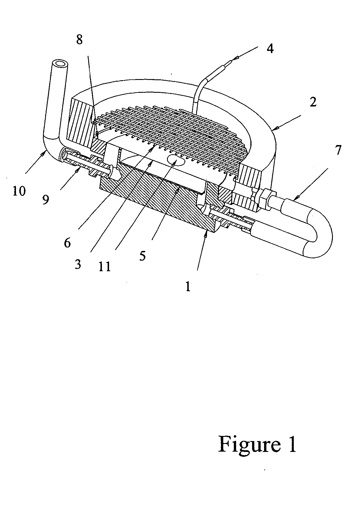
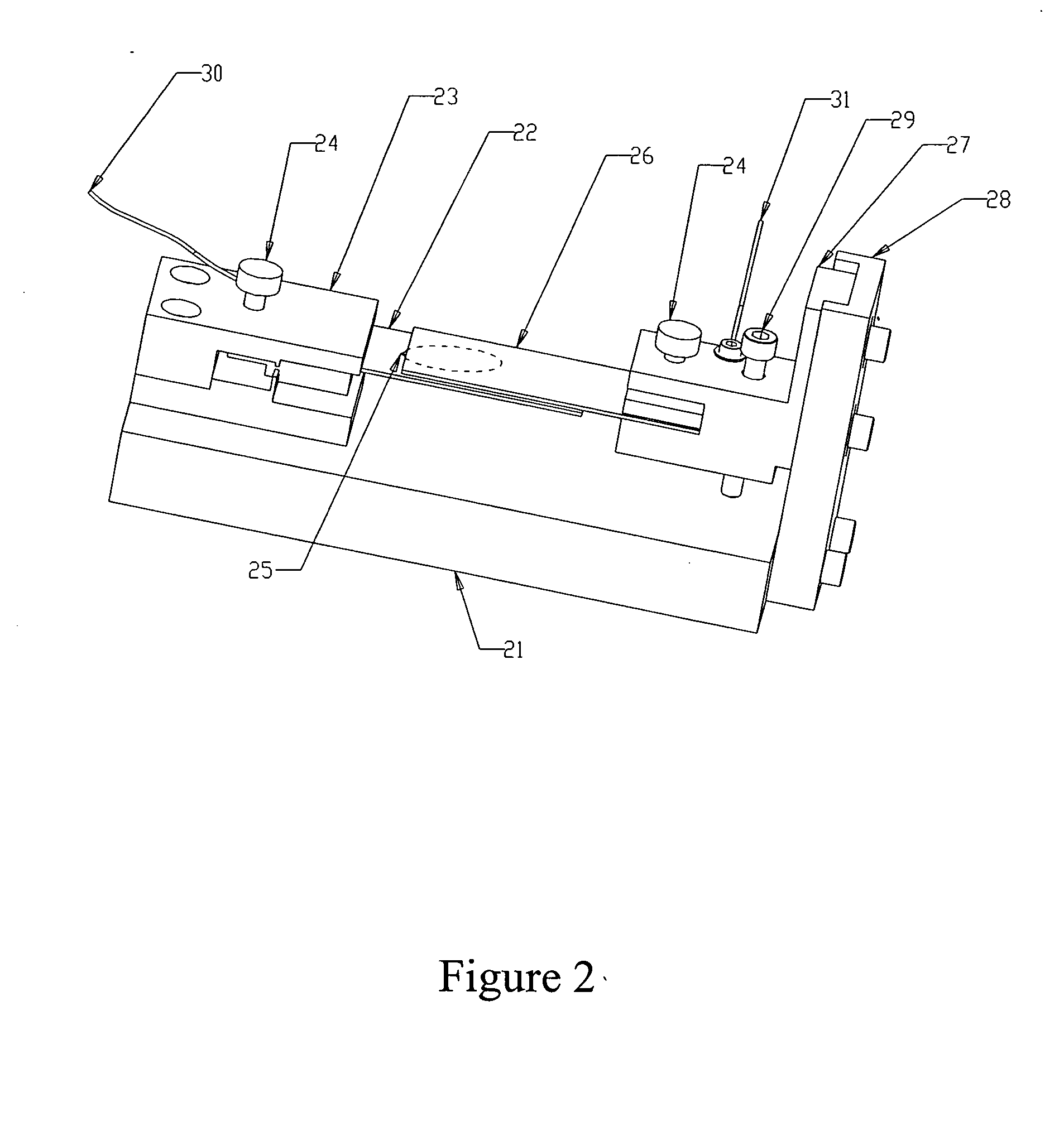
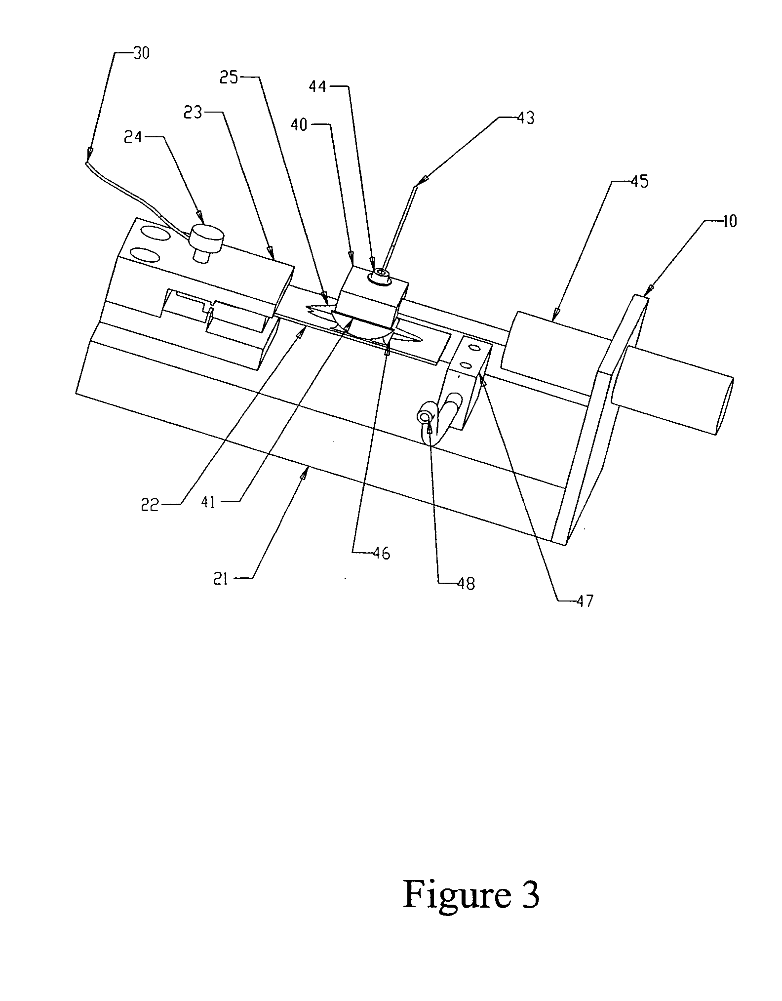
[0037] The following experiment was run to determine if antibody could be introduced electrophoretically into tissue. The tissue was adhered to a hydrophilic polytetrafluoroethylene (PTFE) membrane (TEFLON® Plumber's Tape) to enable manipulation and orientation of the tissue in the gel, and then embedded in an agarose gel for subsequent electrophoresis.

[0038] Procedure: four sections of 5 μm-thick human tonsil were mounted to PVA-treated hydrophilic PTFE membrane, air dried for 48 hours, overnight dried at 60° C., manually de-paraffinized and re-hydrated (standard process of dipping sections sequentially in xylene, then 100% EtOH, 90% EtOH, 80% EtOH, 70% EtOH, and finally 100% H2O). The PTFE membrane was made hydrophilic by wetting in Isopropyl alcohol first, then soaking for several hours in a solution of 0.1% polyvinyl alcohol in phosphate buffer, pH 2.2 and 5% glutaraldehyde, and rinsed in DI water. Any h...


PUM

PropertyMeasurementUnit
Electric fieldaaaaaaaaaa

Abstract

The present invention introduces a radically different way of accelerating biomolecule conjugates into tissue, and hence towards their targets for purposes of tissue staining. The invention provides for an order of magnitude improvement over the prior art diffusion process used to stain tissue. The invention comprises a method of tissue staining by applying an electric field to a tissue sample in the presence of an electrolyte and biomolecular conjugates of interest suspended in the electrolyte. Typical staining times are reduced to seconds as opposed to 30-120 minutes common in the prior art. The invention is also directed to devices for performing the method.

Description

BACKGROUND [0001] 1. Field of the Invention [0002] This invention relates generally to the field of automated tissue staining apparatus, and in particular is a new method of introducing stains into tissue using electrophoresis. [0003] 2. Description of Related Art [0004] Tissue staining is an ancient art by modem standards that goes back over one hundred years. Recently, efforts have been made to automate the procedure of applying different types of chemical and biochemical stains to tissue sections. Instruments that have been invented for this purpose include the Ventana Medical Systems' line of dual carousel-based instruments such as the 320, ES®, NexES®, BENCHMARK®, and the BENCHMARK® XT. Patents that describe these systems include U.S. Pat. Nos. 5,595,707, 5,654,199, 6,093,574, and 6,296,809, all of which are incorporated herein by reference in their entirety. Another type of automated stainer is the TechMate® line of stainers, described in U.S. Pat. Nos. 5,355,439 and 5,737,499...

Claims


Application Information

Patent Timeline
IPC IPC(8): A61N1/30A61N1/32G01N1/30G01N31/22
CPCA61N1/30G01N31/22G01N1/30A61N1/325
Inventor LEMME, CHARLESRICHARDS, WILLIAMBRYANT, DAVIDWOLF, CATHERINEGHUSSON, ANDREWASHBY, AUSTINSHOWALTER, WAYNEHARTMAN, ANTHONYKRAM, BRIAN
Owner VENTANA MEDICAL SYST INC
Learn More