

DNA-dependent MRI contrast agents
- Summary
- Abstract
- Description
- Claims
- Application Information
AI Technical Summary
Benefits of technology
Problems solved by technology
Method used
Image

Examples
example 1
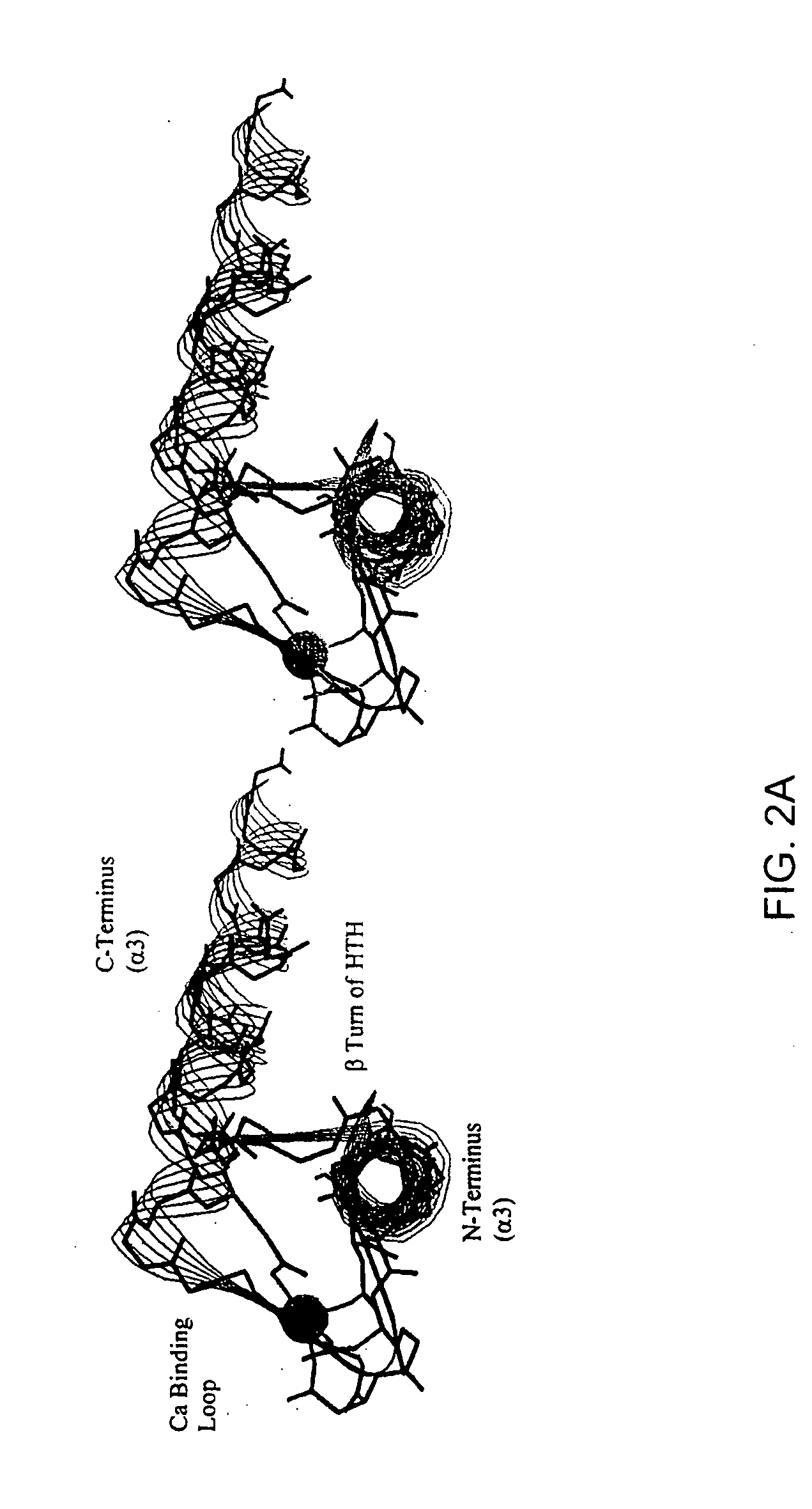
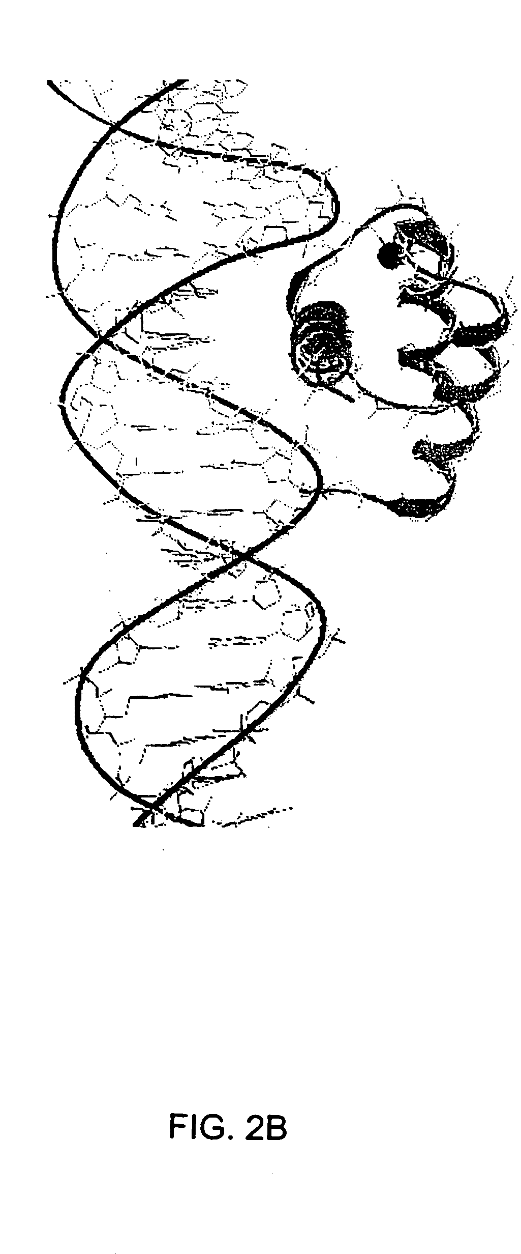
[0112] The remarkable similarity of the intermolecular interactions of many DNA binding proteins, despite little homology in their primary sequence, suggests that certain motifs are particularly well suited to complement the structure of B-form DNA (Brennan et al., 1989; Burley, 1994; Pabo et al., 1984; Patikoglou et al, 1997). The helix-turn-helix (HTH) motif is a well-known DNA binding motif which complements the shape of the DNA major groove. Homeodomains were first discovered in Drosophila, these small proteins bind DNA and regulate growth and development (Gehring et al., 1994; Kornberg, 1993; Treisman et al., 1992). Homeodomains are transcription factors, either activators or repressors (e.g., Cro and λ repressors) which typically have about 60 residues in the homeodomain arranged as three helices, with the amino terminal arm contacting the minor groove (Burley, 1994; Patikoglou et al., 1997; Laughan, 1991). Homeodomains which comprise the HTH motif can specifically bind to DNA...
example 2
[0153] Genetic mutations causing cancer and other diseases could be suppressed if target sequences could be selectively cleaved. Hydrolytic cleavage of DNA by Lewis-acid metal ions occurs with limited sequence discrimination, however, proteins are capable of selectively recognizing given duplex DNA sequences. Transcription factors and repressor proteins containing the helix-turn-helix motif (HTH) bind DNA tightly through a series of positively charged residues, which orient the recognition helix within the major groove of DNA. Sidechains of this helix contact and complement the base pair edges, resulting in the recognition of up to 18 base pairs for homodimeric transcription factors such as cro, lambda, or Trp repressors. As discussed above, the supersecondary structure of the HTH and the EF-hand, a calcium binding motif, are superimposable. This similarity can be exploited to design small synthetic peptides that bind lanthanides, retain the HTH fold and cleave DNA. The sequence spe...
example 3
Methods
[0161] P3W peptide (TERRRQQLDKDGDGTIDEREIKIWFQNKRAKIK, Gd-binding EF-hand site is shaded) was synthesized and purified (>95% by HPLC) by the Caltech Peptide Synthesis Facility, Pasadena, Calif. The gadolinium complex was prepared by dissolving P3W in 50 mM HEPES buffer, pH 7.4, and adding 0.8 equivalents Gd(NO3)3 to give a 500 μM stock solution. Peptide concentration was estimated by absorbance (ε280=7290 M−1cm−1); the absence of uncomplexed Gd3+ was confirmed by colorometric analysis with xylenol orange. All Gd concentrations were determined by ICP-MS (Agilent 7500). Relaxation rates were determined at 20 and 60 MHz, 37° C. using Broker NMS 120 and Brtiker mg60 Minispec spectrometers, respectively. NMRD profiles were recorded on a Koenig-Brown field cycling relaxometer; phantoms were imaged on a GE 1.5T clinical imager.
Results
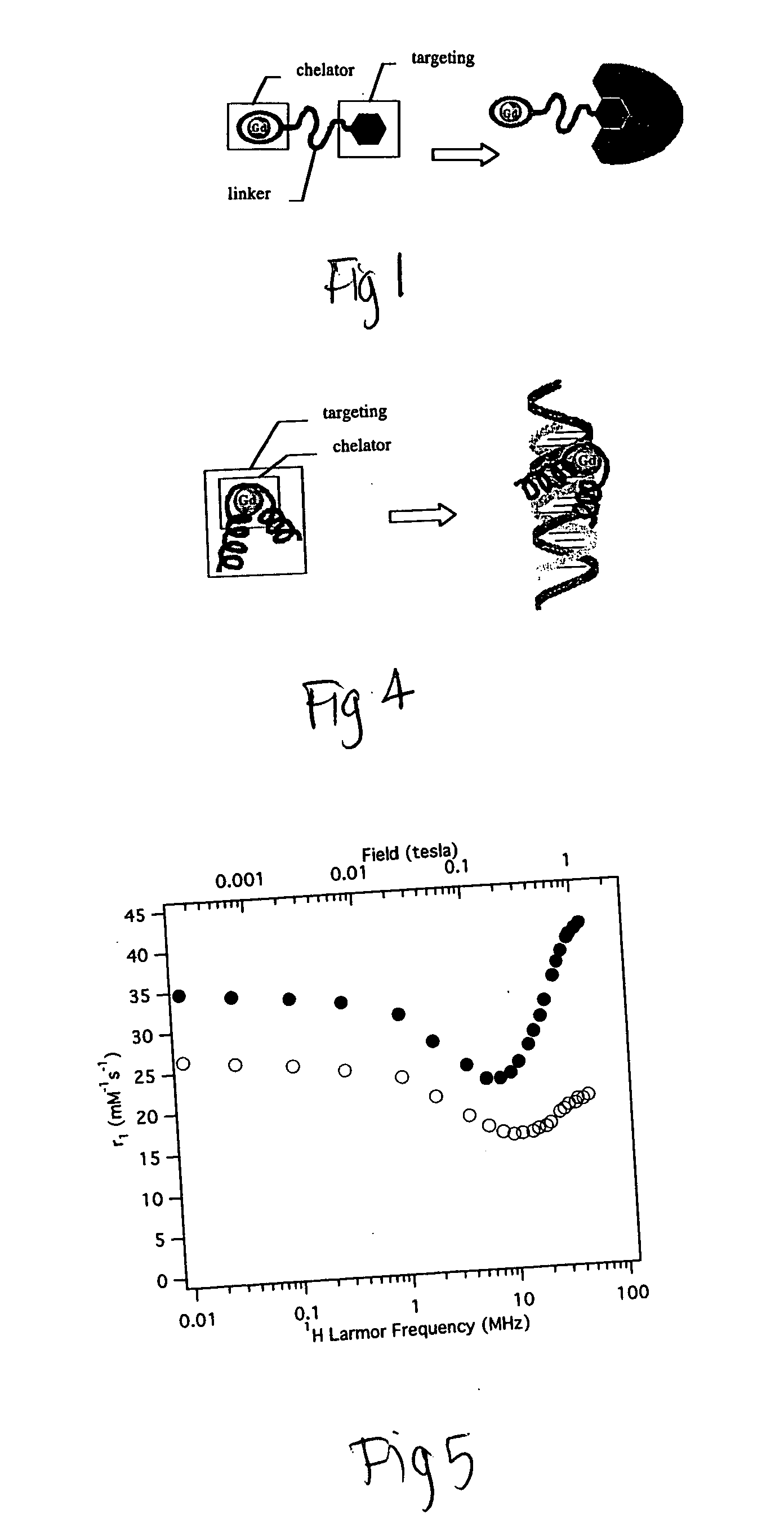
[0162] If a chelating moiety is incorporated within a targeting group, internal motion and rotation flexibility may be reduced and greater relaxiv...
PUM
| Property | Measurement | Unit |
|---|---|---|
| Mass | aaaaa | aaaaa |
| Molality | aaaaa | aaaaa |
| Molecular weight | aaaaa | aaaaa |
Abstract
Description
Claims
Application Information