Arterial diseases and injuries are common and often have severe consequences including death.
Atherosclerosis is a major problem in the aged
population, particularly those in developed countries.
This
disease tends to be progressive in a considerable number of instances and may result in significant morbidity; and, in instances of severe
atherosclerotic disease,
lower limb amputations and / or mortality.
Although a
catheter-based contrast arteriography technique generally provides high quality arterial images, there is a risk of arterial injury or damage by the
catheter and its
insertion.
Furthermore, such a technique may result in a
stroke, loss of a limb,
infarction or other injury to the tissue supplied by the artery.
In addition, hemorrhage at the
catheter insertion or perforation sites may require blood transfusions.
Moreover,
kidney failure and brain injury may result from the toxic effects of the X-
ray contrast.
Further, although catheter arteriography is highly accurate under ideal conditions, it often fails to demonstrate distal vessels suitable for bypass in more than half of the patients with
severe disease.
Additionally, overlapping
cortical bone can make interpretation of overlying vessels difficult.
These type of tests require a skilled and experienced examiner to maintain acceptable accuracy and are often problematic in obese individuals.
Although imaging from the
groin vessels to the level of the mid popliteal artery is possible in many patients, arterial information below the mid popliteal artery is frequently inaccurate.
In addition, because sonography provides indirect information of the arterial characteristics, sonography cannot image stenoses directly in the majority of cases.
As such,
estimation of the degree of luminal
stenosis relies on velocity measurements which can often be inaccurate.
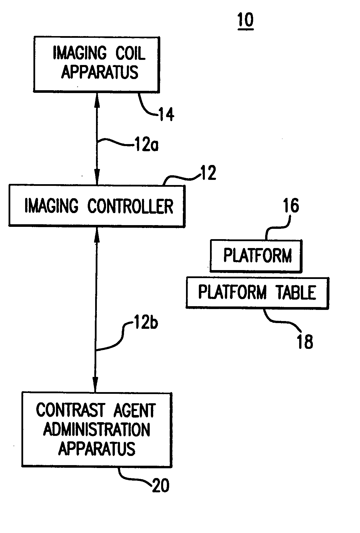
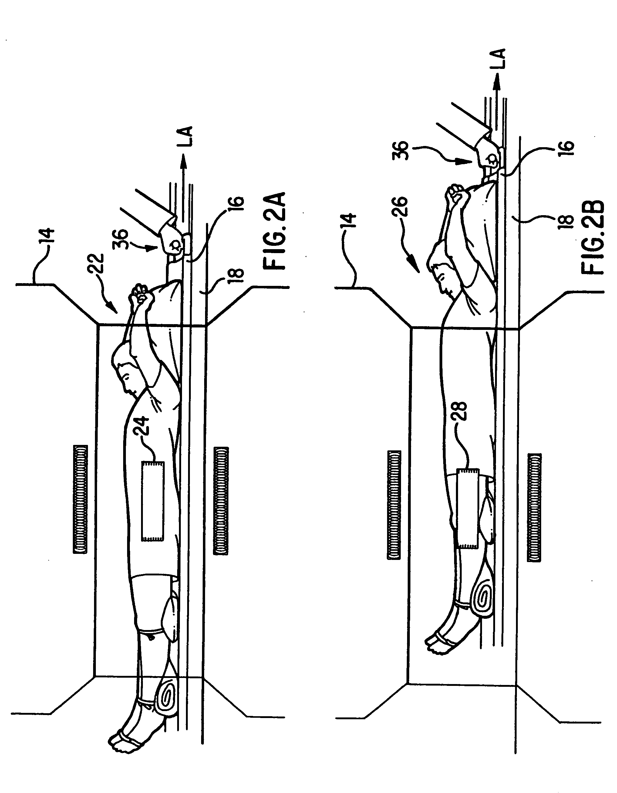
One of the limitations to employing contrast enhanced
magnetic resonance angiography for imaging of the
lower limb vasculature has been the lack of a suitable coil for imaging of a sufficiently large field-of-view which encompasses the lower extremities.
Another potentially more serious problem involves limitations of
magnet imaging region.
In this regard, the length of the
magnet imaging region for conventional
magnet resonance apparatus is insufficient to cover the entire anatomical region-of-interest in one acquisition.
As such, it is necessary to reposition the patient and perform an additional localizer for successive locations down the leg—all of which is time-consuming.
In order to maintain spatial resolution, thin slices must be obtained giving poor spatial “coverage” per unit time.
Although acquisition of the 2-D time-of-flight images in the
coronal plane would significantly reduce imaging time and therefore cost, imaging in this plane would result in saturation of the flowing blood and non-diagnostic studies.
In
spite of the high quality images of the abdominal aortic aneurysms,
thoracic aorta, renal and mesenteric arteries, and
aorta and iliac vessels which have been consistently obtained using contrast enhanced magnetic
resonance arteriography, there still remains several problems with using such imaging techniques to evaluate arteries in lower extremities.
Although this overcomes the problems of both in-plane saturation effects and the necessity for expensive and as yet experimental surface coils, the imaging volume is limited by the largest field-of-view.
Even if this large field-of-view could be used, the increased matrix size necessary to maintain resolution would increase examination time (increased number of phase-encoding steps) and increase
echo time (increased frequency-encoding steps), both of which are undesirable.
Additionally, the
signal-to-
noise from the top and bottom ends of the imaging volume would likely be inadequate for diagnostic purposes; and even if adequate images-were obtained over the entire imaging volume, the anatomical coverage would still be insufficient for adequate evaluation of the lower extremities.
This time
delay in collecting image data for the second, lower level would be of such a magnitude that enhancement of lower limb veins would complicate interpretation of images and probably render lower extremity arterial imaging non-diagnostic.

