Activatable imaging probes

a technology of active imaging and probes, applied in the field of biological and cell biology, can solve the problems of low target/background ratio, many limitations of conventional near infrared fluorescence probes, etc., and achieve the effect of altering the optical properties of chromophores

Inactive Publication Date: 2005-08-04
WEISSLEDER RALPH +2
View PDF43 Cites 69 Cited by
  • Summary
  • Abstract
  • Description
  • Claims
  • Application Information

AI Technical Summary

Benefits of technology

[0034] The invention also features in vivo optical imaging methods for guiding therapeutic, e.g., surgical, interventions by: (a) administering to a subject an imaging probe including a chromophore attachment moiety and a plurality of chromophores wherein the plurality of chromophores are chemically linked to the chromophore attachment moiety so that upon activation of the imaging probe, the optical properties of the chromophores are altered; (b) allowing time for molecules in a target tissue to activate the probe, if the molecules and/or target tissue are present; (d) illuminating the target tissue with light of a wavel

Problems solved by technology

Despite good penetration of biological tissues by light, conventional near infrared fluorescence probes are subje

Method used


Image

  • Activatable imaging probes
  • Activatable imaging probes
  • Activatable imaging probes

Examples


example 1

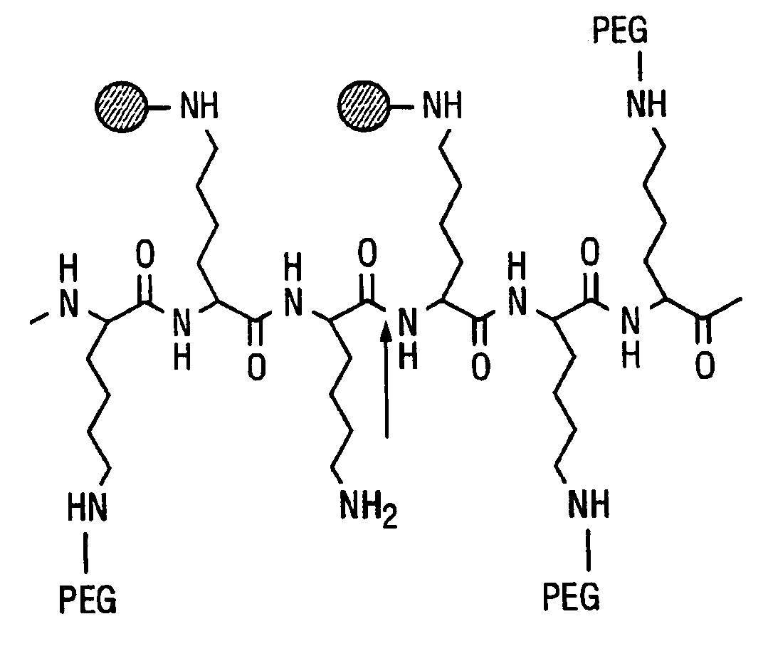
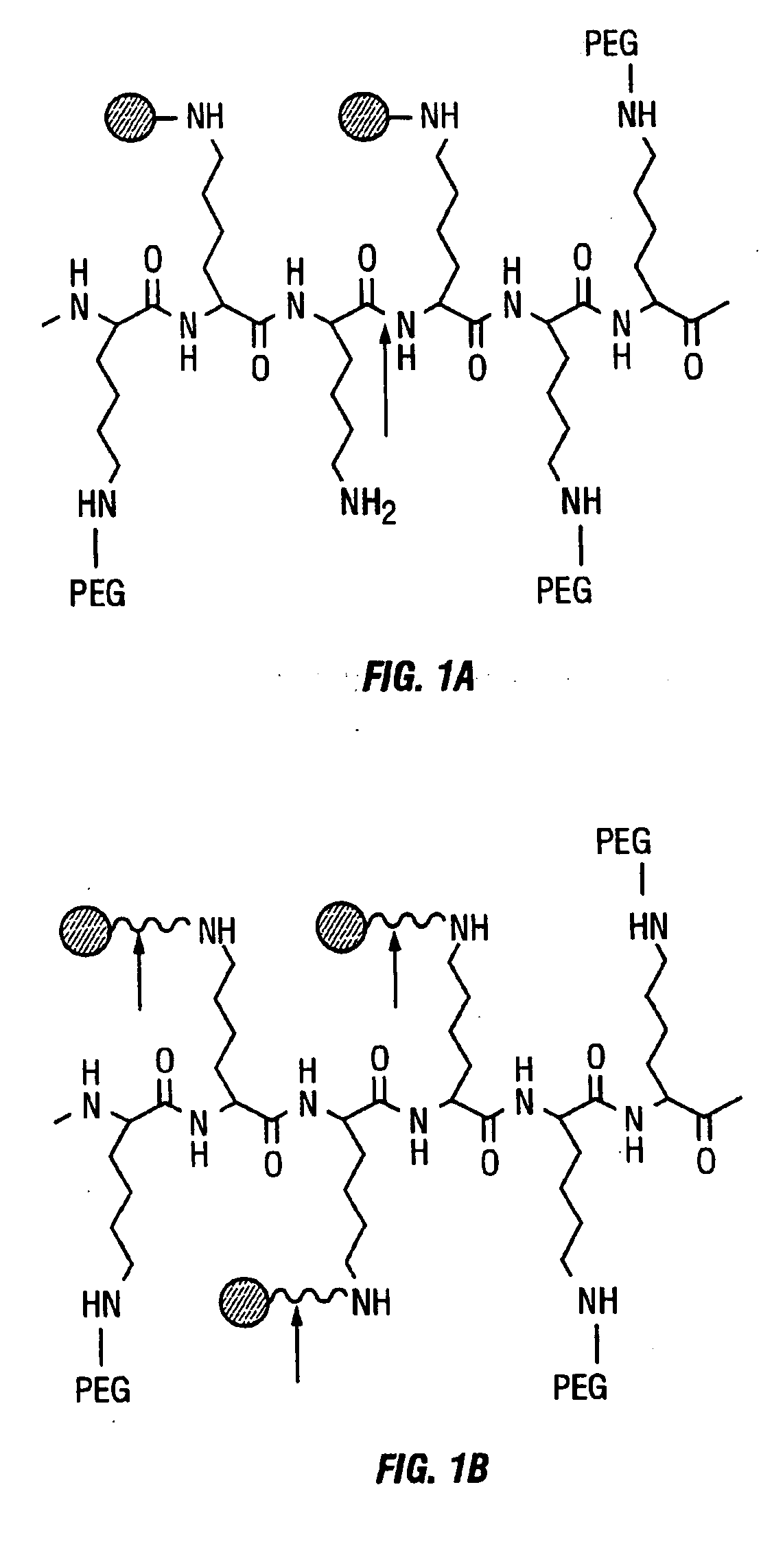
[0144] To demonstrate the ability of the probes to image tumors, we tested the near intramolecularly-quenched infrared imaging probe (Cy11-PL-MPEG; 20% fluorochrome loading) in tumor-bearing mice. Nude mice bearing tumor line 9L or LX1 received 2 nmol of Cy11-PL-MPEG intravenously. The mice were imaged by near infrared light immediately and up to 36 hours after intravenous administration of the probe. The tumor was visible as an area of intense fluorescence, in contrast to the surrounding tissue. An increase in fluorescence signal within tumor was observed as a function of time, as the probe was internalized into tumor cells and became activated by endosomal hydrolases.

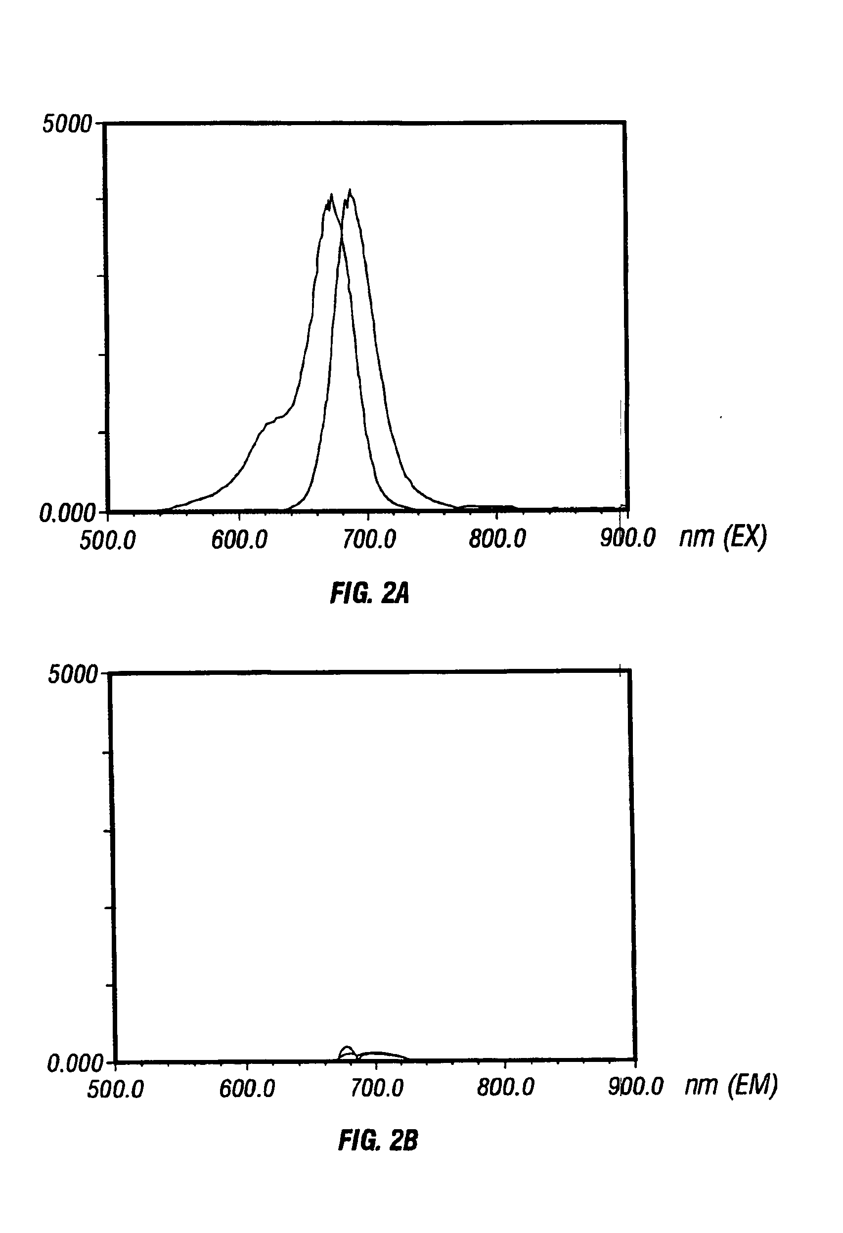
[0145] Using cathepsin D (2000, Cancer Res. 60: 4953-4958) as a model target protease, we synthesized a long circulating, synthetic graft copolymer bearing near infrared (NIR) fluorochrome positioned on cleavable substrate sequences. In its native state, the reporter probe was essentially non-fluorescent at 700 nm du...

example 2

[0147] To demonstrate the ability of fluorescent probes to image colonic polyps, malignant and benign Apc-Min (C57BL / 6J-ApcMin) mice, a strain highly susceptible to spontaneous intestinal adenoma formation, were evaluated after the intravenous injection of 2 nmol per mouse of cathepsin B sensitive probe. Twenty-four hours after probe injection, animals were sacrificed and colons resected. White light and fluorescent images demonstrated the marked difference in fluorescent signal intensity in the polyps as compared to adjacent normal epithelium.

[0148] The resulting marked increase in contrast between normal and abnormal tissue may be exploited during colonoscopy (or endoscopy) to aid in lesion detection.

example 3

[0149] To demonstrate the ability of the probes of the current invention to image ovarian cancer, very small peritoneal tumor deposits using CaD− and CaD+ cell lines (transfected 3Y1 rat embryonic tumor cell line) were implanted into mice intraperitoneally. The Cathepsin D probe described in more detail previously was then administered IV and the peritoneal surfaces were imaged 24 hours later using white light (i.e. as in conventional endoscopy) or at 700 nm (NIRF imaging). Microscopic deposits of 300 μm could be readily detected by NIRF imaging that were not visible by white light imaging.

[0150] The resulting marked increase in detection of minimal residual disease in ovarian cancer may be exploited during laproscopy (or endoscopy) to aid in lesion detection and to monitor therapy.


PUM

PropertyMeasurementUnit
Therapeuticaaaaaaaaaa
Nucleic acid sequenceaaaaaaaaaa
Fluorescenceaaaaaaaaaa

Abstract

The invention relates to activatable imaging probes that includes a chromophore attachment moiety and one or more, e.g., a plurality of, chromophores, such as near-infrared chromophores, chemically linked to the chromophore attachment moiety so that upon activation of the imaging probe the optical properties of the plurality of chromophores are altered. The probe optionally includes protective chains or chromophore spacers, or both. Also disclosed are methods of using the imaging probes for optical imaging.

Description

CROSS-REFERENCE TO RELATED APPLICATIONS [0001] This application claims priority from the following three United States Provisional Patent Application Ser. No. 60 / 260,123, filed on Jan. 5, 2001, 60 / 277,352, filed on Mar. 19, 2001, and Serial Number to be Determined, filed on Nov. 9, 2001, all of which are incorporated herein by reference in their entireties.FIELD OF THE INVENTION [0002] The invention relates to biochemistry, cell biology, and optical imaging. BACKGROUND OF THE INVENTION [0003] Optically based biomedical imaging techniques have advanced over the past decade due to developments in laser technology, sophisticated reconstruction algorithms, and imaging software originally developed for non-optical, tomographic imaging modes such as CT and MRI. Visible wavelengths are used for optical imaging of surface structures by means of endoscopy and microscopy. [0004] Near infrared wavelengths (approx. 600-1000 nm) have been used in optical imaging of internal tissues, because near...

Claims


Application Information

Patent Timeline
IPC IPC(8): C12Q1/68
CPCC12Q1/6816C12Q1/6823C12Q1/6841C12Q2565/107
Inventor WEISSLEDER, RALPHTUNG, CHING-HSUANMAHMOOD, UMAR
Owner WEISSLEDER RALPH
Why Eureka
Try Eureka