Systems and methods for electrosurgical tissue contraction within the spine

a tissue contraction and electrosurgical technology, applied in the field of electrosurgical tissue contraction systems and methods, can solve the problems of back and leg pain, narrowing of the nerve openings in the side of the spine, weakening of the shock absorption properties of the disc, etc., to achieve precise and rapid removal, inhibit the clogging of the lumen, and minimize damage and necrosis of the underlying tissue

Inactive Publication Date: 2005-08-25
ARTHROCARE
View PDF3 Cites 147 Cited by
  • Summary
  • Abstract
  • Description
  • Claims
  • Application Information

AI Technical Summary

Benefits of technology

[0012] In one aspect of the invention, a method is provided for treating herniated discs within a patient's spine. Specifically, the method of the present invention comprises introducing an electrosurgical probe within the spine (either in open or endoscopic procedures) so that one or more electrode terminal(s) are brought into at least partial contact or close proximity with the target herniated disc. High frequency voltage is then applied between the electrode terminal(s) and one or more return electrode(s) to apply sufficient energy to the disc tissue to reduce a volume of the disc, thereby relieving pressure on a spinal nerve. In one embodiment, the high frequency voltage is sufficient to ablate a portion of the nucleus pulposis, either the extruded portion outside of the annulus or a portion or all of the pulposis within the annulus. In another embodiment, the electrode terminal is advanced into the annulus and sufficient high frequency voltage is applied to contract or shrink the collagen fibers within the nucleus pulposis. This causes the pulposis to shrink and withdraw from its impingement on the spinal nerve. In other embodiments, the present invention may be used to both ablate the extruded portion of the nucleus pulposis, and then to contract or shrink the inner disc material to allow repair of the annulus.
[0020] The apparatus will preferably further include a fluid delivery element for delivering electrically conducting fluid to the electrode terminal(s) and the target site. The fluid delivery element may be located on the probe, e.g., a fluid lumen or tube, or it may be part of a separate instrument. Alternatively, an electrically conducting gel or spray, such as a saline electrolyte or other conductive gel, may be applied the target site. In this embodiment, the apparatus may not have a fluid delivery element. In both embodiments, the electrically conducting fluid will preferably generate a current flow path between the electrode terminal(s) and one or more return electrode(s). In an exemplary embodiment, the return electrode is located on the probe and spaced a sufficient distance from the electrode terminal(s) to substantially avoid or minimize current shorting therebetween and to shield the return electrode from tissue at the target site.

Problems solved by technology

This results in a weakening of the shock absorption properties of the disc and a narrowing of the nerve openings in the sides of the spine which may pinch your nerve.
This disc degeneration can eventually cause back and leg pain.
The mere proximity of the nucleus pulposis or a damaged annulus to a nerve can cause direct pressure against the nerve, resulting in numbness and weakness of leg muscles.
In some cases, the disc tissue is irreparably damaged, thereby necessitating removal of a portion of the disc or the entire disc to eliminate the source of inflammation and pressure.
In addition, the risk of instability from ligament and bone removal is generally lower in endoscopic procedures than with open discectomy.
Unfortunately, these mechanical instruments greatly lengthen and increase the complexity of the procedure.
In addition, these instruments sever blood vessels within this tissue, usually causing profuse bleeding that obstructs the surgeon's view of the target site.
In addition to the above problems with mechanical instruments, there are serious concerns because these instruments are not precise, and it is often difficult, during the procedure, to differentiate between the target disc tissue, and other structures within the spine, such as bone, cartilage, ligaments, nerves and non-target tissue.
Unfortunately, lasers are both expensive and somewhat tedious to use in these procedures.
Another disadvantage with lasers is the difficulty in judging the depth of tissue ablation.
These monopolar devices, however, suffer from the disadvantage that the electric current will flow through undefined paths in the patient's body, thereby increasing the risk of unwanted electrical stimulation to portions of the patient's body.
This current, however, may inadvertently flow along body paths having less impedance than the defined electrical path, which will substantially increase the current flowing through these paths, possibly causing damage to or destroying surrounding tissue or neighboring peripheral nerves.

Method used


Image

  • Systems and methods for electrosurgical tissue contraction within the spine
  • Systems and methods for electrosurgical tissue contraction within the spine
  • Systems and methods for electrosurgical tissue contraction within the spine

Examples


Embodiment Construction

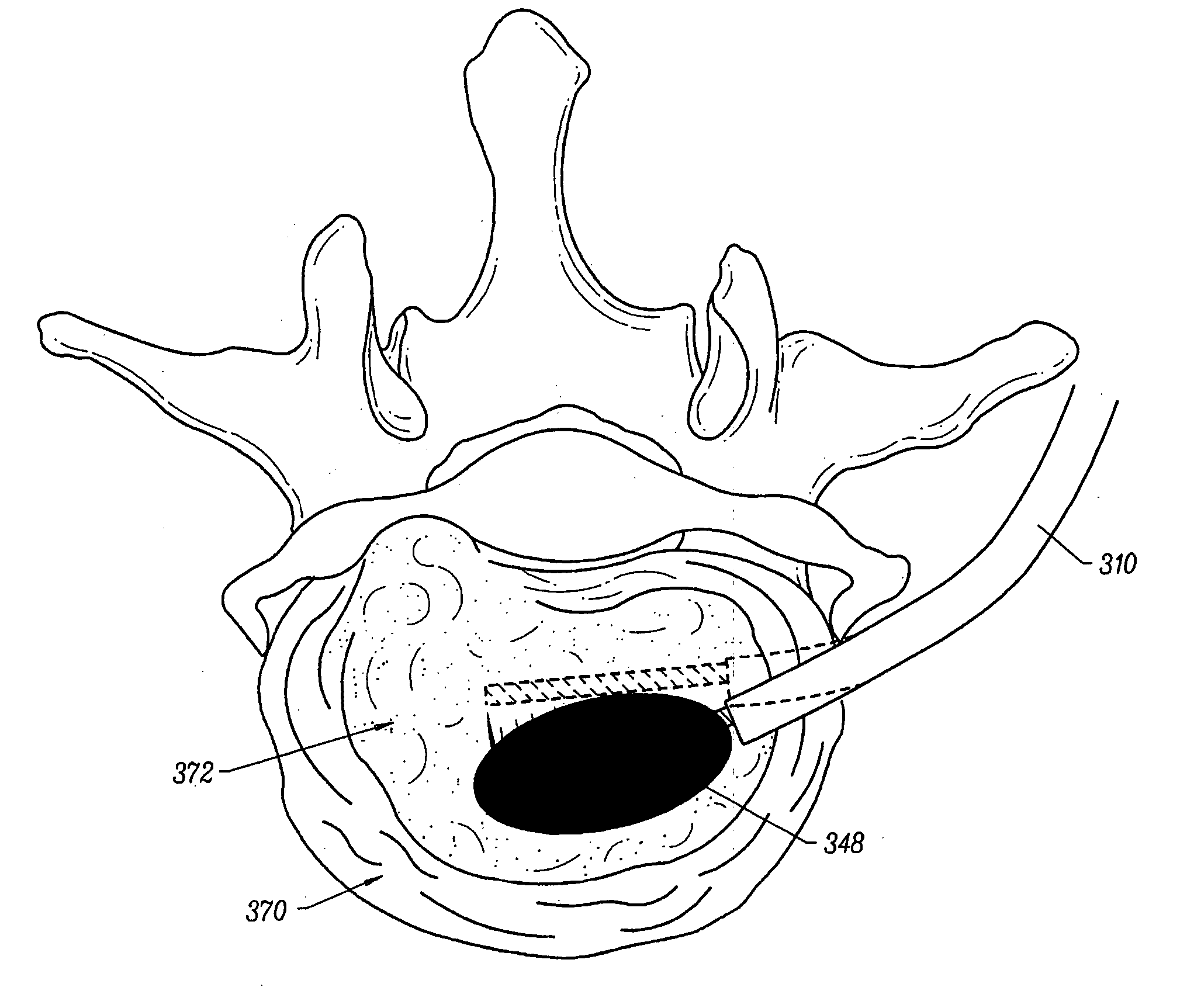
[0051] The present invention provides systems and methods for selectively applying electrical energy to a target location within or on a patient's body, particularly including tissue or other body structures in the spine. These procedures include laminectomy / disketomy procedures for treating herniated disks, decompressive laminectomy for stenosis in the lumbosacral and cervical spine, medial facetectomy, posterior lumbosacral and cervical spine fusions, treatment of scoliosis associated with vertebral disease, foraminotomies to remove the roof of the intervertebral foramina to relieve nerve root compression and anterior cervical and lumbar diskectomies. These procedures may be performed through open procedures, or using minimally invasive techniques, such as thoracoscopy, arthroscopy, laparascopy or the like.

[0052] In the present invention, high frequency (RF) electrical energy is applied to one or more electrode terminals in the presence of electrically conductive fluid to remove ...


PUM

PropertyMeasurementUnit
temperatureaaaaaaaaaa

Abstract

The present invention provides systems and methods for selectively applying electrical energy to a target location within a patient's body, particularly including tissue in the spine. The present invention applies high frequency (RF) electrical energy to one or more electrode terminals in the presence of electrically conductive fluid to contract collagen fibers within the tissue structures. In one aspect of the invention, a system and method is provided for treating herniated or swollen discs within a patient's spine by applying sufficient electrical energy to the disc tissue to contract or shrink the collagen fibers within the nucleus pulposis. This causes the pulposis to shrink and withdraw from its impingement on the spinal nerve.

Description

RELATED APPLICATIONS [0001] This application is a continuation of U.S. patent application Ser. No. 09 / 512,742, filed Feb. 24, 2000, which is a continuation-in-part of U.S. patent application Ser. No. 08 / 690,159, filed Jul. 18, 1996, the complete disclosure of which is incorporated herein by reference for all purposes. This application also derives priority from U.S. patent application Ser. No. 08 / 942,580 entitled “Systems and Methods for Electrosurgical Tissue Contraction”, filed on Oct. 2, 1997 and U.S. patent application entitled “Systems and Methods for Endoscopic Sinus Surgery”, filed on Dec. 15, 1997, the complete disclosures of which are incorporated herein by reference for all purposes. [0002] The present invention is related to commonly assigned co-pending Provisional Patent Application Nos. 60 / 062,996 and 60 / 062,997, non-provisional U.S. patent application Ser. No. 08 / 970,239 entitled “Electrosurgical Systems and Methods for Treating the Spine”, filed Nov. 14, 1997, and Ser...

Claims


Application Information

Patent Timeline
Patent Type & Authority Applications(United States)
IPC IPC(8): A61B18/12A61B17/00A61B17/22A61B18/00A61B18/14A61B19/00A61F2/02A61M1/00A61M3/02A61M25/01
CPCA61B18/12A61M25/0158A61B18/14A61B18/1402A61B18/148A61B18/1482A61B18/1485A61B18/149A61B18/1492A61B2017/00026A61B2017/00084A61B2017/00097A61B2017/00101A61B2017/00247A61B2017/00292A61B2017/003A61B2017/22001A61B2017/22038A61B2018/00029A61B2018/00083A61B2018/00119A61B2018/0016A61B2018/00178A61B2018/00196A61B2018/00327A61B2018/00392A61B2018/00434A61B2018/00505A61B2018/00577A61B2018/00583A61B2018/00642A61B2018/0066A61B2018/00678A61B2018/00702A61B2018/00726A61B2018/00761A61B2018/00791A61B2018/00797A61B2018/00821A61B2018/00827A61B2018/00875A61B2018/00886A61B2018/1213A61B2018/124A61B2018/1253A61B2018/126A61B2018/1273A61B2018/1467A61B2018/1472A61B2018/1497A61B2018/162A61B2018/165A61B2019/5276A61B2218/002A61M3/0283A61M25/0133A61M25/0147A61B18/1206A61B2090/378A61F2/2493A61B2218/007
Inventor UNDERWOOD, RONALD A.DAVISON, TERRY S.THAPLIYAL, HIRA V.EGGERS, PHILIP E.
Owner ARTHROCARE
Why Eureka
Try Eureka