Targeted transscleral controlled release drug delivery to the retina and choroid

a technology of transscleral controlled release and drug delivery, which is applied in the field can solve the problems of affecting the treatment effect of retinal and choroidal diseases, affecting the ability of the retina to absorb biologic agents, so as to achieve the effect of minimal systemic absorption and toxicity

Inactive Publication Date: 2005-09-22
ADAMIS ANTHONY P +2
View PDF99 Cites 71 Cited by
  • Summary
  • Abstract
  • Description
  • Claims
  • Application Information

AI Technical Summary

Benefits of technology

[0014] We have developed a minimally invasive transscleral drug delivery modality that can target and unidirectionally deliver therapeutic concentrations of bioactive proteins to the choroid and retina without significant systemic absorption or tissue damage. These methods may be used to treat a number of diseases affecting the retina and choroid.
[0028] As used herein, by “facilitating” is meant enhancing the efficacy of the delivery of a diagnostic or therapeutic agent to the sclera.
[0030] By “treat” is meant to submit or subject an animal, tissue, cell, lysate or extract derived from a cell tissue, or molecule derived from a cell tissue to a compound in order to lessen the effects of a retinal or choroid disease.
[0035] The present invention provides a means by which to treat macular degeneration, diabetic retinopathy, retinitis pigmentosa, retinal vein occlusions, sickle cell retinopathy, and other diseases of the choroidal and retinal tissues. Defined amounts of the agent can be delivered for prolonged periods of time (weeks to years). The risk of systemic absorption and toxicity is minimal with this method, and intraocular injections, with the concomitant problems of retinal detachment and enthopthalmitis are avoided.

Problems solved by technology

The development of strategies to treat retinal and choroidal diseases is an ongoing therapeutic challenge.
Delivery of biologic agents to the retina and choroid is rendered difficult by the fact that the internal limiting membrane (ILM) of the retina is impermeable to linear molecules larger than 40 kDa and globular molecules greater than 70 kDa, precluding intravitreous or topical transcorneal delivery (Smelser et al., In Structure of the eye, II.
Abstract number 3761, 1999) Thus, one of the major problems in the treatment of retinal and choroidal diseases is the delivery of therapeutic levels of medications to target tissues.
Although a variety of local delivery systems for the treatment of posterior segment eye conditions have evolved over the years, each system has limitations.
Topical administration is widely utilized in clinical practice but is inefficient for treating posterior segment conditions due to a long diffusional path length, counter-directional intraocular convection, lacrimation, and corneal impermeability to large molecules, and thus requires frequent dosing (Lang, Adv.
Depot injections, by either subconjunctival or retro-orbital routes, are a relatively simple and effective means of achieving local concentrations of medications (Baum, 1973, supra; Baum, 1976, supra) but are limited to medications such as antibiotics and corticosteroids and can spill over into the systemic circulation.
Intravitreal injection is effective for directed intraocular delivery, but at the same time increases the risk for complications such as vitreous hemorrhage, retinal detachment, and endophthalmitis.
Yet, transscleral iontophoresis can be accompanied by deleterious retinal necrosis and gliosis, making this method undesirable (Lim, Opthalmology, 100:373-6, 1993).
Since these polymers and liposomal spheres are placed into the vitreous for intraocular release, these methods have inherent limitations, such as the need for repeated implantation subsequent to drug delivery, and the risk of intraocular injury if the devices are not fixed to the sclera.
Photoactivated liposomes or caged-molecules may hold promise for selective delivery (Asrani et al., Invest Ophthalmol. Vis. Sci. 38:2702-2710, 1997; Arroyo et al., Thromb. Haemost. 78:791-793, 1997); however, radiational and thermal damage associated with these modalities, as well as the limited repertoire of drugs that can be enveloped limit the clinical utility of these approaches at present.

Method used


Image

  • Targeted transscleral controlled release drug delivery to the retina and choroid
  • Targeted transscleral controlled release drug delivery to the retina and choroid
  • Targeted transscleral controlled release drug delivery to the retina and choroid

Examples


example 1

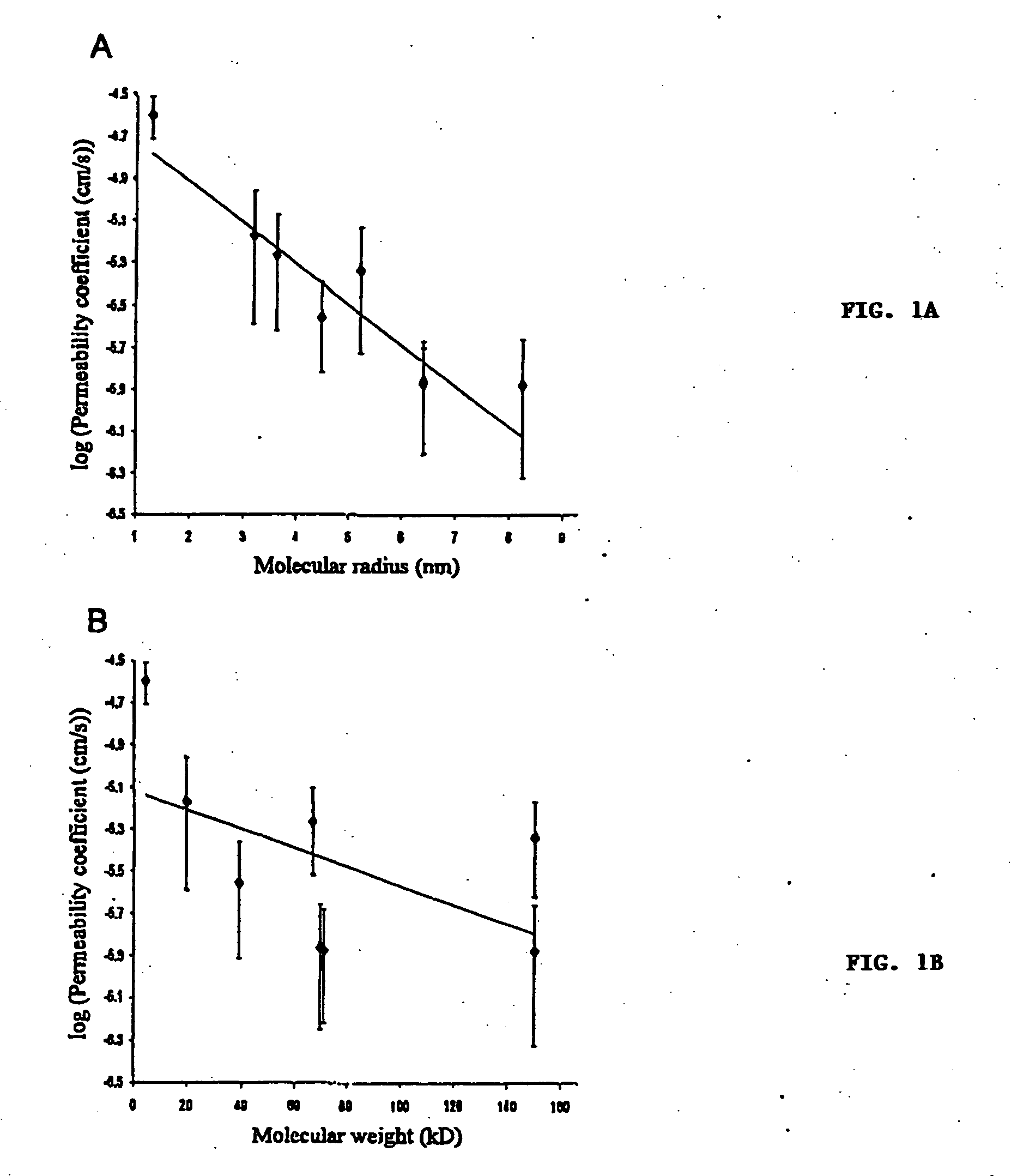
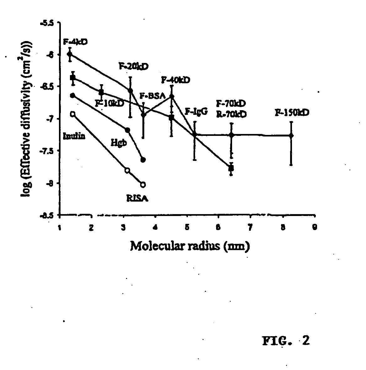
In Vitro Diffusion of High Molecular Weight Compounds Through Sclera

Isolation and Preparation of Fresh Rabbit Sclera

[0054] Dutch-belted rabbits (Pine Acres Rabbitry, Vermont, Mass.), each weighing 2-3 kg, were anesthetized with intramuscular 40 mg / kg ketamine (Abbott, N. Chicago, Ill.) and 10 mg / kg xylazine (Bayer, Shawnee Mission, Kans.). Scleral thickness was measured using a RK-5000 ultrasound pachymeter (KMI Surgical Products, West Chester, Pa.). The eyes were enucleated immediately before sacrifice and immersed in Unisol (Alcon, Ft. Worth, Tex.) for 10 minutes or less. The adherent muscles were excised and episcleral tissue was removed with a sterile gauze sponge. Areas free of nerve and vessel emissaries were used to obtain 7×12 mm slices of sclera under microscopic caliper guidance. Each piece of sclera was used on the day of isolation.

In Vitro Diffusion Apparatus

[0055] A 5×10 mm window, 2 mm from the bottom, was created on one face of a spectrophotometry polystyrene c...

example 2

Osmotic Pump Implantation

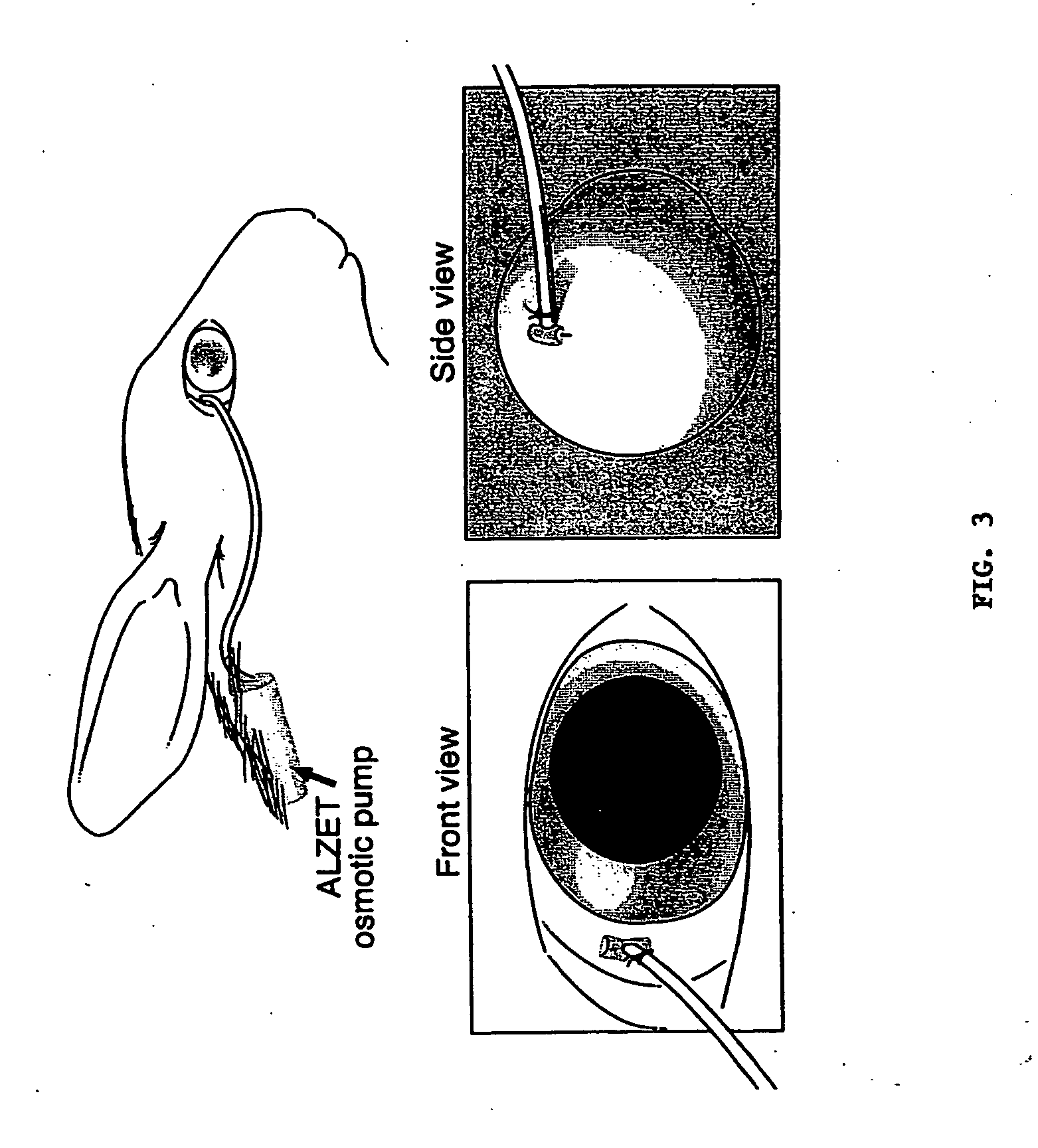
[0074] Dutch-belted rabbits were anesthetized with intramuscular ketamine (40 mg / kg; Abbott, N. Chicago, Ill.) and xylazine (10 mg / kg; Bayer, Shawnee Mission, Kans.). Osmotic pumps (ALZET, ALZA, Palo Alto, Calif.) were loaded with drug and incubated at 37° C. prior to implantation. The osmotic pump was implanted subcutaneously between the scapulae and connected to a brain infusion kit (ALZA), which was modified so that the tip could be secured to, and face, the orbital surface of the sclera with a single biodegradable polyglactin 910 suture (Ethicon, Somerville, N.J.) in the superotemporal quadrant of the eye, 14 to 16 mm posterior to the limbus (near the equator) (FIG. 3). Care was taken to make a partial thickness pass through the sclera. If uvea, blood or vitreous was observed during the procedure, the experiment was terminated.

example 3

Collection of Ocular Tissue and Blood

[0075] Blood was collected by cardiac puncture prior to surgical enucleation of the eyes under deep anesthesia. Aqueous humor of each eye was collected using a 30-gauge needle. Vitreous humor, retina, choroid, and orbital tissue of both eyes were dissected and isolated under a microscope. The choroid of the treated eye was separated into two hemispheres, proximal (in which the tip of the pump was centered) and distal to the tip of the pump. Animals were sacrificed with intracardiac pentobarbital (100 mg / kg) (Vortech, Dearborn, Mich.).


PUM

PropertyMeasurementUnit
molecular weightaaaaaaaaaa

Abstract

The invention provides methods for delivering a therapeutic or diagnostic agent to the eye of a mammal. The method involves contacting sclera with a therapeutic or diagnostic agent so as to permit its passage through the sclera into the choroidal and retinal tissues. The sclera may be contacted with a therapeutic or diagnostic agent together with a device for enhancing transport of the agent through the sclera.

Description

CROSS REFERENCE TO RELATED APPLICATIONS [0001] This application is a continuation-in-part of U.S. Ser. No. 60 / 114,905, filed Jan. 5, 1999.FIELD OF THE INVENTION [0002] The field of the invention is treatment of retinal and choroidal diseases. BACKGROUND OF THE INVENTION [0003] The development of strategies to treat retinal and choroidal diseases is an ongoing therapeutic challenge. Highly specific biologic reagents, which include proteins of relatively high molecular weight, are under development for the treatment of ocular diseases. For example, the overexpression of vascular endothelial growth factor (VEGF) is required for retinal-ischemia associated intraocular neovascularization, leading to proliferative diabetic retinopathy, while mutations in tissue inhibitor of metalloproteinase-3 (TIMP-3) result in Sorsby's macular dystrophy. [0004] Delivery of biologic agents to the retina and choroid is rendered difficult by the fact that the internal limiting membrane (ILM) of the retina ...

Claims


Application Information

Patent Timeline
Patent Type & Authority Applications(United States)
IPC IPC(8): A61F9/007A61D7/00A61K31/711A61K31/721A61K38/00A61K38/16A61K39/395A61K45/00A61P27/02A61P43/00C07K16/24
CPCA61K38/16C07K16/24A61K2039/505A61P27/02A61P43/00
Inventor ADAMIS, ANTHONY P.GRAGOUDAS, EVANGELOS S.MILLER, JOAN W.
Owner ADAMIS ANTHONY P
Why Eureka
Try Eureka