Methods, compositions, and kits for analysis of enzyme activity in cells

a technology of enzyme activity and kit, which is applied in the field of methods, compositions, and kits for analysis of enzyme activity in cells, can solve the problems of inability to accurately reflect the activity of enzymes, disruption and death of cells, and the destruction of cells that are typically used to extract or purify enzymes, etc., to achieve the effect of facilitating intracellular delivery of substrates and greater yield

Inactive Publication Date: 2005-11-03
APPL BIOSYSTEMS INC
View PDF11 Cites 16 Cited by
  • Summary
  • Abstract
  • Description
  • Claims
  • Application Information

AI Technical Summary

Benefits of technology

[0005] In one aspect, methods are provided for detecting an activity of an enzyme in a cell. In some embodiments, the method involves contacting the cell with a liposome that contains (i.e. encapsulates) at least one substrate that is capable of producing a detectable light signal when acted on by the enzyme, and detecting the amount of a light signal in the cell wherein the amount indicates a level of the enzyme activity in the cell. Encapsulation in a liposome facilitates intracellular delivery of substrate. Delivery is faster and / or in greater yield (on a concentration basis) than when the unencapsulated substrate is used. The detectable light signal can be a fluorescent signal or a chemiluminescent signal, for example.

Problems solved by technology

However, the preparation of extracts or purified enzymes typically involves destruction of the cell.
Because enzymes are frequently bound in highly organized enzyme pathways, the disruption and death of the cell can greatly affect enzyme activity.
Such methods are indirect, and may not accurately reflect the activity of the enzyme.
However, such fluorescent substrates are usually of considerable size and very few are able to penetrate the outer membrane of the cell.
These techniques can damage the cell membrane and alter its permeability, thus allowing the substrate, and other molecules to leak from the cell.
Moreover, these methods are physically disruptive to the cell and can lead to drastic alteration its overall biochemistry or, in many cases, cell death.

Method used


Image

  • Methods, compositions, and kits for analysis of enzyme activity in cells
  • Methods, compositions, and kits for analysis of enzyme activity in cells
  • Methods, compositions, and kits for analysis of enzyme activity in cells

Examples


example 1

7.1 Example 1

Preparation of 100 nm Cationic Liposomes

[0092] Large unilamellar vesicles (LUV) of diameter 100 nm were prepared by the extrusion method essentially as described by Chatterjee, et al. (in Methods in Molecular Biology: Liposome Methods and Protocols (S. Basu and M. Basu eds.), Humana Press, 2002, vol. 199, chapter 1). Sterile techniques were used throughout this procedure to prevent bacterial contamination of the liposomes. 1,2-Dioleoyl-sn-Glycero-3-Phosphocholine (DOPC) (12.5 mg, Avanti Polar Lipids, catalog no. 850375), and 1,2-Dioleoyl-sn-Glycero-3-Ethylphosphocholine (EDOPC) (12.5 mg, Avanti Polar Lipids, catalog no. 890704) were dissolved in chloroform (5 ml) in a 25 ml recovery flask. The solvent was thoroughly evaporated under high vacuum to leave a thin film. Sterile filtered PBS buffer (2 ml, pH 7.2) was added and the suspension was subjected to five cycles of freezing (−78° C., dry ice acetone bath) under argon and thawing (40° C.) to hydrate the lipids. The r...

example 2

7.2 Example 2

Preparation of 100 nm Cationic Liposomes Containing DDAO-gal or DDAO

[0093] DOPC and EDOPC were dissolved in chloroform and the solvent was evaporated to leave a thin film, as described in Example 1. 9H-(1,3-dichloro-9,9-dimethylacridin-2-one-7-yl) β-D-galactopyranoside (DDAO-gal) (5 mg, Molecular Probes, catalog no. D-6488) or 7-hydroxy-9H-(1,3-dichloro-9,9-dimethylacridin-2-one) (DDAO) (5 mg, Molecular Probes, catalog no. H-6482) was dissolved in DMSO (20 μl) and added to sterile filtered PBS buffer (2 ml, pH 7.2). The DDAO-gal or DDAO in PBS was added to the lipids and the suspension was subjected to five cycles of freezing (−78° C., dry ice acetone bath) under argon and thawing (40° C.) to hydrate the lipids. The resulting LMV were extruded and purified as described in Example 1. The concentration of DDAO-gal or DDAO within the liposomes was estimated to be 0.25 mg / ml.

example 3

7.3 Example 3

Cell Staining with Liposomes Containing DDAO-gal

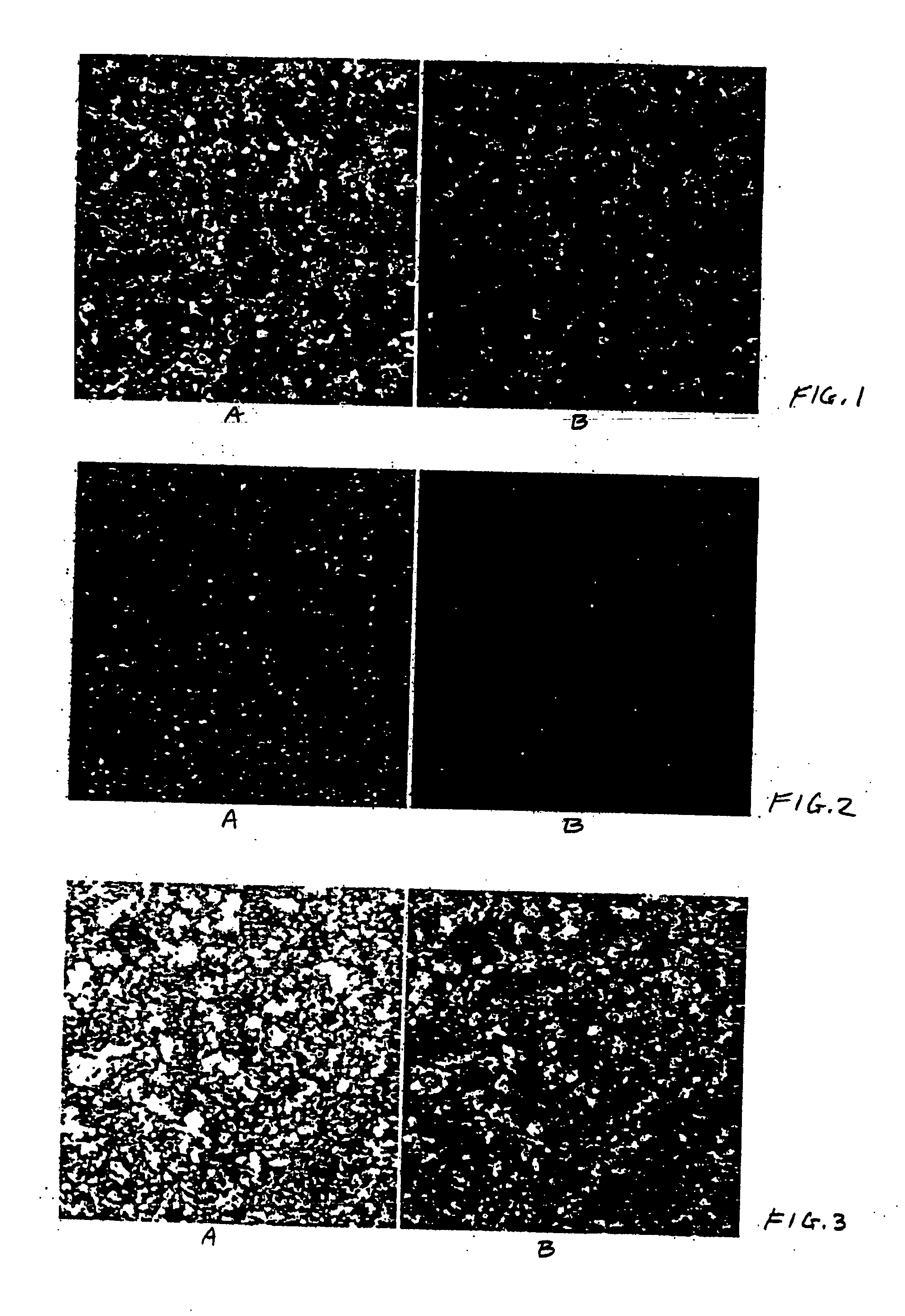
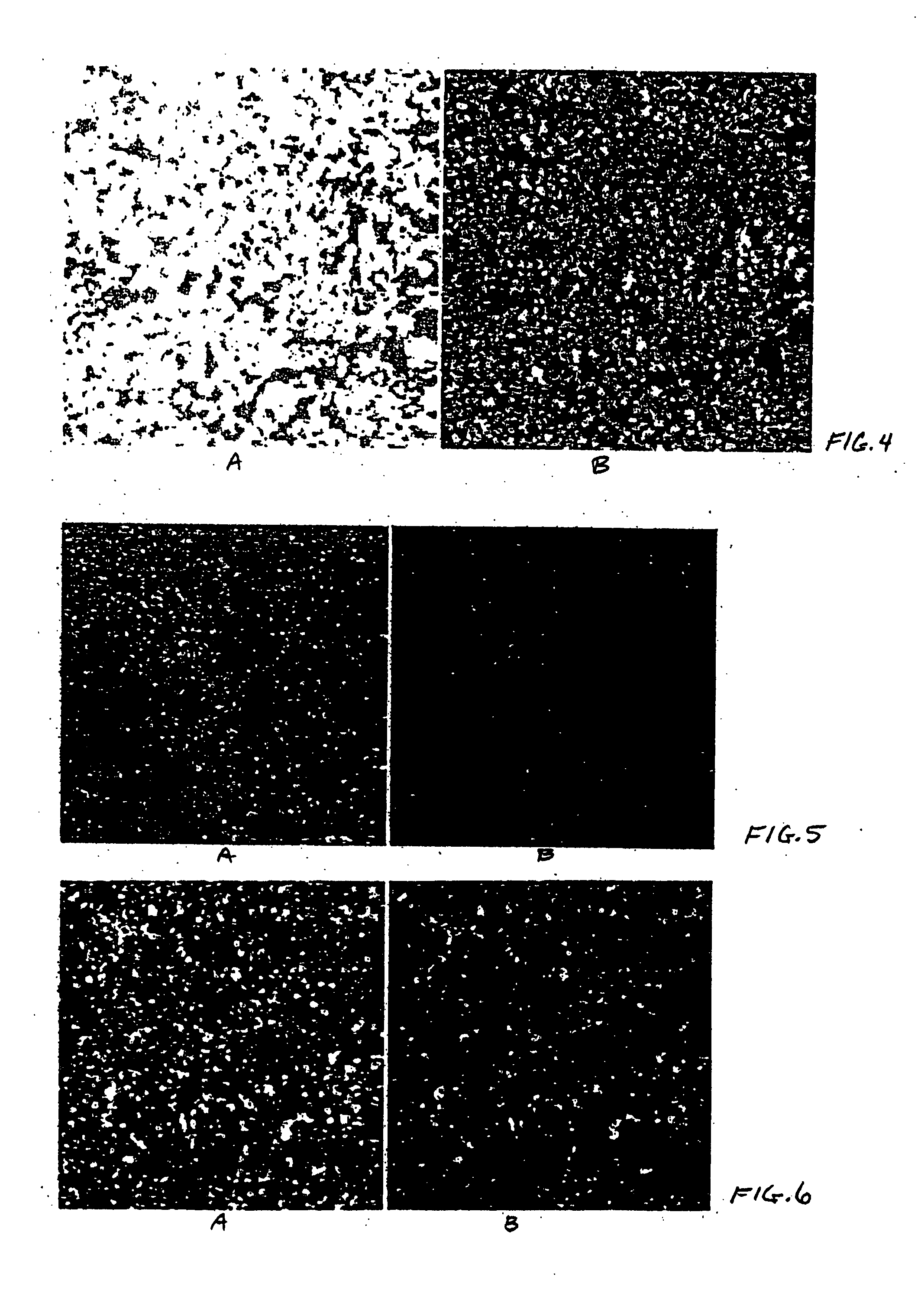
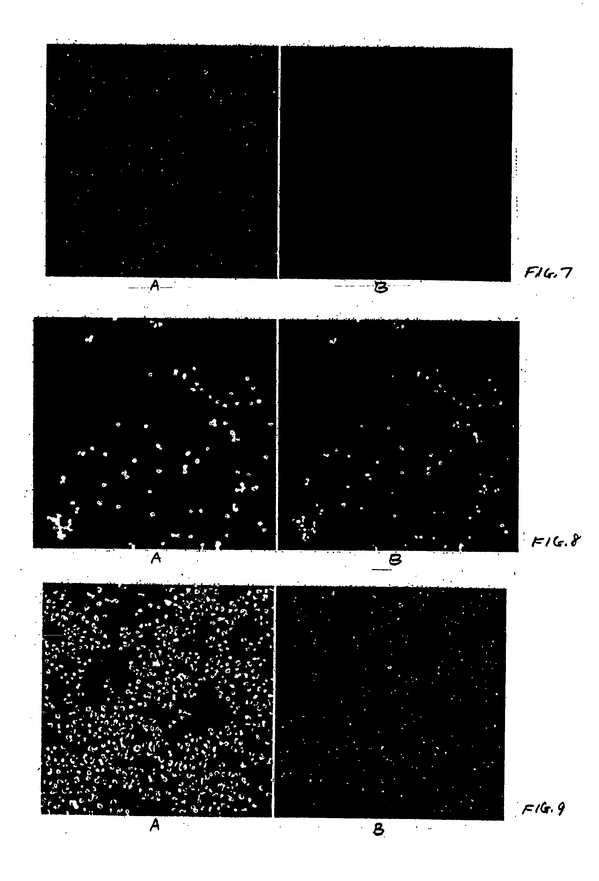
[0094] Psi 2 BAGα cells (ATCC, catalog no. CRL-9560) in Dulbecco's modified Eagle's medium (ATCC, catalog no. 30-2002) containing 5% Fetal Bovine Serum (FBS, HyClone, catalog no. SH30071.03) and 1% penicillin-streptomycin solution (pen / strep, ATCC, catalog no. 30-2300) were seeded (200 μl / well, 10,000 cell / well) in black 96 well microtiter plates (ABI, catalog no. 4308776). HeLa cells (ATCC, catalog no. CCL-2) in Eagle's minimum essential medium (ATCC, catalog no. 30-2003) containing 5% FBS and 1% pen / strep were seeded (200 μl / well, 10,000 cell / well) in black 96 well microtiter plates (ABI, catalog no. 4308776). CHO cells (ATCC, catalog no. CCL-61) in F-12K medium (ATCC, catalog no. 30-2004) containing 5% FBS and 1% pen / strep were seeded (200 μl / well, 10,000 cell / well) in black 96 well microtiter plates (ABI, catalog no. 4308776). After overnight incubation at 37° C. under 5% CO2 the supernatants were removed carefully so...


PUM

PropertyMeasurementUnit
diameteraaaaaaaaaa
temperatureaaaaaaaaaa
wavelengthaaaaaaaaaa

Abstract

In one aspect, the present disclosure relates to methods for detecting an activity of an enzyme in a cell. In some embodiments, the methods include contacting a cell with a liposome containing at least one substrate thereby facilitating introduction of the substrate into the cell. The substrate is capable of producing a detectable light signal when acted on by the enzyme, and the signal is detected. The methods can be used in screening agents that can inhibit or activate an enzyme activity. The methods can also be used in various downstream assays such the detection of interactions between intracellular proteins, screening for variants of an enzyme, and detection of various diseases. Compositions and kits for carrying out the various methods are also provided.

Description

1. CROSS-REFERENCE TO RELATED APPLICATIONS [0001] This application claims benefit under 35 U.S.C. §119(e) to application Ser. No. 60 / 529,953, entitled “Methods and Compositions for Analysis of Enzyme Activity in Cells,” filed Dec. 15, 2003 and application Ser. No. 60 / 542,425, entitled “Methods, Compositions, and Kits for Analysis of Enzyme Activity in Cells,” filed Jan. 6, 2004.2. FIELD [0002] The disclosure generally concerns methods and compositions useful in the identification and / or analysis of enzyme activity in cells. 3. INTRODUCTION [0003] The study of enzyme activity within cells has been conducted using a variety of methods. In one method, the cell membrane is broken to create a cytosolic extract of cellular components including the enzyme which is the object of study. Various tests are performed on the cytosolic extract or on the purified enzyme to determine the enzyme activity. However, the preparation of extracts or purified enzymes typically involves destruction of the ...

Claims


Application Information

Patent Timeline
Patent Type & Authority Applications(United States)
IPC IPC(8): C12Q1/00C12Q1/34C12Q1/37C12Q1/42C12Q1/44C12Q1/48C12Q1/66C12Q1/68G01N33/50
CPCC12Q1/00C12Q1/34C12Q1/37C12Q1/42C12Q1/44G01N2500/10C12Q1/485G01N33/5008G01N33/502G01N33/5091C12Q1/48
Inventor GRAHAM, RONALD J.SEKAR, MICHAELBARBISIN, MAURA
Owner APPL BIOSYSTEMS INC
Why Eureka
Try Eureka