Gastrointestinal stem cells and uses thereof

Inactive Publication Date: 2005-11-17
BAYLOR COLLEGE OF MEDICINE
View PDF1 Cites 17 Cited by
  • Summary
  • Abstract
  • Description
  • Claims
  • Application Information

AI Technical Summary

Benefits of technology

[0015] Amongst the mammalian tissues that display continuous cell turnover (bone marrow, skin and gastro-intestinal epithelium, for example), the epithelium of the small intestine has by far the highest rate of turnover. In both rodents and humans, the majority of cells within the epithelium are replaced every 3-4 days, although certain cell types exhibit slower turnover. As such, the present invention exploits the nature of these highly proliferative cells in contemplating their use for therapeutic purposes.
[0016] The four principal cell lineages found in the epithelium (enterocytes, goblet cells, Paneth cells and enteroendocrine cells) are all known to arise from stem cells located deep in the crypts. Despite extensive studies on the kinetics of intestinal stem cells, in both physiological and pathological (e.g., after radiation injury) states, to date there are no published methods for isolation of the gastrointestinal stem cells of the present invention. A related issue is that for any preparation of putative stem cells, characterization of their “stemness” comprises demonstration of the capacity for extensive proliferation and/or the capacity for the production of differentiated progeny. Even cell aggregates (that include epithelial stem cells as well as other cells, including mesenchymal cells, for example) have so far proven refractory to differentiation in vitro. In contrast, such aggregates grafted either subcutaneously or mucosally have been demonstrated to differentiate into all four principal cell lineages of the small intestinal epithelium. Thus, in vivo cell transplant models provide, in some embodiments, conditions for demonstrating “sternness”. In addition, such models are preferable to characterize the desired therapeutic potential of preparations of intestinal epithelial stem cells described herein.
[0017] In a specific embodiment of the present invention, intestinal epithelial stem cells share certain characteristics with bone marrow (and other) stem cells through their capacity to be identified and isolated by established flow cytometric methods. Flow cytometric methods are described herein for isolation of viable stem cells from the epithelium of the intestine, such as the exemplary mouse small intestine. For example, intestinal epithelial cells are subjected to sorting procedures that have been successfully used with bone marrow cells to identify a side population (“SP”) fraction that is highly enriched in stem cells. The SP cells are then analyzed for the presence of markers of “stemness” and/or absence of differentiation markers. An additional or alternative sorting approach may utilize assessment of one or more markers with antibodies. Pursuant to these embodiments, a mouse model having local, limited sterilization of intestinal crypts is described such that an ideal denuded region is generated for subs

Problems solved by technology

A related issue is that for any preparation of putative stem cells, characterization of their “stemness” comprises demonstration of the capacity for extensive proliferation and/or the capacity for the producti

Method used


Image

  • Gastrointestinal stem cells and uses thereof
  • Gastrointestinal stem cells and uses thereof
  • Gastrointestinal stem cells and uses thereof

Examples


example 1

Methods to Generate Single Cell Suspensions of Intestinal Tissue Suitable for Flow Cytometry

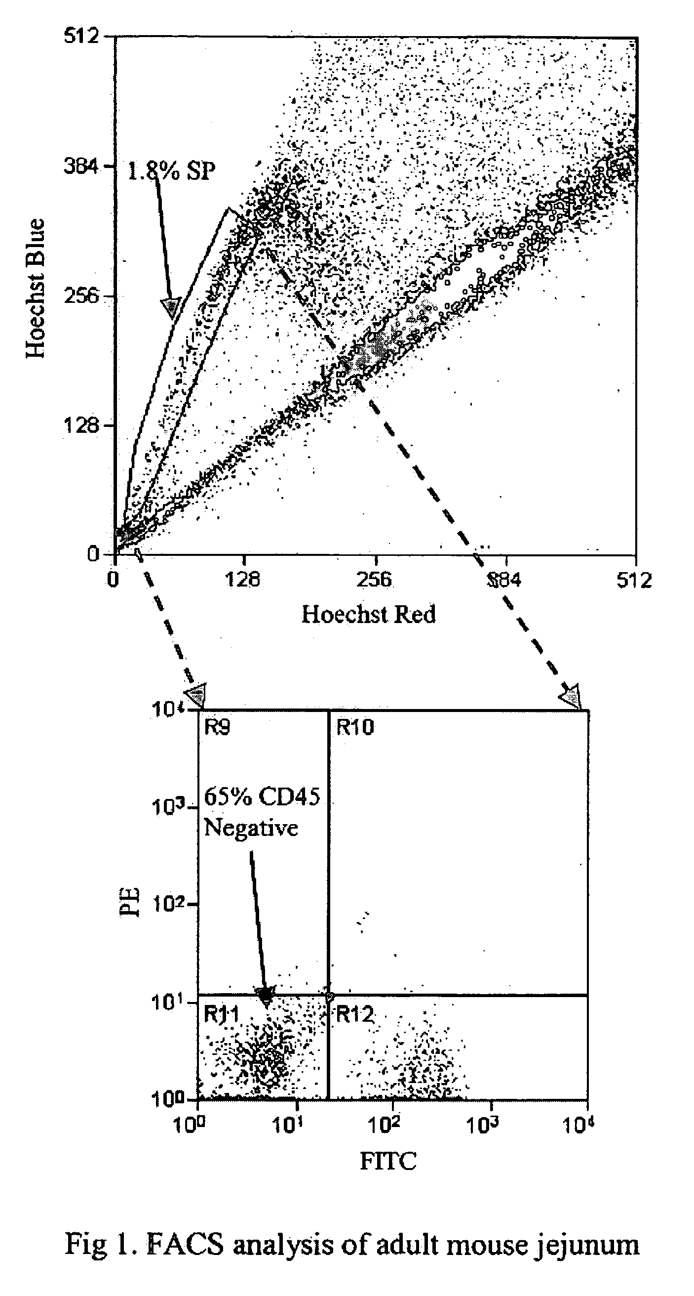
[0107] Although flow cytometry has been used for cell cycle analyses of the intestinal epithelium (Cheng and Bjerknes, 1985; Pallavicini et al., 1984), such studies typically use glutaraldehyde fixed cells (Cheng and Bjerknes, 1983). To date, there are no reports on the use of flow cytometry to separate viable cells from the epithelium (the exception being intraepithelial lymphocytes). Thus, in a specific embodiment of the invention, mucous-free single cell suspensions of mouse intestinal epithelium are produced, and the cells remain viable through the manipulations required for preparation, staining and sorting. In an additional specific aspect of the invention, staining with Hoechst 33342 yields a side population (SP), as in other tissues.

[0108] To pursue both these goals, adult mouse intestine was dissociated, such as by a modification of the method of Evans, et al. (1992) and then subje...

example 2

Methods to Generate Single Cell Suspensions of Colon and / or Stomach Tissue Suitable for Flow Cytometry

[0128] As with the small intestine, there is a method that yields a viable single cell preparation suitable for flow cytometry from the colon and / or stomach. Two studies were performed with mouse colon using the same digestion method as was used for the jejunum, yet significant clumping was generated, presumably due to mucus from the goblet cells that are more numerous in the epithelium of the colon than of the jejunum. Logical modifications to the method include use of a mucolytic agent such as dithiothreitol and / or N-acetyl-cysteine. In specific embodiments, modifications to the Evans et al. method (1992) are utilized, or a mannitol-based method described by Perret et al. for rat colon (1977) is utilized. Interestingly, this method requires no proteases and yet apparently gives epithelial cells that are totally free from the underlying stroma. The preparations have high viability...

example 3

General Methods for Sorting

[0129] Intestinal epithelial cells are suspended at 106 cells per ml in DME containing 2% fetal calf serum (FCS) and 5 μg / ml Hoechst 33342 (Sigma). After incubation at 37° C. for 90 min, they are spun down and resuspended in cold Hank's balanced salt solution containing 2% FCS. Staining with appropriate dilutions of FITC-labeled anti-CD3 and FITC-labeled anti-CD-45 (BD Pharmingen) is performed on ice for 10 min. After washing, cells will be resuspended in Hank's salt solution containing 2 μg / ml propidium iodide to exclude dead cells, and kept at 4° C. until sorting. A 350 nm argon-laser is used to excite Hoechst 33342 and propidium iodide. Analysis is performed on a triple-laser instrument (Cytomation, Fort Collins, USA) at 405 / 30 and 670 / 30 nm, as described for bone marrow (Goodell et al., 1996). The side population (SP) is identified and selected by gating on the characteristic emission fluorescence profile of SP cells. A second argon laser, tuned to 48...


PUM

PropertyMeasurementUnit
Compositionaaaaaaaaaa
Mechanical propertiesaaaaaaaaaa
Disorderaaaaaaaaaa

Abstract

The present invention relates to compositions and methods concerning isolated gastrointestinal stem cells. Particularly, the invention provides isolated gastrointestinal stem cells comprising a CD45 negative marker, a collagen IV negative marker, and that is Msi-1 positive. In particular embodiments the isolated cells are comprised in a single cell suspension. In other particular embodiments, the isolated cells are utilized for therapeutic purposes, such as for a gene therapy vector and/or for replenishing stem cells in a gastrointestinal tract in need thereof.

Description

[0001] The present invention claims priority to U.S. Provisional Patent Application Ser. No. 60 / 557,797, filed Mar. 30, 2004, which is incorporated by reference herein in its entirety.STATEMENT REGARDING FEDERALLY SPONSORED RESEARCH OR DEVELOPMENT [0002] The present invention was developed at least in part from funding provided by the National Institutes of Health Grant No. R21 DK61132, by the National Institutes of Health Grant No. T32DK07664, and by the National Institutes of Health Grant No. P30DK56338. The United States Government may have certain rights in the invention.FIELD OF THE INVENTION [0003] The present invention regards the fields of cell biology, molecular biology, and medicine. In particular, the invention is related to the field of intestinal stem cells and therapeutic uses thereof. BACKGROUND OF THE INVENTION [0004] The gastrointestinal tract comprises a useful structure conducive to its digestive and absorptive functions, and it also provides a source of highly pr...

Claims


Application Information

Patent Timeline
IPC IPC(8): A61K35/12A61K48/00C12N5/074
CPCA61K35/12C12N2509/00C12N5/068
Inventor HENNING, SUSAN J.
Owner BAYLOR COLLEGE OF MEDICINE
Why Eureka
Try Eureka