
Emboli filter export system

a filter and emboli technology, applied in the field of emboli filter export system, can solve the problems of localized cell death or microinfarct, myocardial infarct, paralysis, etc., and achieve meaningful long-term clinical benefit, prevent ischemic complications, and reduce platelet aggregation

Active Publication Date: 2005-12-15
MEDTRONIC INC
View PDF63 Cites 159 Cited by
  • Summary
  • Abstract
  • Description
  • Claims
  • Application Information

AI Technical Summary

Benefits of technology

[0008] A significant reason for ischemic injury during percutaneous procedures can be generation of emboli which block smaller distal vessels. One approach to curb this complication has been to use pharmacological therapies during the time of the intervention. Limited therapeutic success has been reported with the use of calcium channel blockers, adenosine, and sodium nitroprusside (Webb, J G, Carere, R G, Virmani, R, Baim, D, Teirstein, P S, Whitlow, P, McQueen, C, Kolodgie, F D, Buller, E, Dodek, A, Mancini, G B, & Oesterle, S: Retrieval and analysis of particulate debris after saphenous vein graft intervention. J Am Coll Cardiol 2000, 34:468-475, incorporation herein by reference.). Glyoprotein IIb/IIIa inhibitors have been used for percutaneous coronary interventions to reduce platelet aggregation, but also fail to show meaningful long term clinical benefit. (Mathew, V, Grill, D E, Scott, C G, G

Problems solved by technology

Loss of blood flow to surrounding tissue causes localized cell death or microinfarcts.
Cerebral microinfarcts can cause stroke leading to confusion, disturbance of speech, paralysis, visual disturbances, balance disturbances and even death.
In the heart, emboli can cause myocardial infarcts, i.e. heart attacks.
Blockage of capillaries associated with the coronary arteries can result in corresponding microinfarctions / microinfarcs.
Anything that loosens or breaks up this plaque can generate emboli.
The clinical ramifications of emboli are staggering.
As a result, blood flow to the distal vascular bed can be diminished and periprocedural end-organ ischemia and infarction can result.
Distal embolization of large particles produced at the time of such interventions as balloon inflation or stent deployment may obstruct large, epicardial vessels, and smaller particles (as small as 15-100 microns) can cause microinfarcts and / or myocardial infarctions and left ventricular dysfunction.
This is a significant clinical problem.
These complications are due in large measure to CPB-generated emboli.
Ironically, the surgical interventions used to remove or bypass the plaque of arteriosclerosis (e.g., balloon dilatation angioplasty, endarterectomy, bypass grafting and stenting) can themselves disrupt plaque.
But CABG is not the only procedure with potential to generate emboli.
Of the over 1.8 million intervention procedures performed annually, greater than 10% result in neurocognitive disturbance and / or ischemic event.
While daunting, cost figures fail to include the social and financial burden placed on family members upon hospital release.
In summary, embolic events complicating percutanuous endovascular procedures cause high rates of clinically observed neurological disturbances and cardiovascular disease, decreased quality of life and increased patient care spending.
Glyoprotein IIb / IIIa inhibitors have been used for percutaneous coronary interventions to reduce platelet aggregation, but also fail to show meaningful long term clinical benefit.
Surgical procedures for the treatment of renal artery stenosis can also generate emboli.
Foreign material in the stream of flow can cause turbulence or low flow.
Such flow conditions have been shown to increase rates of infection.
Thrombus not only generates emboli, but also increases the risk of infection.
It is evident that a wide variety of embolic events cause high rates of clinically observed symptoms, decreased quality of life and increased patient care spending.
However, in most cases, the device is removed at some point from the flow, generally after the causes of an embolic event are no longer present.
The removal of the device from the patient can disrupt the device in a way that can result in release of some of the emboli from the device.
Any released emboli can flow down stream and pose a risk to the patient.

Method used


Image

  • Emboli filter export system
  • Emboli filter export system
  • Emboli filter export system

Examples


Embodiment Construction

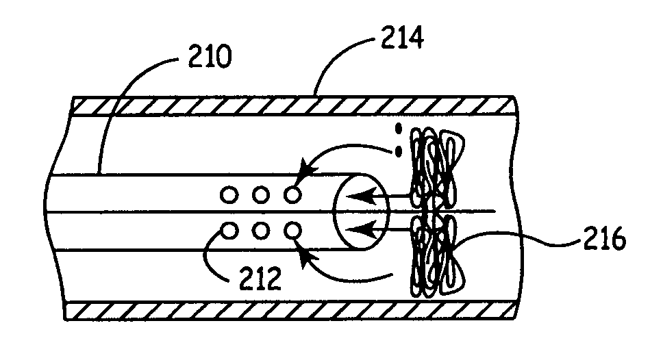
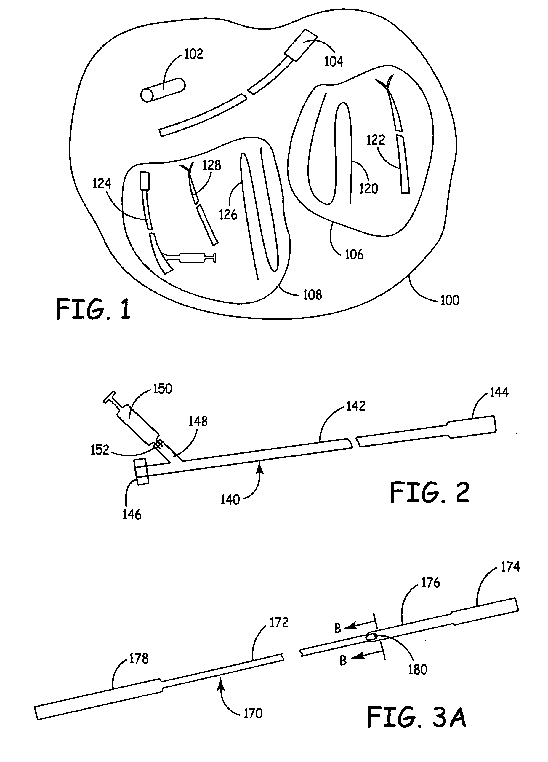
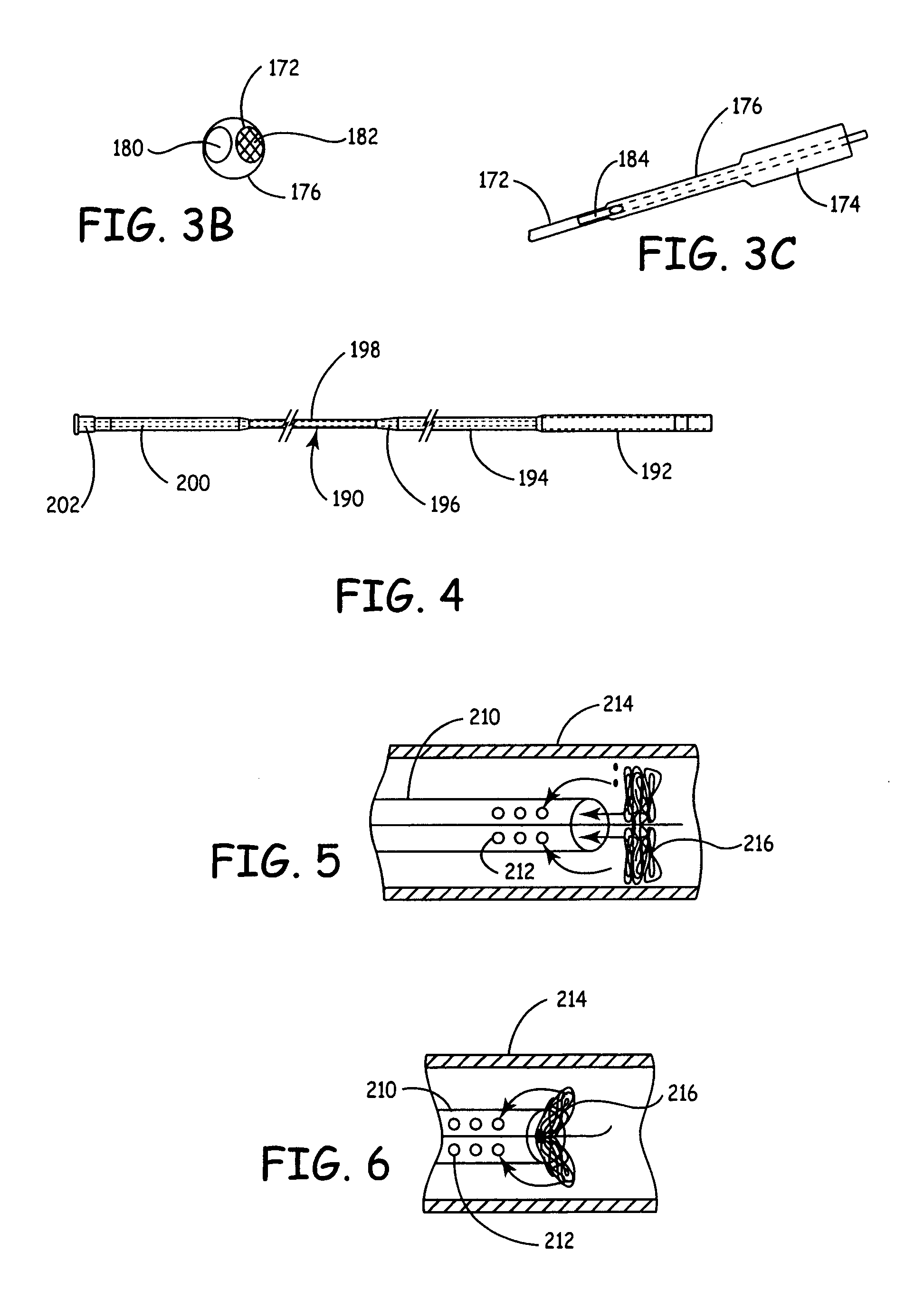
[0041] An improved system for trapping and removing emboli from a patient involves an aspiration catheter that, in some embodiments, both transmits suction to the distal end of the catheter to aspirate liquid into the catheter as well as provides a protective lumen in which to draw the embolism protection device for removal from the patient. Thus, for removal, the embolism protection device can be drawn within the catheter while aspiration is applied. The aspiration catheter then functions as a vehicle for the removal of the embolism protection device. The use of suction along with the sheath catheter system can significantly reduce or eliminate the release of any emboli during the removal of the embolism protection device. In some embodiments, the aspiration catheter comprises an expanded compartment into which the embolism protection device is drawn in a recovery configuration. In some embodiments, the embolism protection device comprises a three dimensional filtering matrix that ...


PUM


Abstract

Methods for the removal of an embolism protection device use aspiration during the drawing of the embolism protection device into the catheter. Generally, the embolism protection device comprises a three dimensional filtering matrix that provides improved filtering without blocking the flow through the patient's vessel. In some embodiments, the embolism protection device can be actuated between a deployed configuration and a removal configuration with a reduced area across the cross section of the vessel lumen. The embolism protection device in the removal configuration can be drawn within the aspiration catheter. The aspiration catheter can have a distal portion with an expanded compartment with an average diameter at least about 20 percent larger than the average diameter of the shaft of the catheter within about 10 centimeters of the expanded compartment. In a rapid exchange version, the rapid exchange segment can have a length of at least about 10 centimeters.

Description

FIELD OF THE INVENTION [0001] The invention relates to catheters for the removal of emboli traps, i.e. embolism protection devices. In particular, the invention relates to aspiration catheters that facilitate the removal of an embolism protection device, which can have a three-dimensional filtering matrix, from a patient's vessels with reduced or eliminated release of emboli. The invention further relates to improved aspiration catheters that are capable of rapid exchange, which can form a protective lumen for a three-dimensional filtering matrix during removal from the patient. BACKGROUND OF THE INVENTION [0002] An embolus can be any particle comprising a foreign and / or native material, which enters the vascular system or other vessel of the body with potential to cause occlusion of blood flow. Emboli can be formed from aggregates of fibrin, blood cells or fragments thereof, collagen, cholesterol, plaque, fat, calcified plaque, bubbles, arterial tissue, and / or other miscellaneous f...

Claims


Application Information

Patent Timeline
IPC IPC(8): A61B17/22A61F2/01A61M1/00A61M29/00
CPCA61B17/22A61B2017/22034A61B2017/22079A61B2217/005A61F2/01A61B2017/22035
Inventor GALDONIK, JASON A.OGLE, MATTHEW F.POKORNEY, JAMES
Owner MEDTRONIC INC
Learn More