Methods, device and system for in vivo diagnosis

a technology diagnostic method, applied in the field of in vivo diagnosis, can solve the problems of high cost of endoscopic examination, poor prognosis, stress, etc., and achieve the effect of facilitating the differen

Inactive Publication Date: 2005-12-29
GIVEN IMAGING LTD
View PDF24 Cites 139 Cited by
  • Summary
  • Abstract
  • Description
  • Claims
  • Application Information

AI Technical Summary

Benefits of technology

[0008] According to embodiments of the invention optically based techniques are used to facilitate the difference between normal and pathological (e.g., malignant) cells in a body lumen. Embodiments of the invention relate to a typically non-invasive autonomous ingestible device, which enables in situ visualization and detection of, for example, neoplastic or malignant or damaged cells or tissue even in areas that are inaccessible to endoscopes.

Problems solved by technology

Gastric cancer is a major cause of death worldwide especially in developing countries.
Delayed detection of GI tract cancers is a major factor contributing to an overall poor prognosis.
The deep anatomical location of organs and parts of the GI tract may also add to the low yield of early detection.
Endoscopic examinations are typically expensive and stressful for patients, leading to very low patient compliance and to low yield of early detection.
Additionally, a large part of the GI tract (for example, most of the small intestine) is inacessible to endoscopes.
Thus, endoscopic examinations provide only a partial answer to the needs of early detection and they are not perceived as beneficial in wide screening for cancers.

Method used


Image

  • Methods, device and system for in vivo diagnosis
  • Methods, device and system for in vivo diagnosis
  • Methods, device and system for in vivo diagnosis

Examples


example 1

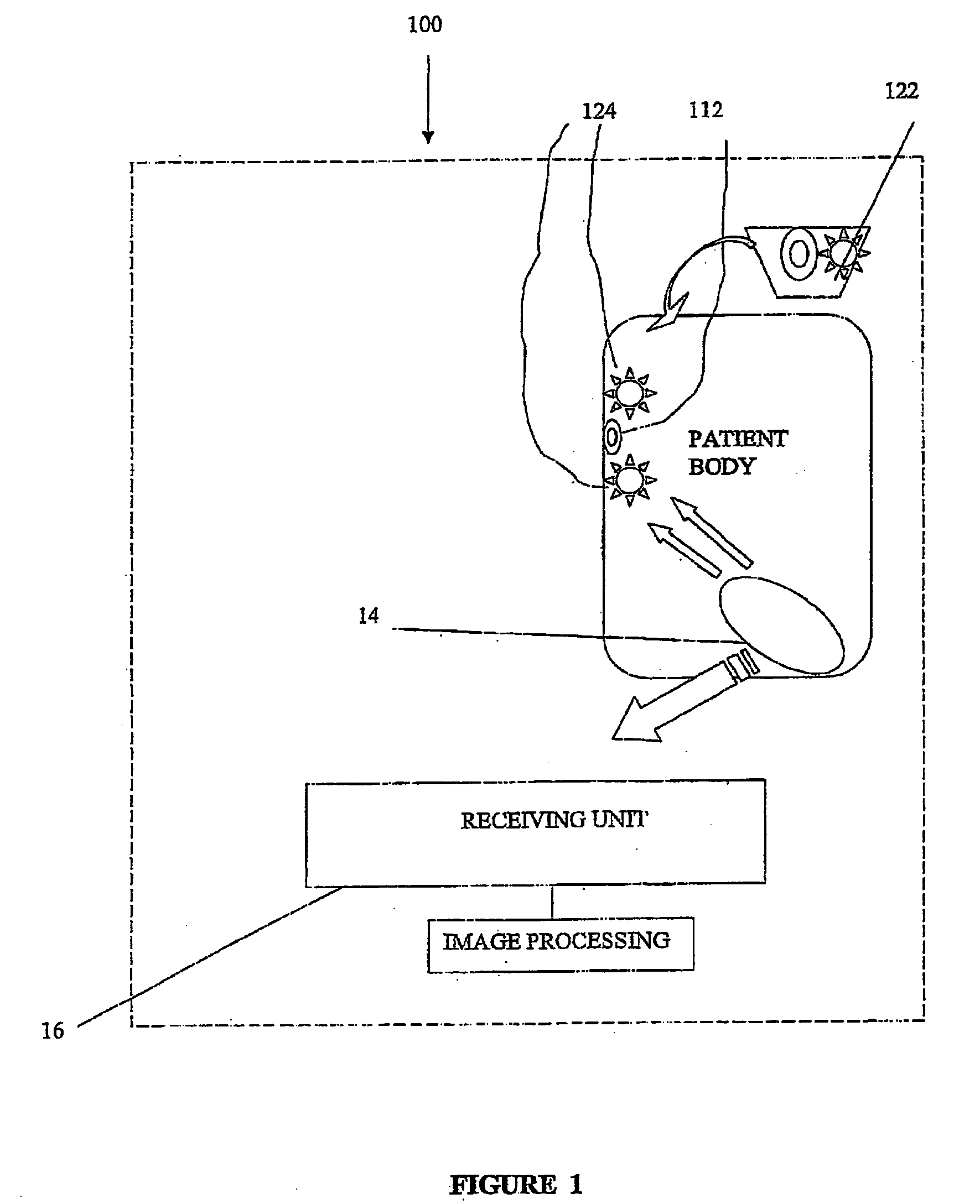
[0039] Procedure for PPD Using an Ingestible Imaging Capsule [0040] 1. A patient after a 12 hour fast is given 0.5 liter water; [0041] 2. half an hour later 5-ALA is administered to the patient in the form of a powder of more than 99% purity (commercially available, e.g. from Medac GmbH, Hamburg, Germany). A water / juice solution is prepared at a dose of 15-60 mg / Kg body weight, and is orally ingested by the patient; [0042] 3. 4-7 hours after oral ingestion the patient ingests 0.5 liter of water to wash the GI tract tissue from excess 5-ALA; [0043] 4. 10 minutes after the ingestion of water the patients ingest a Given™ capsule which includes at least one blue LED and a red filter over parts of the CMOS image sensor; [0044] 5. images of the GI tract are obtained in the usual manner (recorded and down loaded to the RAPID™ work station) as well as fluorescent images obtained by the CMOS image sensor. For view of the entire GI tract images are collected for a period of at least 8 hours; ...

example 2

[0051] Procedure I for Chromo-Capsule Endoscopy [0052] 1. 1% sterile solution of methylene blue is sprayed onto the esophagus mocosa; [0053] 2. 10% acetylcysteine (mucolytic agent) is applied to the esophagus mucosa within minutes of the methylene blue application; [0054] 3. A horizontally positioned patient immediately ingests a Given™ M2A™ capsule; [0055] 4. The patient may be slowly rotated; [0056] 5. images of the GI tract are obtained in the usual manner (recorded and down loaded to the RAPID™ work station). For view of the entire GI tract images are collected for a period of at least 8 hours.

example 3

[0057] Procedure II for Chromo-Capsule Endoscopy [0058] 1. 0.5% to 0.8% solution of indigo carmine is sprayed onto the esophagus mucosa; [0059] 2. A horizontally positioned patient immediately ingests a Given™ M2A™ capsule which includes at least one blue LED; [0060] 3. images of the GI tract are obtained in the usual manner (recorded and down loaded to the RAPID™ work station). For view of the entire GI tract images are collected for a period of at least 8 hours.

[0061] It will be appreciated that the positioning and rotating of the patient may enhance positioning and coverage by the capsule of the lumen being inspected. For example, swallowing a capsule while being horizontal positioned ensures the capsule stays in the esophagus for a desired amount of time.

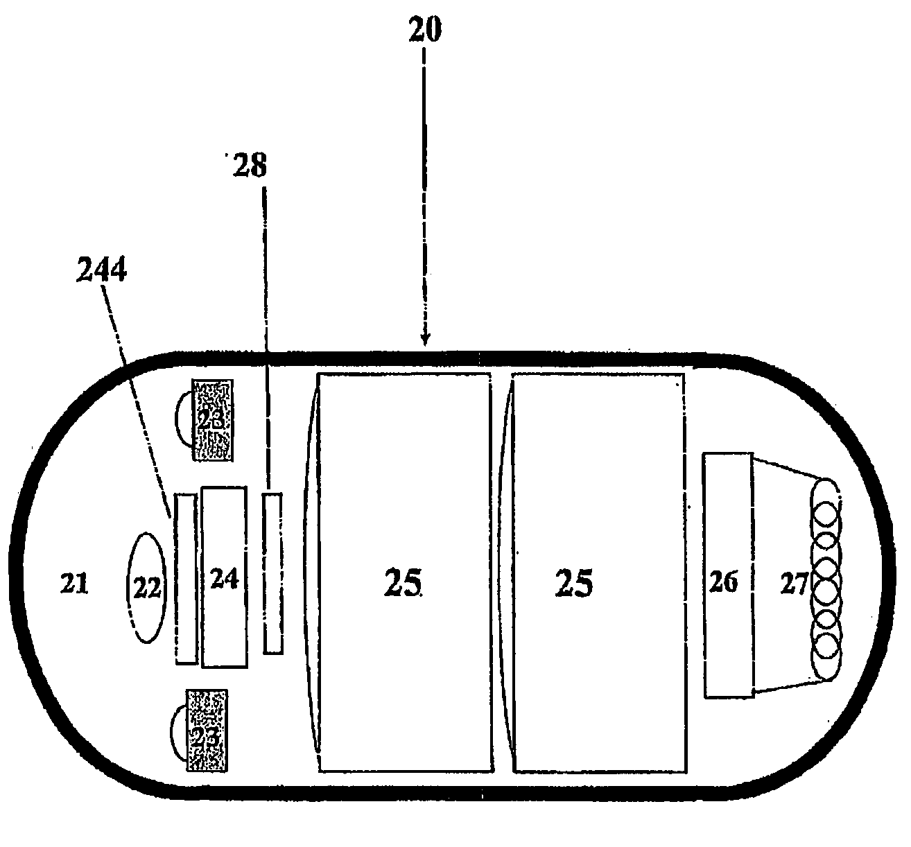
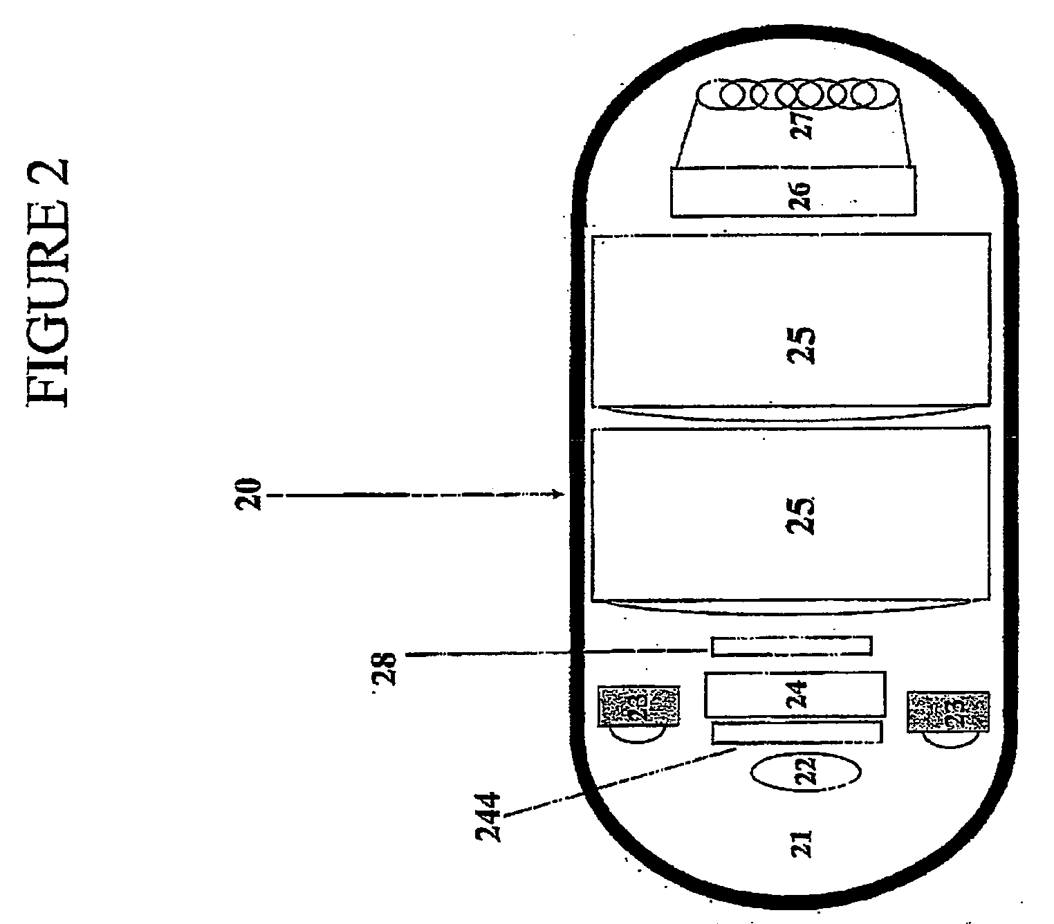
[0062] For efficient view of areas such as the esophagus a specifically designed imaging capsule may be used. For example, a capsule having a plurality of optical pathways may be used for obtaining a wide angle of view. Such a...


PUM


Abstract

A system and method for in vivo diagnosis are provided. A composition consisting of at least a marking agent and a pharmaceutically acceptable carrier is administered to a patient and an autonomous in vivo device, which includes an illumination source and an image sensor, is used to obtain endo luminal images of the patient.

Description

FIELD OF THE INVENTION [0001] The present invention relates to in vivo diagnosis. More specifically, the invention relates to optically based methods, devices and systems for detection of pathologies or other medical conditions in body lumens, for example, in the gastrointestinal (GI) tract. BACKGROUND OF THE INVENTION [0002] Pathologies of the GI tract (which may consist for example of the pharynx, esophagus, stomach, duodenum, small bowel, and colon), include, among others, esophageal carcinoma (e.g., Barrett's esophagus), peptic ulcer diseases, colorectal carcinoma and inflammatory bowel diseases (e.g., ulcerative colitis and Crohn's disease). Early pancreatic cancer may manifest itself through neoplasms released into the small intestine by the pancreas. Gastric cancer is a major cause of death worldwide especially in developing countries. The major type of gastric cancer is adenocarcinoma, which can be further categorized into an intestinal type and a diffuse type. Intestinal ty...

Claims


Application Information

Patent Timeline
Patent Type & Authority Applications(United States)
IPC IPC(8): A61B1/04A61B5/00
CPCA61B1/041A61B1/042A61B5/0084A61B5/0071A61B1/043
Inventor LEWKOWICZ, SHLOMOMERON, GAVRIEL
Owner GIVEN IMAGING LTD
Why Eureka
Try Eureka