Systems for identifying, displaying, marking, and treating suspect regions of tissue

a tissue sample and suspect technology, applied in the field of tissue sample suspect identification, display, marking and treatment, can solve the problems of indeterminate diagnosis, insufficient colposcopic technique, insufficient direct visual observation alone, etc., and achieve the effect of accurate identification and localization of neoplasia

Inactive Publication Date: 2006-01-19
MEDISPECTRA
View PDF3 Cites 168 Cited by
  • Summary
  • Abstract
  • Description
  • Claims
  • Application Information

AI Technical Summary

Benefits of technology

[0017] The invention provides systems for automatically localizing suspect areas of a tissue sample. The invention further provides systems for displaying, marking, and treating the identified areas so that healthy tissue suffers minimal impact upon treatment and / or excision of the diseased tissue. The invention facilitates minimally-invasive treatment of diseased tissue by accurately directing treatment to only those areas that are identified as diseased, or in which there is a suspicion of pathology.
[0018] Thus, the invention provides a diagnostic method that includes examining an in-vivo tissue sample with an optical signal detection system that automatically assigns a classification to each of a plurality of regions of the tissue; creating an overlay map indicating the assigned classifications; and displaying the overlay map to facilitate identification of suspect portions of the tissue sample.
[0022] The invention also provides methods of treating suspect portions following their identification. Various treatment methods that are compatible with methods of the invention include, for example, photodynamic therapy, cryotherapy, and direct chemical treatment. Photodynamic therapy can involve the use of one or more photosensitive agents, including exogenous agents and / or endogenous agents. The photosensitive agent can include a hematoporphyrin, a phthalocyanine, and / or a chlorin. Examples include dihematoporphyrin, 5-aminolevulinic acid, protoporphyrin IX, temoporfin, and mesotetrahydroxyphenylchlorin. The photosensitive agent is generally activated by nonionizing optical energy directed at the tissue of interest, and the photoactivated drug molecule then typically reacts with oxygen to produce chemicals that kill nearby cells. Methods of the invention allow application of the photosensitive agent only on areas in which there is high suspicion of disease, thereby having minimal damaging impact on surrounding healthy tissue.
[0023] The treatment step can also include removing tissue from a suspect portion using laser ablation. Again, methods of the invention allow treatment directed specifically within diseased areas, thereby limiting damage of healthy tissue.
[0025] In another aspect, the invention provides a system for detection of suspect portions of a tissue sample including an optical signal detection apparatus for obtaining one or more optical signals from each of a plurality of regions of a tissue sample; a memory that stores code and a processor that executes the code for producing an overlay map, and a display for facilitating identification of suspect portions of the tissue according to the overlay map. The processor executes software (1) to identify a characteristic of each of the regions based at least in part on the one or more optical signals from the region and (2) to define the overlay map for visually indicating the identified characteristics.
[0032] In yet another aspect, the invention is directed to the use of biomarkers and optical probes for both diagnostic identification of neoplasias, as well as for treatment. For example, in one embodiment, the invention is directed to a method for identifying a characteristic of each of a plurality of regions of a tissue sample, including the steps of obtaining one or more optical signals from each of a plurality of regions of a tissue sample and automatically identifying a characteristic of each region based at least in part on the one or more optical signals from the region, where the optical signals are obtained following application of an optical probe and / or a biological-responsive probe. Linkage of a neoplasia-specific biomarker with an optical reporter group can be used in combination with the optical detection methods of the invention to more accurately identify and localize neoplasia in vivo. Thus, the optical detection system used in combination with a fluorescent-labeled biomarker, for example, can produce an optical map of the cervix in which areas targeted by the biomarker are enhanced by the added fluorescent biomarker signal.

Problems solved by technology

However, direct visual observation alone may be inadequate for proper identification of an abnormal tissue sample, particularly in the early stages of disease.
Colposcopic techniques are not perfect.
Colposcopic images may contain complex and confusing patterns and may be affected by glare, shadow, or the presence of blood or other obstruction, rendering an indeterminate diagnosis.
However, examinations using spectral analysis may be adversely affected by glare, shadow, or the presence of blood or other obstruction, rendering an indeterminate diagnosis.
Some artifacts may not be detectable by analysis of the spectral data alone; hence, erroneous spectral data may be inseparable from valid spectral data.
Also, the surface of a tissue sample under spectral examination is generally not homogeneous.
Areas of disease may be interspersed among neighboring healthy tissue, rendering overly-diffuse spectral data erroneous.
Furthermore, current methods of displaying data based on tissue classification algorithms do not facilitate quick, accurate, or clear communication of diagnostic results.
Current techniques generally require interpretation by a skilled medical professional for meaningful and accurate conveyance of diagnostic information, due, in part, to the unfiltered nature of the diagnostic data
Additionally, current in-vivo tissue analysis methods generally cannot provide results immediately following examination.
Similarly, biopsy and / or treatment of tissue cannot be performed immediately following an in-vivo tissue examination.
Furthermore, current treatment methods for cancers, pre-cancers (such as cervical neoplasia), and other diseases are limited in that they not only kill diseased cells, they also damage nearby healthy tissue.
While PDT is effective at killing undesired cells, it can also damage nearby healthy tissue, and, thus, its utility is limited.
In this method, liquid nitrogen or other extremely cold agent is used to freeze cells, thereby destroying them.
However, this method destroys surrounding healthy tissue as well.
These chemical agents can also destroy nearby healthy tissue.
Furthermore, these chemical agents can be very expensive to produce, and diffuse and / or unnecessary use should be avoided.

Method used


Image

  • Systems for identifying, displaying, marking, and treating suspect regions of tissue
  • Systems for identifying, displaying, marking, and treating suspect regions of tissue
  • Systems for identifying, displaying, marking, and treating suspect regions of tissue

Examples


Embodiment Construction

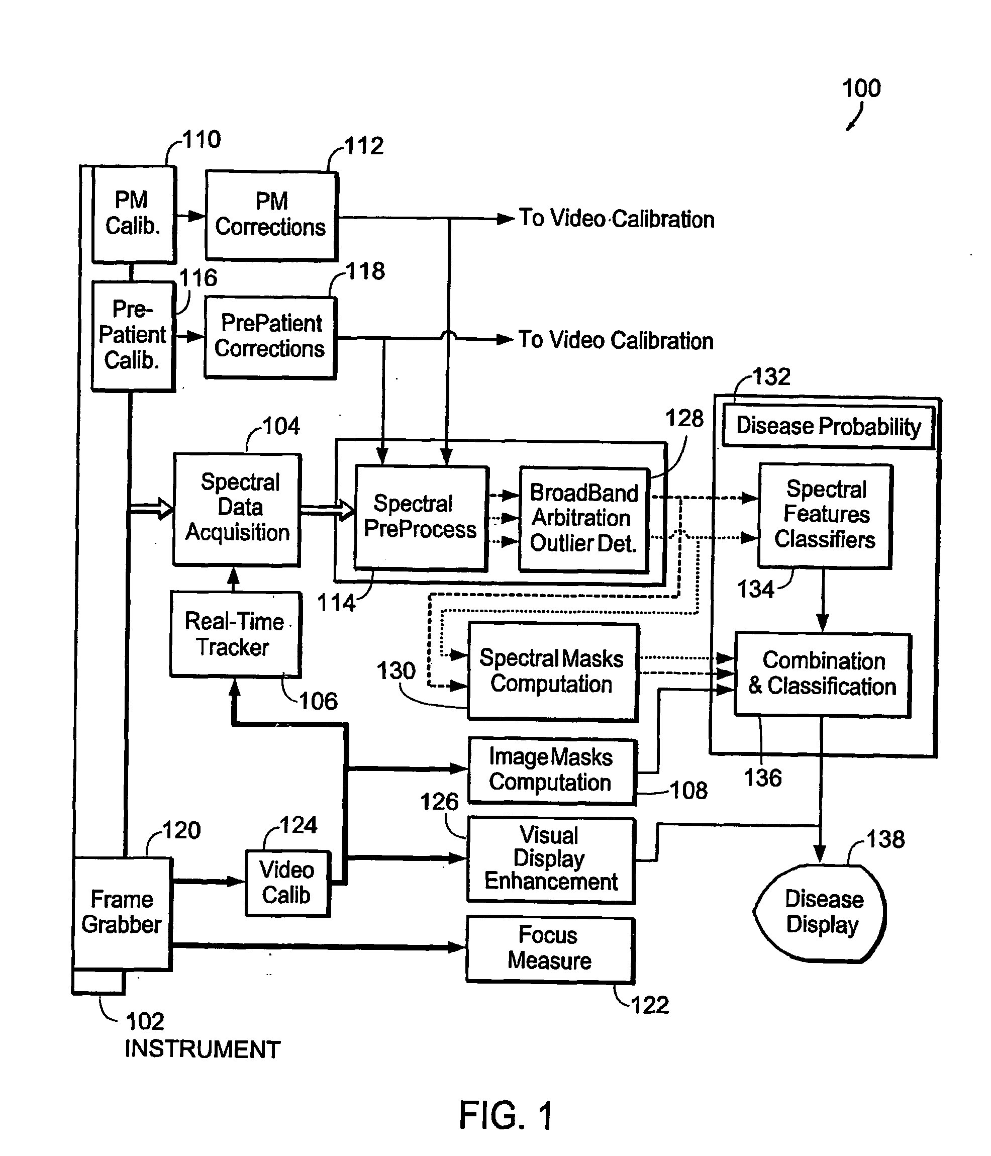
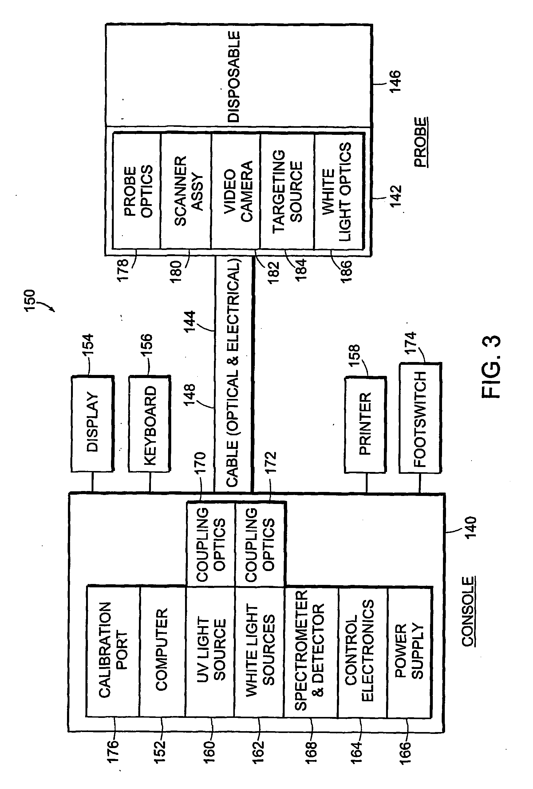
[0181] The invention provides systems and methods for obtaining spectral data and image data from a tissue sample, for processing the data, and for using the data to diagnose, mark, and / or treat suspect regions of the tissue sample. Methods of the invention include obtaining data from a tissue sample, determining which data are of diagnostic value, processing the useful data to obtain a prediction of disease state, and displaying the results in a meaningful way. In one embodiment, spectral data and image data are obtained from a tissue sample and are used to create a diagnostic map of the tissue sample indicating regions in which there is a high probability of disease. The diagnostic map is used to mark and / or treat suspect regions of tissue.

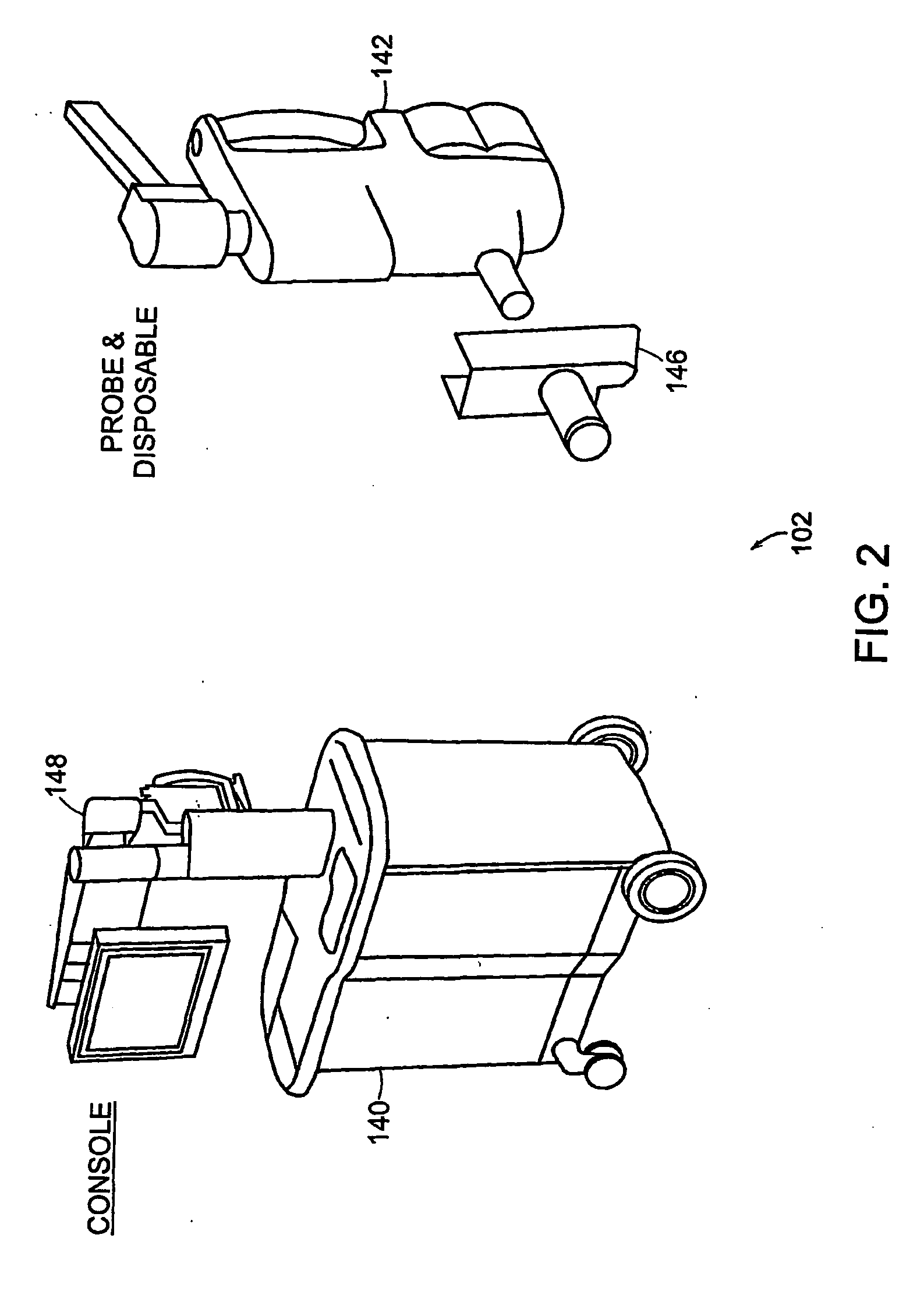
[0182] The systems and methods of the invention can be used to perform an examination of in situ and / or in-vivo tissue. The invention provides for the identification of suspect regions of tissue, which are then further analyzed, excised, and / or...


PUM

PropertyMeasurementUnit
optical signal detectionaaaaaaaaaa
real-time colposcopicaaaaaaaaaa
photosensitiveaaaaaaaaaa

Abstract

The invention provides systems for automatically localizing areas of disease within a tissue sample using fluorescence spectra, reflectance spectra, and video images obtained from the sample. The invention further provides systems for displaying, marking, and treating the identified areas so that healthy surrounding tissue suffers minimal impact upon treatment and / or excision of the diseased tissue. The invention facilitates minimally-invasive treatment by accurately directing treatment to only those areas that are identified as diseased.

Description

RELATED APPLICATIONS [0001] This application claims as priority documents U.S. patent application Ser. No. 10 / 418,902, filed Apr. 18, 2003, and U.S. Provisional Patent Application No. 60 / 560,384, filed Apr. 7, 2004.FIELD OF THE INVENTION [0002] This invention relates generally to systems, methods, and apparati that facilitate diagnostic evaluation of a tissue sample. More particularly, in certain embodiments, the invention relates to the identification, display, marking, and treatment of suspect regions of a tissue sample. BACKGROUND OF THE INVENTION [0003] It is common in the field of medicine to perform visual examination to diagnose disease. For example, visual examination of the cervix can discern areas where there is a suspicion of pathology. However, direct visual observation alone may be inadequate for proper identification of an abnormal tissue sample, particularly in the early stages of disease. [0004] In some procedures, such as colposcopic examinations, a chemical agent, ...

Claims


Application Information

Patent Timeline
Patent Type & Authority Applications(United States)
IPC IPC(8): G06K9/00A61B1/00A61B1/04A61B5/00G01N21/27G01N21/64G06T7/00G06T11/00G06V10/56
CPCA61B1/00142G06K9/4652A61B1/043A61B5/0071A61B5/0075A61B5/0084A61B5/6886A61B5/7257A61B2560/0233G01N21/274G01N21/31G01N21/6428G06T7/0012G06T11/00G06T2207/30024A61B5/7264A61B5/7267A61B1/0005A61B1/042A61B5/0068G16H50/20G06V10/56
Inventor FLEWELLING, ROSSFGRIFFIN, CHRISTOPHER E.JIANG, CHUNSHENGSCHOTT, JEAN-PIERRESCHOMACKER, KEVIN T.
Owner MEDISPECTRA
Why Eureka
Try Eureka