
Methods for treating HIV infected subjects

a technology for infected subjects and hiv, applied in the field of methods for treating hiv infected subjects, can solve the problems of contamination of the entire t cell population during long-term culture, the inability to supply a renewable supply of accessory cells, and the significant problem of long-term culture systems for mhc-matched antigen presenting cells as accessory cells

Inactive Publication Date: 2006-01-19
GENETICS INST INC
View PDF23 Cites 9 Cited by
  • Summary
  • Abstract
  • Description
  • Claims
  • Application Information

AI Technical Summary

Benefits of technology

This patent describes a method for expanding T cells in the absence of exogenous growth factors and accessory cells. The method involves activating T cells through a primary activation signal and a costimulatory molecule on the surface of the T cells. The T cells are then stimulated with a second agent that provides a secondary activation signal. The resulting T cell population is polyclonal with respect to antigen reactivity. The method can be used to treat infectious diseases or cancer by expanding specific T cell populations and restoring them to the individual. The method also provides a renewable source of T cells for immunotherapy or in vitro analysis of infectious agents.

Problems solved by technology

The requirement for MHC-matched antigen presenting cells as accessory cells presents a significant problem for long-term culture systems.
The necessity for a renewable supply of accessory cells is problematic for treatment of immunodeficiencies in which accessory cells are affected.
In addition, when treating viral infection, accessory cells which may carry the virus may result in contamination of the entire T cell population during long term culture.

Method used


Image

  • Methods for treating HIV infected subjects
  • Methods for treating HIV infected subjects
  • Methods for treating HIV infected subjects

Examples


example 1

Long Term Growth of CD4+ T cells With Anti-CD3 and Anti-CD28 Antibodies

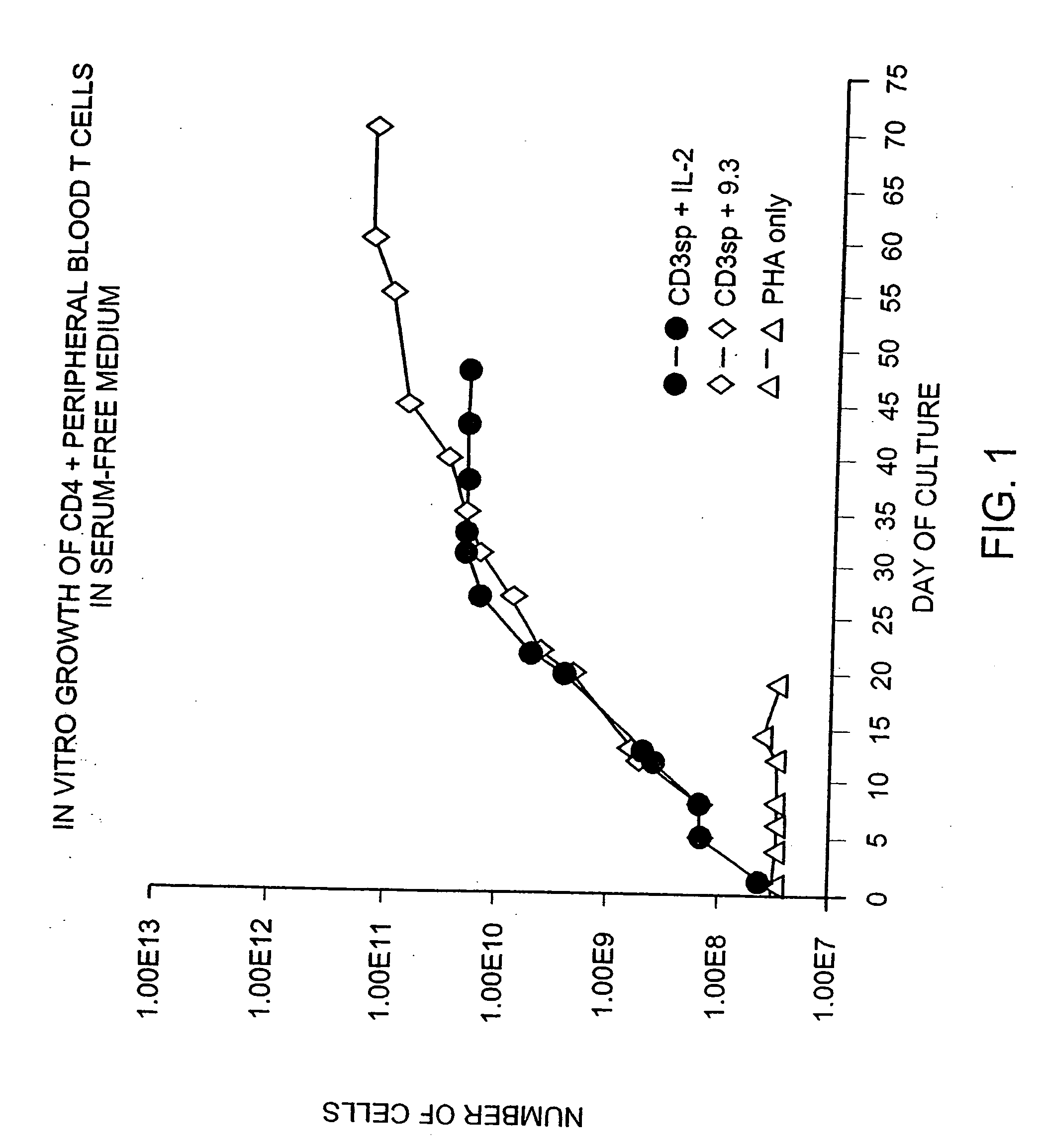
[0169] Previous known methods to culture T cells in vitro require the addition of exogenous feeder cells or cellular growth factors (such as interleukin 2 or 4) and a source of antigen or mitogenic plant lectin. Peripheral blood CD28+ T cells were isolated by negative selection using magnetic immunobeads and monoclonal antibodies as described in the Methods and Materials section above. CD4+ cells were further isolated from the T cell population by treating the cells with anti-CD8 monoclonal antibody and removing the CD8+ cells with magnetic immunobeads. Briefly, T cells were obtained from leukopheresis of a normal donor, and purified with FICOLLdensity gradient centrifugation, followed by magnetic immunobead sorting. The resulting CD28+, CD4+ T cells were cultured in defined medium (X-Vivo10 containing gentamicin and L-glutamine (Whittaker Bioproducts) at an initial density of 2.0×10⁶ / ml by adding cells to cul...

example 2

Long Term Growth of Anti-CD28-Treated T cells In Medium Containing Fetal Calf Serum

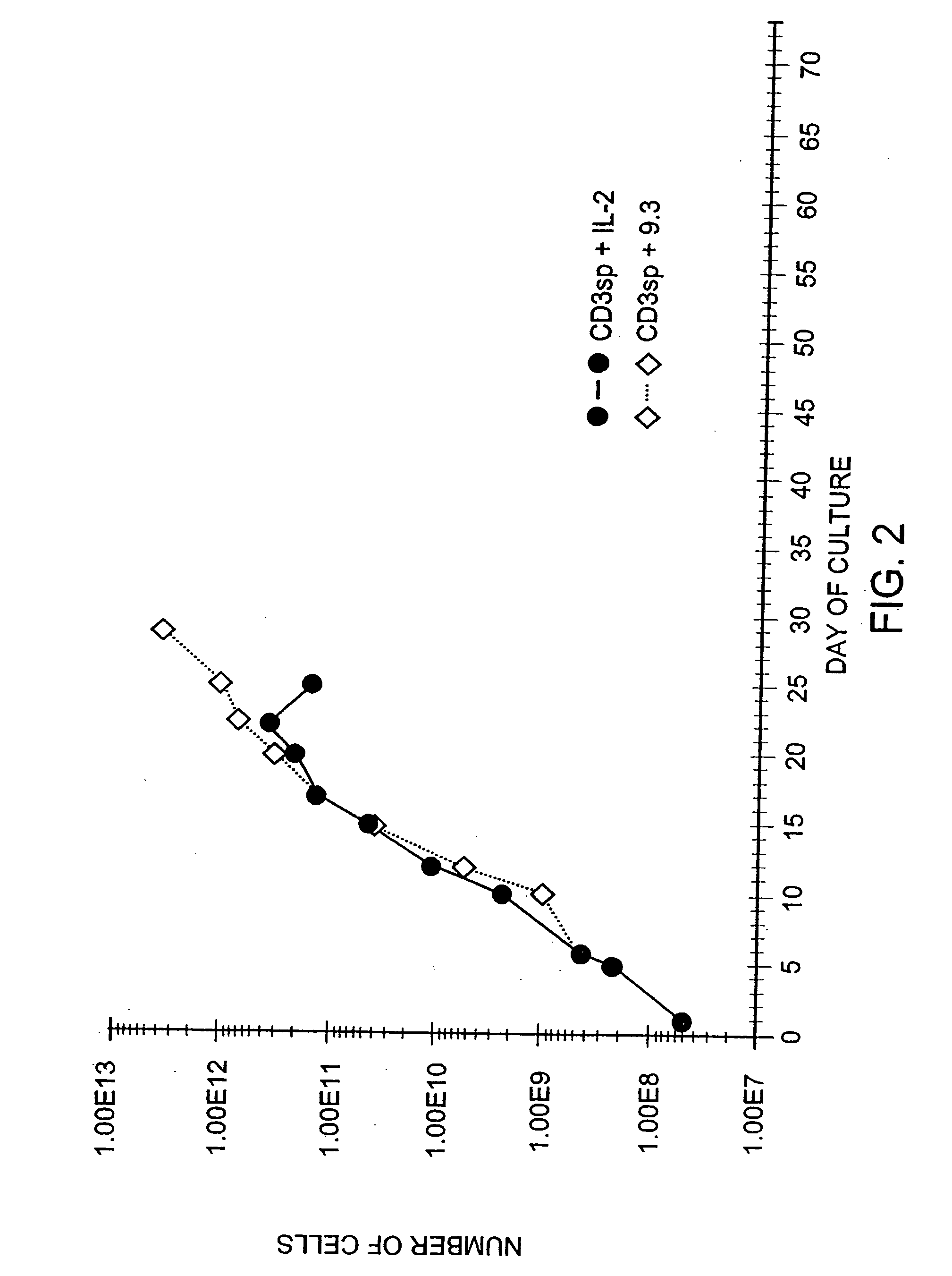
[0171] Another series of experiments tested whether the growth advantage of CD28 receptor stimulation was due to replacement of factors normally present in fetal calf serum. T cells were obtained from leukopheresis of a normal donor, and purified with FICOLLdensity gradient centrifugations, followed by magnetic immunobead sorting. The resulting CD28+, CD4+ T cells were cultured at an initial density of 2.0×10⁶ / ml in medium (RPMI-1640 containing 10% heat-inactivated fetal calf serum [Hyclone, Logan, Utah] and gentamicin and L-glutamine) by adding cells to culture dishes containing plastic-adsorbed OKT3. After 48 hours, the cells were removed and placed in flasks containing either hIL-2 (10% final concentration, CalBiochem) or anti-CD28 mAb 9.3 (800 ng / ml). The cells were fed with fresh medium as required to maintain a cell density of 0.5×10⁶ / ml, and restimulated at approximately weekly intervals by ...

example 3

Long Term Growth of T cells in Phorbol Ester, lonomycin and Anti-CD28-Stimulated T cells

[0173] Further experiments tested whether alternative methods of activating T cells would also permit CD28 stimulated growth. Pharmacologic activation of T cells with PMA and ionomycin is thought to mimic antigen receptor triggering of T cells via the TCR / CD3 complex. T cells were obtained from leukopheresis of a normal donor, and purified with sequential FICOLL™ and PERCOLLdensity gradient centrifugations, followed by magnetic immunobead sorting. The resulting CD28+, CD4+ T cells were cultured at an initial density of 2.0×10⁶ / ml by adding cells to culture dishes containing phorbol myristic acid (PMA 3 ng / ml, Sigma) and ionomycin (120 ng / ml, Calbiochem, lot #3710232). After 24 hours, the cells were diluted to 0.5×10⁶ / ml and placed in flasks containing either rIL-2 (50 IU / ml, Boerhinger Mannheim, lot #11844900)) or anti-CD28 mAb (1 ug / ml). The cells were fed with fresh medium as required to mai...


PUM

PropertyMeasurementUnit
densityaaaaaaaaaa
pHaaaaaaaaaa
temperatureaaaaaaaaaa

Abstract

Methods for inducing a population of T cells to proliferate by activating the population of T cells and stimulating an accessory molecule on the surface of the T cells with a ligand which binds the accessory molecule are described. T cell proliferation occurs in the absence of exogenous growth factors or accessory cells. T cell activation is accomplished by stimulating the T cell receptor (TCR) / CD3 complex or the CD2 surface protein. To induce proliferation of an activated population T cells, an accessory molecule on the surface of the T cells, such as CD28, is stimulated with a ligand which binds the accessory molecule. The T cell population expanded by the method of the invention can be genetically transduced and used for immunotherapy or can be used in methods of diagnosis.

Description

RELATED APPLICATIONS [0001] This application is a continuation-in-part of the following U.S. applications: U.S. Ser. No. 08 / 435,816, filed May 4, 1995 entitled “Methods for Selectively Stimulating Proliferation of T cells”; U.S. Ser. No. 08 / 403,253, filed Mar. 10, 1995 entitled “Methods for Selectively Stimulating Proliferation of T cells”; U.S. Ser. No. 08 / 253,964, filed Jun. 3, 1994, entitled “Methods for Selectively Stimulating Proliferation of T cells”; U.S. Ser. No. 08 / 073,223, filed Jun. 4, 1993, entitled “Methods for Selectively Stimulating Proliferation of T cells”; U.S. Ser. No. 864,805, filed Apr. 7, 1992, entitled “CD28 Pathway Immunoregulation”; U.S. Ser. No. 864,866, filed Apr. 7, 1992, entitled “Enhancement of CD28-Related Immune Response”; and U.S. Ser. No. 864,807, filed Apr. 7, 1992, entitled “Immunotherapy Involving Stimulation of Th CD28 Lymphokine Production”. Each of these applications is a continuation-in-part of U.S. Ser. No. 275,433, filed Nov. 23, 1988, enti...

Claims


Application Information

Patent Timeline
Patent Type & Authority Applications(United States)
IPC IPC(8): A61K39/21C12N15/86A61K35/14A61K35/17A61K35/28A61K38/00A61K38/17A61K38/46A61K39/00A61K39/02A61K39/12A61K48/00C07K14/705C07K16/00C07K16/28C12N5/0783
CPCA61K35/17C12N2740/16011A61K39/02A61K39/12A61K48/00A61K2039/505A61K2039/507A61K2039/5158C07K14/705C07K14/70521C07K14/70532C07K16/00C07K16/2806C07K16/2809C07K16/2812C07K16/2815C07K16/2818C07K16/2833C07K16/2866C07K16/289C07K16/2896C07K2317/24C07K2317/34C07K2317/54C07K2317/55C07K2317/73C07K2317/74C07K2319/00C07K2319/30C12N5/0636C12N2501/51C12N2501/515C12N2501/599C12N2710/00088A61K38/00A61K39/4611A61K39/464838
Inventor JUNE, CARLTHOMPSON, CRAIGNABEL, GARYGRAY, GARYRENNERT, PAUL
Owner GENETICS INST INC
Learn More