Microplates containing microsphere fluorescence standards, microsphere standards, and methods for their use

a microsphere fluorescence and microsphere technology, applied in the field of microsphere fluorescence standards for microspheres, can solve the problems of ineffective cell-free systems, inconvenient cellular assays, and prohibitively expensive and time-consuming serial assays of each material,

Inactive Publication Date: 2006-03-02
LIFE TECH CORP
View PDF10 Cites 7 Cited by
  • Summary
  • Abstract
  • Description
  • Claims
  • Application Information

AI Technical Summary

Benefits of technology

[0013] Microplates containing spherical bead standards, and methods for their preparation are disclosed. Preparation of monodisperse distributions of beads in microplate wells can be achieved by various methods including airbrushing, application with an inkjet printer, or controlled evaporation. Spherical bead standards containing two or more regions stained with dyes of different fluorescence lifetimes, and methods for their preparation are also disclosed.

Problems solved by technology

Serial assays of each material would be prohibitively expensive and time consuming.
Cell-free systems do not effectively model the complex interactions, signaling, and feedback that are present in intact cells and tissues.
Despite the widespread usage of microplate assay systems, they are not well suited for cellular assays.
HCS systems are inherently complicated.
Despite this need, there is no widely used or accepted calibration standard for HCS systems.
However, cells are relatively unstable, non-uniform, and difficult to handle in a reliable manner.
The biological diversity of a cell population makes this a less useful standard, and would be a less effective calibration tool.
In addition, the residual dye molecules in the wells after staining process are difficult to remove completely, leading to increased background fluorescence.

Method used


Image

  • Microplates containing microsphere fluorescence standards, microsphere standards, and methods for their use
  • Microplates containing microsphere fluorescence standards, microsphere standards, and methods for their use
  • Microplates containing microsphere fluorescence standards, microsphere standards, and methods for their use

Examples


example 1

Preparation of Microspheres

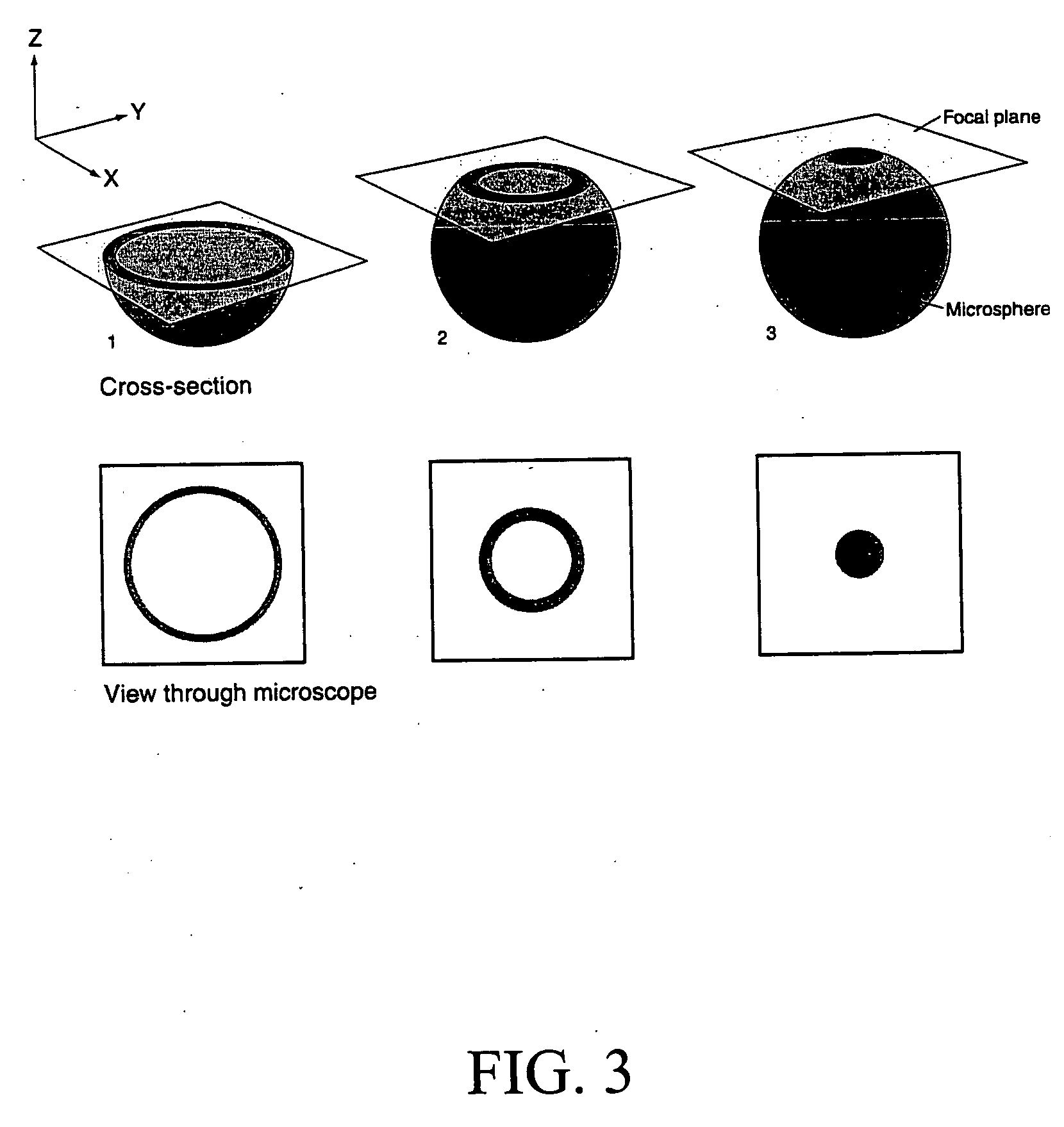
[0059] Microspheres having concentric fluorescent spherical zones can be prepared using the methods described in U.S. Pat. No. 5,786,219 (issued Jul. 28, 1998). The microspheres have at least two distinct concentric zones, labeled with one or more fluorescent dyes.

[0060] FocalCheck microspheres of various micrometer diameters (e.g., 1, 6, and 15 micrometers) are commercially available from Molecular Probes, Inc. (Eugene, Oreg.).

example 2

Preparation of Time-Resolved Microspheres

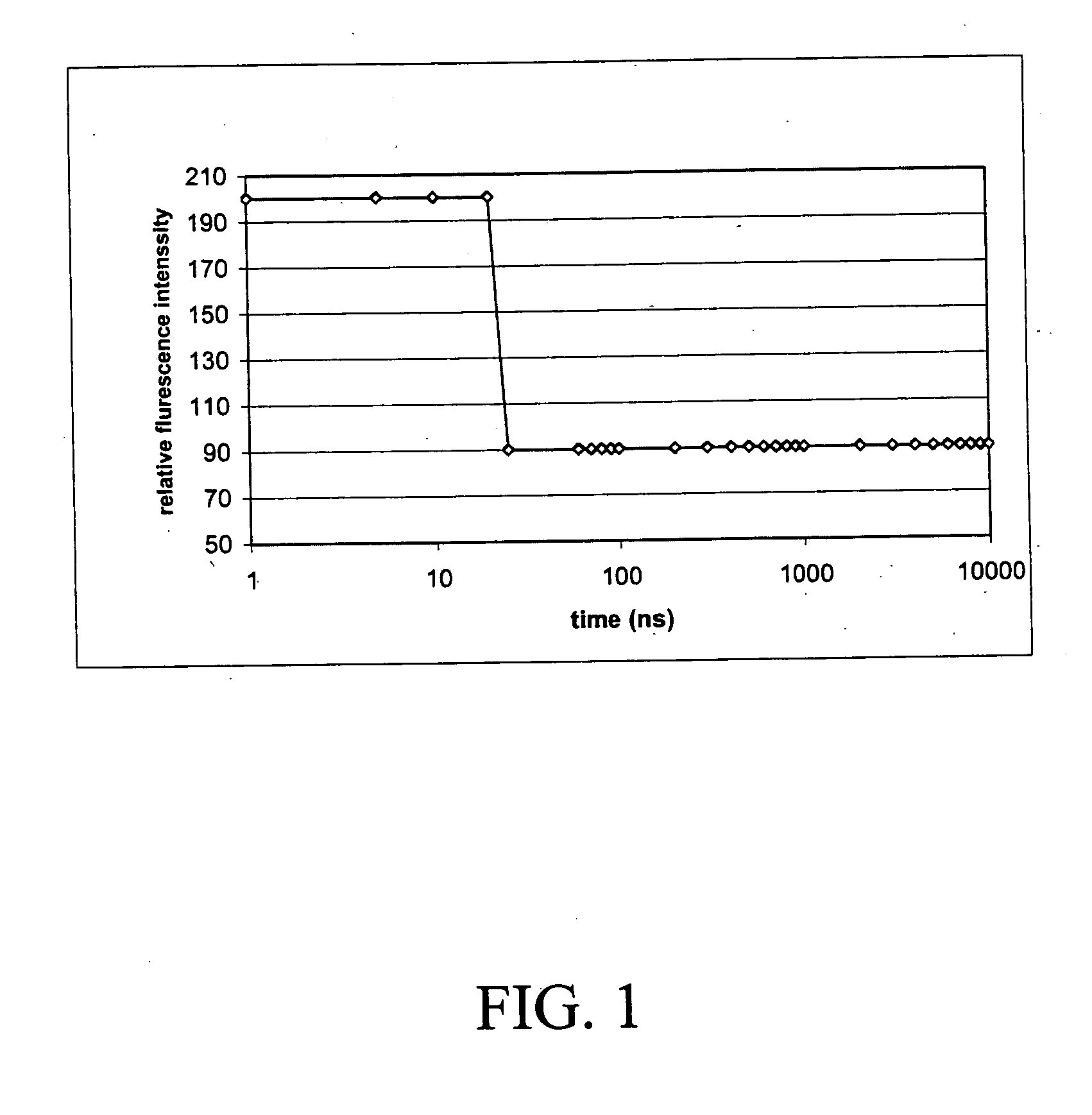
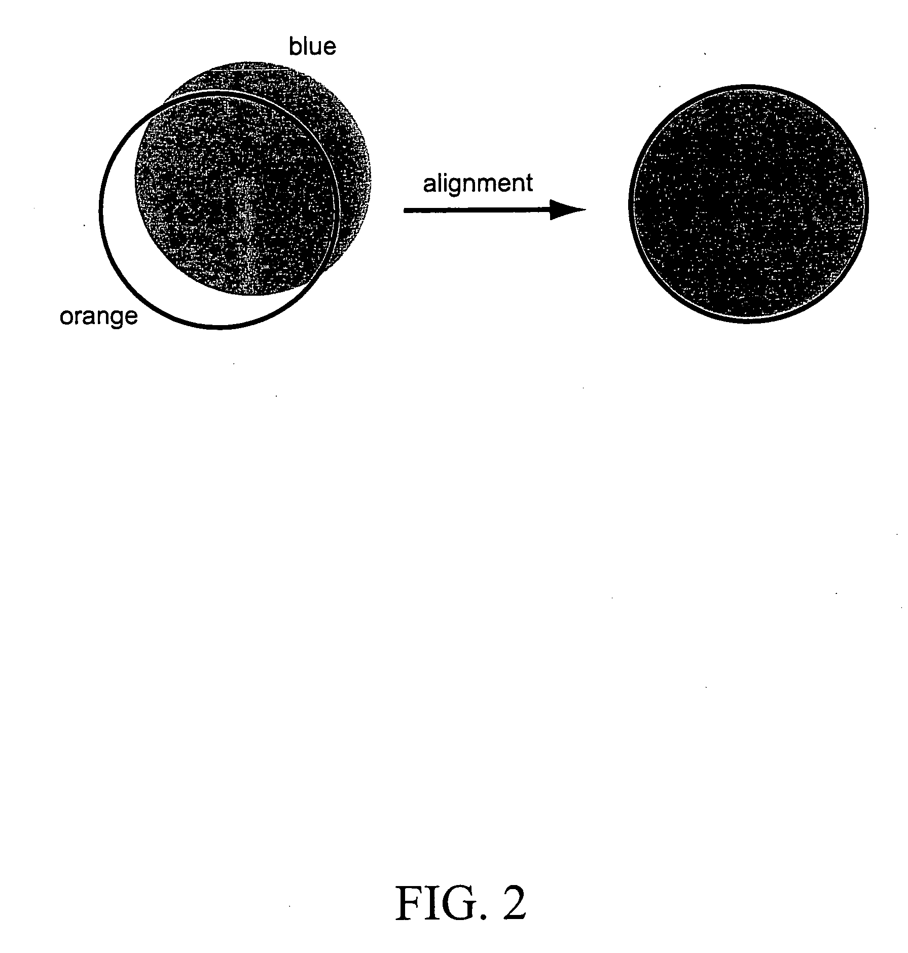
[0061] Microspheres having a uniform orange stain (with short fluorescence life time, about 18 ns) and a deep red ring stain (with long fluorescence life time, about 300 μs) were prepared according to the following method.

[0062] The following stock solutions were prepared: 4,4-difluoro-1,3-diphenyl-5,7-dipropyl-4-bora-3a,4a-diaza-s-indacene (5.0 mg; Molecular Probes Inc.; Eugene, Oreg.) was dissolved in methylene chloride to give a stock solution having a concentration of 2.0 mg / mL (Stock solution A). Pt (II) Meso-tetra (pentafluorophenyl) porphine (4.0 mg, Frontier Scientific, Inc., Logan, Utah) was dissolved in methylene chloride to give a stock solution having a concentration of 4.0 mg / mL (Stock solution B).

[0063] A 1.0 mL suspension (10% solids) of 15.0 μm microspheres (polystyrene / 2% divinylbenzene; Bangs Laboratories) was placed in a test tube. Approximately 3 mL of ethanol was added to the test tube, and the microspheres were resusp...

example 3

Determining Acceptability of Microspheres Coated onto Microplates

[0067] Microplates coated with microspheres were examined using a Zeiss Axioplan fluorescence microscope to assay the coating acceptability. Microscope objectives and stages were thoroughly cleaned to prevent the transfer of any oil or dirt to the microplates. The plates were handled with cotton-gloved hands, touching only the edges of the plate. The plates were examined using proper objective and fluorescence excitation / emission filter combinations (such as UV, FITC, PE or Cy5).

[0068] The plates were examined for the presence or absence of the following three desirable qualities. First, all of the beads should be firmly attached to the bottom of the micro-wells (i.e. no free moving microspheres were observed) and be at same focal plane. Second, there should be several hundreds to thousands of beads distributed throughout the well. The distribution should be random and even. Ideally, more than 95% of the beads are mo...


PUM


Abstract

Microplates containing spherical “microsphere” fluorescence standards are disclosed. The microplates can be prepared using several methods including airbrushing, application with an inkjet printer, or controlled evaporation. Spherical bead standards containing two or more regions stained with dyes of different fluorescence lifetimes, and methods for their preparation are also disclosed. The microplates can be used as calibration standards for fluorescence and confocal microscopes, and as calibration tools for microscope-based high content screening (“HCS”) instruments.

Description

CROSS REFERENCE TO RELATED APPLICATIONS [0001] The present application claims priority to U.S. Provisional Patent Application Ser. No. 60 / 606,694 filed Sep. 1, 2004, the contents of which are incorporated herein by reference.FIELD OF THE INVENTION [0002] The invention relates to microplates containing multicolor fluorescent microspheres and their use as calibration standards for microscope-based high content screening instruments. DESCRIPTION OF RELATED ART [0003] High throughput screening (“HTS”) assays are becoming increasingly common in research and development laboratories. These assays allow researchers to screen a large quantity of materials in a relatively short period of time. Serial assays of each material would be prohibitively expensive and time consuming. [0004] Conventional microplate assay systems are typically performed using cell-free liquid systems, such as ELISA immunoassays or enzyme activity assays. The resulting signal is typically detected resulting from fluoro...

Claims


Application Information

Patent Timeline
Patent Type & Authority Applications(United States)
IPC IPC(8): B01L9/00
CPCB01L3/5085B01L2200/148G02B21/0076B01L2300/0829B01L2300/0609
Inventor ZHANG, YU-ZHONG
Owner LIFE TECH CORP
Why Eureka
Try Eureka