
Methods and kits for isolating sperm cells

Active Publication Date: 2006-03-16
PROMEGA
View PDF23 Cites 5 Cited by
  • Summary
  • Abstract
  • Description
  • Claims
  • Application Information

AI Technical Summary

Benefits of technology

[0003] Techniques currently used to isolate sperm cells from other cells in forensic samples are time consuming and labor intensive, and there is currently a backlog of unprocessed samples. Because of this backlog, some jurisdictions have a policy against processing samples unless a suspect has been identified. Consequently, many unprocessed samples are ultimately discarded, and genetic information contained in the sample is never compared with or entered into the national database, which reduces the ability of law enforcement to identify and apprehend repeat sex offenders.

Problems solved by technology

However, a vaginal swab obtained from a sexual assault victim typically contains relatively few sperm cells and large numbers of epithelial cells from the victim.
As a consequence, unless the sperm cells are first separated from other cells in the sample, DNA purified from a forensic sample is susceptible to overwhelming contamination with epithelial cell DNA.
Contamination with epithelial cell DNA interferes with the ability to establish a match between the genetic profile of DNA from the sample and that of the suspect or a member of the database.
Techniques currently used to isolate sperm cells from other cells in forensic samples are time consuming and labor intensive, and there is currently a backlog of unprocessed samples.
Because of this backlog, some jurisdictions have a policy against processing samples unless a suspect has been identified.
Consequently, many unprocessed samples are ultimately discarded, and genetic information contained in the sample is never compared with or entered into the national database, which reduces the ability of law enforcement to identify and apprehend repeat sex offenders.
This process frequently results in the loss of sperm cells.
This method requires a large amount of antibodies and is therefore relatively expensive.
Furthermore, sperm cells are lost during the wash steps, resulting in reduced yield.
In addition, antibodies may not bind efficiently because of variations or mutations in sperm cell surface antigens in certain individuals, resulting in poor sperm cell yields.
This method is problematic because sperm cells tend to become trapped among the epithelial cells, the sperm cells form clumps that are too large to pass through the membrane, and the membrane tends to clog, which ultimately may result in low yields of sperm cells contaminated with epithelial cells or epithelial cell DNA.
However, the method suffers from disadvantages, including clogging of the membrane, low sperm cell yields and contamination of the sperm cells with epithelial cell DNA.
In the field of reproductive medicine, sperm cells have been isolated from fresh semen using a cell sorter, which although effective, is not practical in the forensic context because it is costly, time consuming, and does not address how to effectively recover sperm cells and epithelial cells from a forensic sample (e.g., a swab or clothing).

Method used


Image

  • Methods and kits for isolating sperm cells
  • Methods and kits for isolating sperm cells

Examples


example 1

Isolation of Sperm Cells from Samples Containing Epithelial Cells

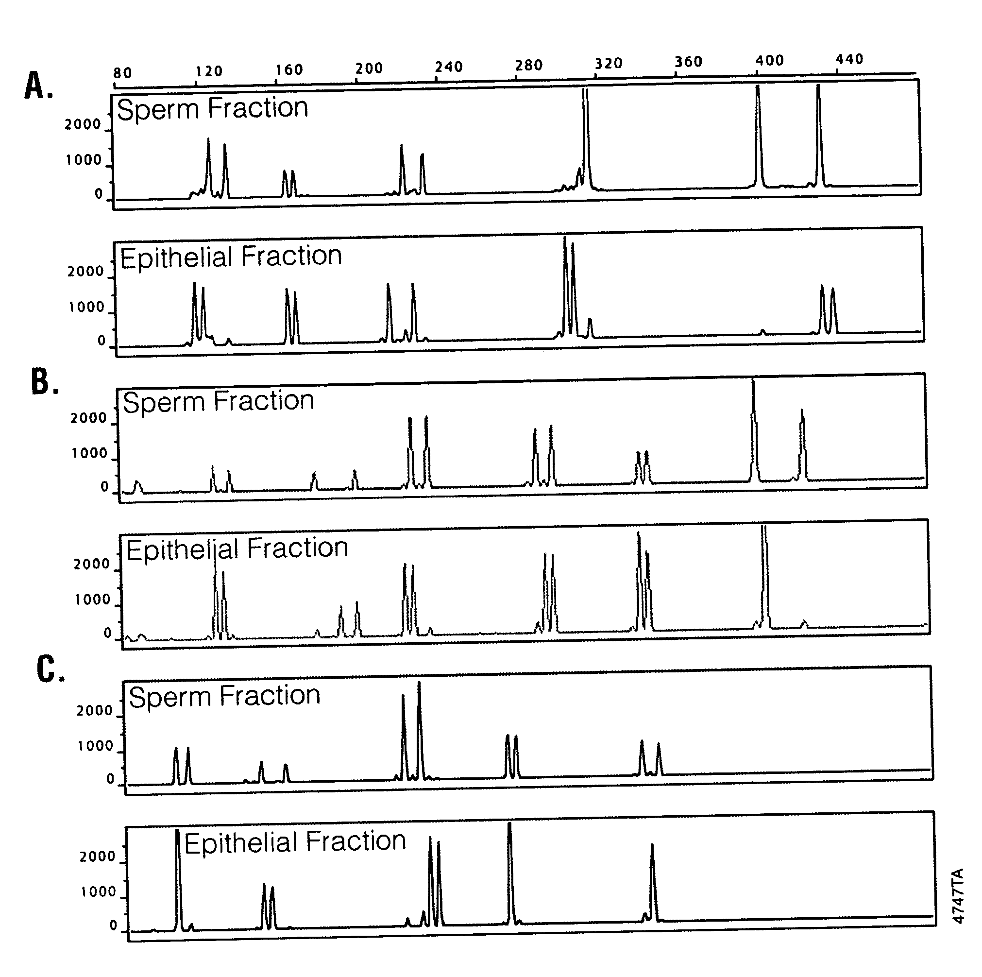
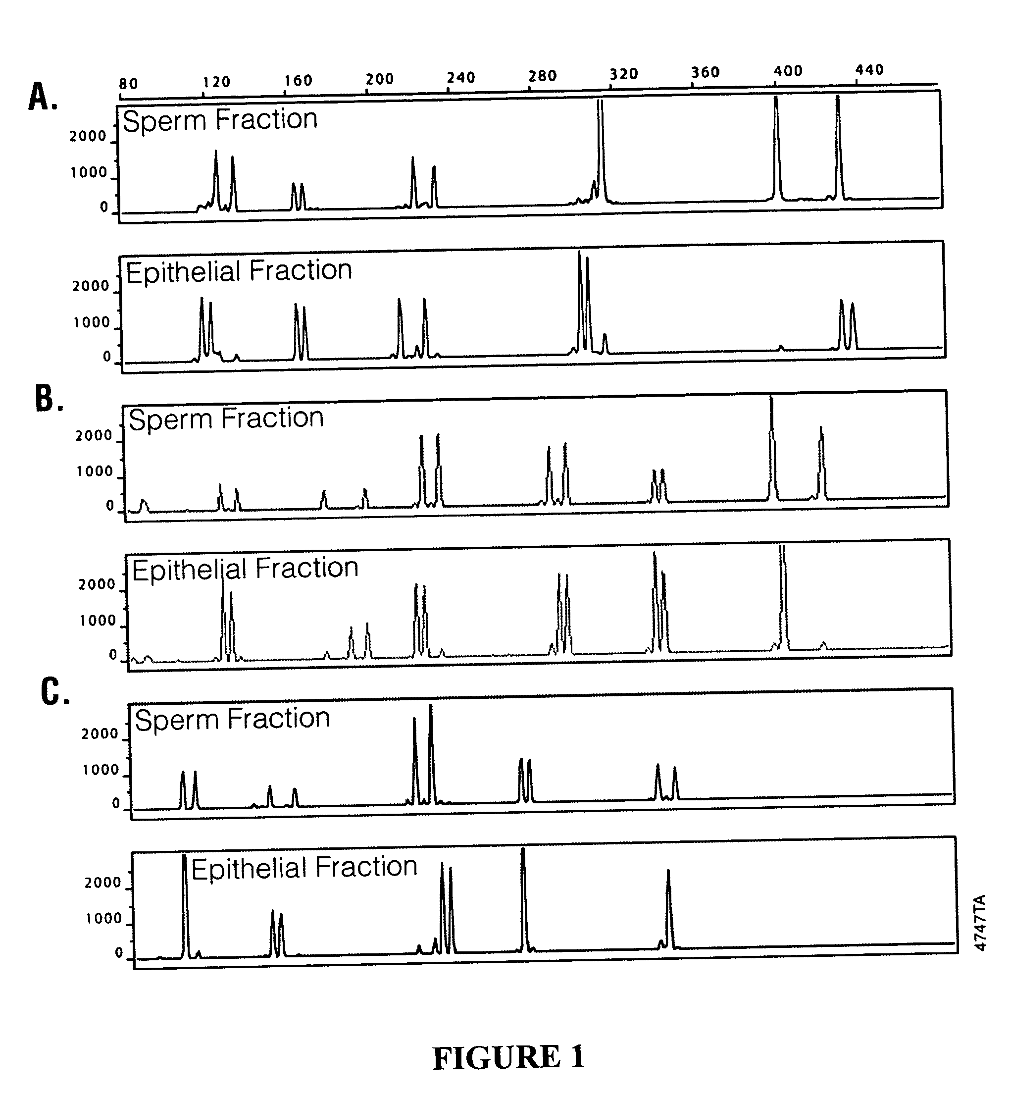
[0030] Samples used in evaluating sperm cell isolation included a fresh buccal swab containing added semen, fresh or four year old vaginal samples containing added semen, and a four year old 11-hour post coital vaginal sample. The solid support (e.g., a swab) containing the samples were placed in a microcentrifuge tube. A 0.5 ml aliquot of a digestion solution containing 50 mM NaCl, 10 mM Tris, pH 8.0, 10 mM EDTA, 0.2% SDS, FD&C Yellow dye, and 270 μg / ml Proteinase K was added to each sample. The tubes containing the samples were vortexed for 30 seconds and incubated at 56° C. for 1 hour. Diethyl glutarate and dimethyl glutarate were combined at ratios of 100:0, 50:50, 40:60, 30:70, 20:80, and 0:100 DEG:DMG to form mixed non-aqueous liquids. The ratio of 50:50 formed a liquid with a density of about 1.055 g / cm3. A 100 μl aliquot of the non-aqueous liquid including ratios of 100:0, 50:50, 40:60, 30:70, 20:80, or 0:100 ...

example 2

Isolation of DNA from Sperm Cells or Lysed Epithelial Cells Using Chaotropic Salt and Reducing Conditions

[0032] The DNA from the sperm cell pellet of Example 1 was isolated according to DNA isolation methods described in Promega Technical Bulletin TB296 using components provided in the DNA IQ™ system (Promega Corp., Madison, Wis. Cat. No. DC6701). A 200 μl aliquot of DNA IQ™ lysis buffer containing 4.5 M guanidine thiocyanate (GTC) and 10 mM dithiothreitol (DTT) was added to the microfuge tube containing the non-aqueous liquid and sperm cell pellet, and vortexed briefly to disrupt the sperm cell pellet and to form a homogenous mixture of the non-aqueous phase and the lysis buffer.

[0033] A 7 μl aliquot of DNA IQ™ Resin was added to the solution and mixed by vortexing at high speed for 3 seconds and incubated at room temperature for 5 minutes. The mixture was vortexed for 2 seconds at high speed, the tube was placed in a magnetic stand, and after the paramagnetic resin was attracted...

example 3

Isolation of DNA from Sperm Cells Using a Detergent and Phenol:Chloroform Extraction

[0035] The DNA from the sperm cell pellet of Example 1 was isolated by first removing the non-aqueous liquid to leave a firm sperm cell pellet. A 300 μl solution containing 10 mM Tris, pH 8.0, 1 mM EDTA, 10 mM DTT, and 1% SDS was added to the sperm cell pellet, and the tube was vortexed to lyse the sperm cells and dissolve the DNA. An equal volume of phenol:chloroform (1:1, v / v) was added and vortexed to denature the protein. The aqueous DNA-containing solution was removed, concentrated in a Microcon® apparatus, washed with 200 μl of a buffer containing 10 mM Tris, pH 8.0, 0.1 mM EDTA, and concentrated again in the Microcon® apparatus to about 40 μl. The purified DNA was transferred to a clean container.


PUM


Abstract

Disclosed are methods for isolating sperm cells from an aqueous sample and kits for isolating sperm cells from an aqueous sample.

Description

[0001] Genetic material obtained from forensic samples can be used to identify perpetrators of sexual assaults or to exonerate innocent suspects. Purified DNA obtained from sperm cells isolated from forensic samples can be used in subsequent genetic identity testing. The genetic profile of sperm cell DNA can be compared to that of a known suspect or to databases containing genetic information about a large number of convicted felons. [0002] In sexual assault cases, a vaginal or rectal swab, or clothing containing a semen stain, is obtained from the victim for forensic analysis. If sperm cells are present in the sample, DNA from the sperm cells can be isolated and used in genetic identity testing. However, a vaginal swab obtained from a sexual assault victim typically contains relatively few sperm cells and large numbers of epithelial cells from the victim. As a consequence, unless the sperm cells are first separated from other cells in the sample, DNA purified from a forensic sample...

Claims


Application Information

Patent Timeline
IPC IPC(8): C12N5/06C12N5/071
CPCC12N15/1003C12N5/0612
Inventor TEREBA, ALLANFLANAGAN, LAURAMANDREKAR, PARAJOLSON, RYAN
Owner PROMEGA
Learn More