Methods for detecting chromosome aberrations

Active Publication Date: 2006-04-13
AGILENT TECH INC
View PDF22 Cites 1 Cited by
  • Summary
  • Abstract
  • Description
  • Claims
  • Application Information

AI Technical Summary

Problems solved by technology

A caveat of ssFISH, however, lies in the fact that a false negative reading can emerge.
Methods based on in situ hybridization of probes to a target nucleic acid sequence are also fraught with problems of high background due to large probes binding to non-target sequences.
The repetitive units of the intermediate component are interspersed within the genome and is the major reason that large genomic nucleic acid probes (i.e. >100 bp) derived from genomic nucleic acid may not be well suited for hybridization analysis.
As stated above however,

Method used


Image

  • Methods for detecting chromosome aberrations
  • Methods for detecting chromosome aberrations
  • Methods for detecting chromosome aberrations

Examples


example 1

[0085] A slide containing metaphase spreads and interphase nuclei was pretreated in TBS (50 mmol / L Tris, 150 mmol / L NaCl, pH 7.6) with 3.7% formaldehyde for 2 minutes at room temperature, as described in Human Cytogenetics, a Practical Approach, Volume 1, 2nd ed., D. E. Rooney and B. H. Czepulkowski editors (1992). The slide was then rinsed twice in phosphate buffered saline (PBS) for 5 minutes per wash at room temperature. After rinsing, the slide was dehydrated in a cold (4° C.) series of ethanol by incubating the slide in 70% ethanol at 4° C. for 2 minutes; incubating the slide in 85% ethanol at 4° C. for 2 minutes; and then incubating the slide in 96% ethanol at 4° C. for 2 minutes, followed by air drying.

[0086] On each target area, 10 μL of DNA Hybridization Buffer (100 ng fluorescein labelled probe, 100 ng Texas Red labelled probe, 5 μM PNA Oligo Mix, 45% formamide, 300 mM NaCl, 5 mM NaPO4, 10% Dextran sulphate) was added and an 18×18 mm coverslip was applied to cover the hyb...

example 2

[0089] Slides containing metaphase spreads and interphase nuclei from normal cells and test sample cells are pretreated in TBS as described above. The slides are then rinsed twice in PBS for 5 minutes per wash at room temperature. After rinsing, the slide are dehydrated in a cold (4° C.) series of 70%, 85%, and 96% ethanol as described above, followed by air drying.

[0090] On each target area, 10 μL of DNA Hybridization Buffer (100 ng fluorescein labeled probe, 100 ng Texas Red labeled probe, 100 ng AMCA (7-amino-4-methylcoumarin-3-acitic acid) labeled probe, 5 μM PNA Oligo Mix, 45% formamide, 300 mM NaCl, 5 mM NaPO4, 10% Dextran sulphate) is added and an 18×18 mm coverslip is applied to cover the hybridization area on the slides. The edges of the coverslip are sealed with rubber cement before denaturation at 82° C. for 5 minutes. The slides are hybridized overnight at 45° C. After hybridization, the coverslip is removed and the slides are washed for 10 minutes in a stringent wash b...

example 3

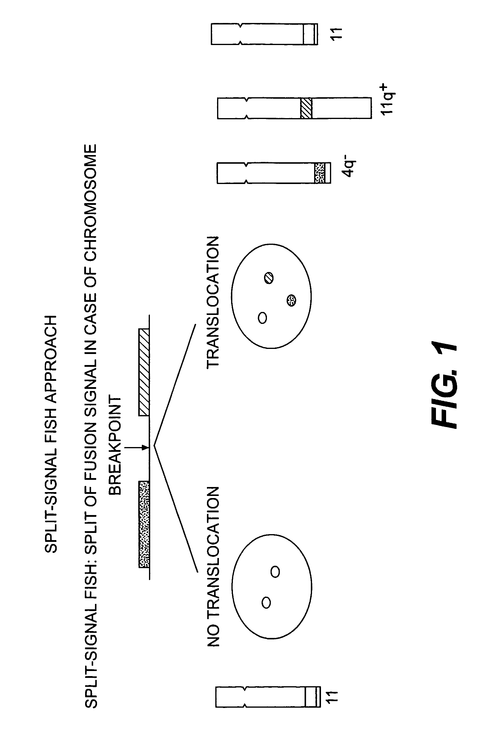
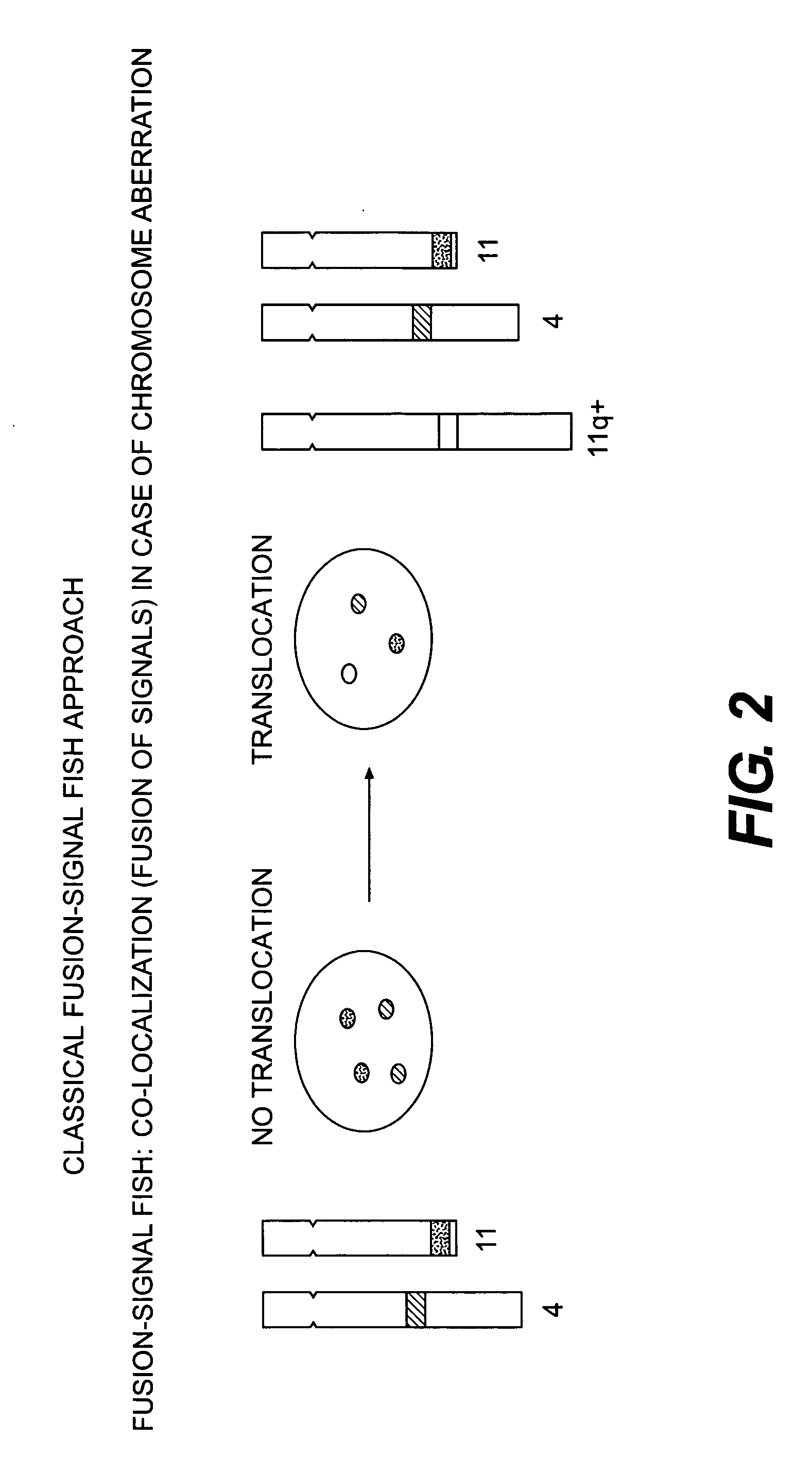
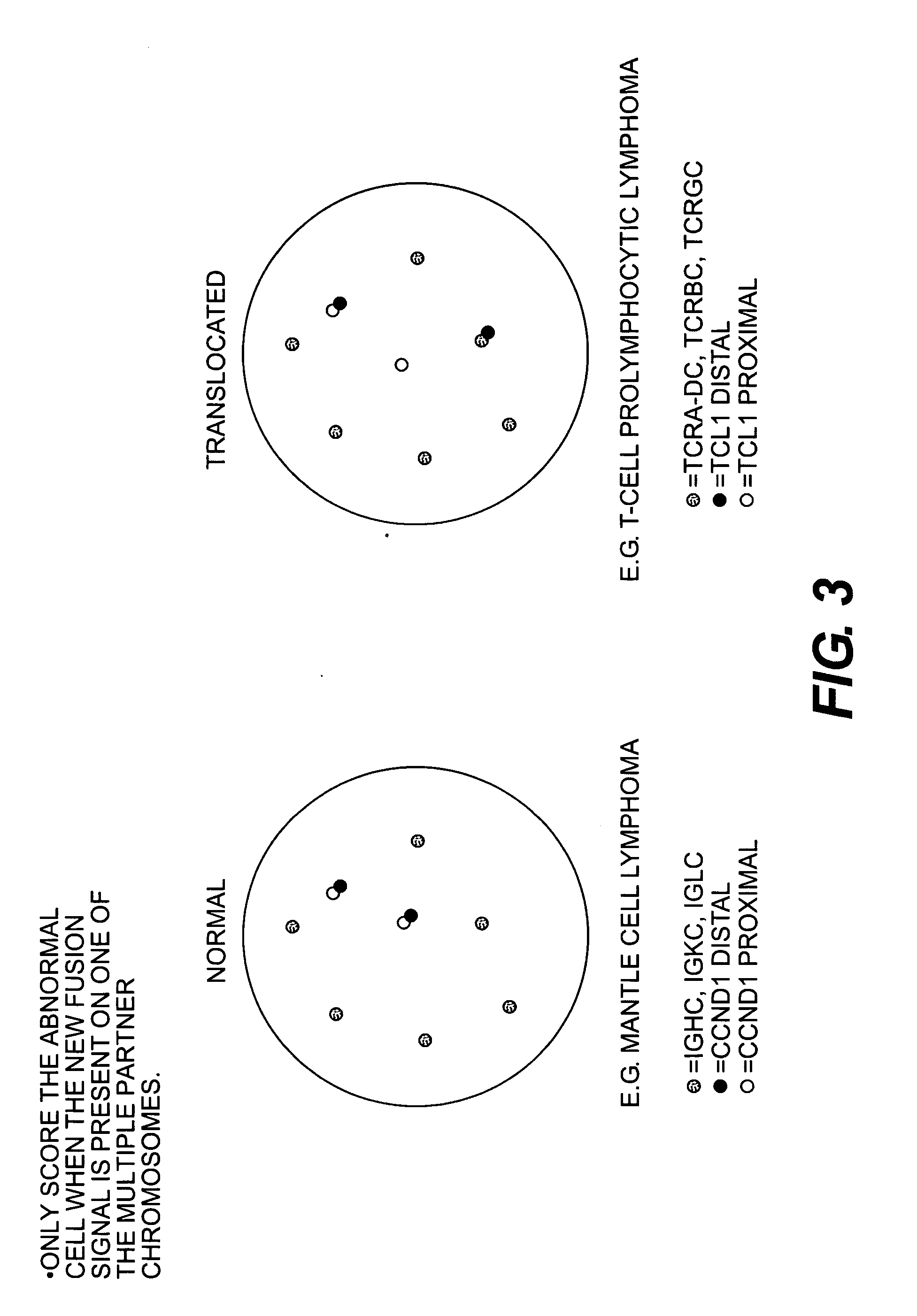
[0093] PDGFRA probes were hybridized to metaphase chromosomes from metaphase spreads from normal tissue and from the cell line ELO-1. A normal PDGFRA locus on chromosome 4 was detected in normal cells as either a yellow dot or by co-localized green / red signals due to the juxtaposition of the red probes and green probes hybridizing to regions upstream and downstream from an anticipated disease-related change in chromosomal structure. An additional chromosome 4 was also detected as a result of an amplification of the derivative chromosome 4.


PUM

PropertyMeasurementUnit
Coloraaaaaaaaaa
Structureaaaaaaaaaa
Chemiluminescenceaaaaaaaaaa

Abstract

The present invention relates to methods for detecting a change in chromosomal structure. These methods employ labeled probes that bind nucleic acids. For example, these probes may be comprised of nucleic acids or nucleic acid analogs and a detectable label.

Description

[0001] This application claims priority to U.S. Provisional Patent Application Nos. 60 / 567,440, and 60 / 567,570, both of which were filed May 4, 2004, and which are both incorporated herein by reference in their entirety.[0002] The present invention, in one embodiment, relates to methods for detecting a change in chromosomal structure. These methods employ probes that bind nucleic acids. For example, these probes may be comprised of nucleic acids or nucleic acid analogs and a detectable label. [0003] Detection of nucleic acid sequences by in situ hybridization, involving fundamental processes in molecular biology, using non-radioactive labels, has been applied for several years (Speel, Histochem. Cell Biol., 112:89-113 (1999)). Probe-based assays are useful in the detection, identification, quantitation or analysis of nucleic acids. Nucleic acid probes have long been used to analyze samples for the presence of nucleic acid from bacteria, fungi, virus or other organisms and are also u...

Claims


Application Information

Patent Timeline
IPC IPC(8): C12Q1/68C07H21/04
CPCB82Y5/00B82Y10/00C12Q1/6841C12Q1/6876C12Q2565/102C12Q2545/113C12Q2600/156
Inventor POULSEN, TIM SVENSTRUPPOULSEN, SUSAN MEDOMPETERSEN, KENNETH HEESCHE
Owner AGILENT TECH INC
Why Eureka
Try Eureka