X-ray needle apparatus and method for radiation treatment

a needle xray and needle xray technology, applied in the direction of radiation beam directing means, radiation therapy, nuclear engineering, etc., can solve the problems of the environment, affecting the treatment effect, and affecting the safety of the individual handler. , to achieve the effect of easy manipulation, high dose rate, shape and energy of the x-ray beam produced, and easy manipulation

Inactive Publication Date: 2006-06-22
ADVANCED X RAY TECH
View PDF17 Cites 28 Cited by
  • Summary
  • Abstract
  • Description
  • Claims
  • Application Information

AI Technical Summary

Benefits of technology

[0021] Briefly, the present invention includes an easily manipulated, portable, high dose rate apparatus having as an x-ray source, an x-ray tube of adjustable intensity, a collimator with one or more incorporated capillary lenses and an implantable needle. The x-ray tube is conjugated with the collimator which, in turn is conjugated with the implantable needle, which has an output window at its terminating end through which a treatment x-ray beam passes. The implantable needle may be fully or partially positioned into a patient to irradiate a desired region with x-rays.
[0022] The output window of the needle is made of an x-ray transparent material such as a plastic, a metal (such as beryllium) or a ceramic, such as boron carbide or boron nitride. By applying a metallic layer on the inside or outside (or both) of the output window, the shape and the energy of the x-ray beam produced can be changed, provided that the energy of the absorption edge (K-edge) of the deposited metal is lower than the energy of the x-ray beam passing through the output window. Thus, the needle x-ray device of the present invention allows adjustment in dose rate and shape of the x-rays delivered to an anatomical site of interest.
[0023] The needle x-ray device of the present invention avoids damaging the healthy tissue surrounding the area of radiation treatment. To achieve a desired radiation pattern over a desired region, while minimally irradiating other regions, the x-ray beam is emitted from a nominal position of the implantable needle located within or adjacent to the desired region to be irradiated. The disclosed invention can provide the required dose by irradiating any part of the desired region, either continuously or periodically, over extended periods of time.

Problems solved by technology

Since present radiation therapy machines apply x-ray radiation to target regions internal to a patient from a source external to the target, substantial damage can be done to healthy tissue surrounding the area of treatment.
While such use of radioisotopes may be effective in treating certain types of tumors, there is little ability to provide selective control of exposure (turn-on and turn-off) treatment radiation parameters.
Handling and disposal of such radioisotopes involves hazards to both the individual handler and the environment.
Although direct irradiation of tumors looks promising, the known insertable miniaturized x-ray tubes have many limitations.
The major disadvantages include: (a) difficulties with focusing and steering the electron beam to the target due to the collision of electrons en route to the target (such background x-rays could be as large as 40% of the primary dose rate at the needle tip)—also the tube must be shielded to avoid external electric and magnetic fields deflecting the beam to cause unwanted “leakage” lateral radiation that may impinge upon surrounding healthy tissue; (b) a necessity to cool the target to increase x-ray production efficiency; (c) impracticability of reducing the diameter of the miniaturized x-ray tube diameter below 4 mm (the requirement to decrease the diameter of the x-ray tube and the need to have a cooling system for the target are mutually exclusive); (d) significant limitations in changing shape of the emitted radiation, and (e) a high operating voltage of about 50-100 kV that is dangerous for the human body.

Method used


Image

  • X-ray needle apparatus and method for radiation treatment
  • X-ray needle apparatus and method for radiation treatment
  • X-ray needle apparatus and method for radiation treatment

Examples


Embodiment Construction

)

[0035] 1. The Apparatus

[0036] The invention includes an insertable needle-based x-ray system that is capable of administering an elevated dose rate. The system includes conditioning optics that is incorporated into the x-ray system in order to provide a high intensity x-ray beam. The x-ray system delivers radiation with a predetermined energy, intensity, and spatial distribution to or towards a selected area of the anatomy—for example, a tumor.

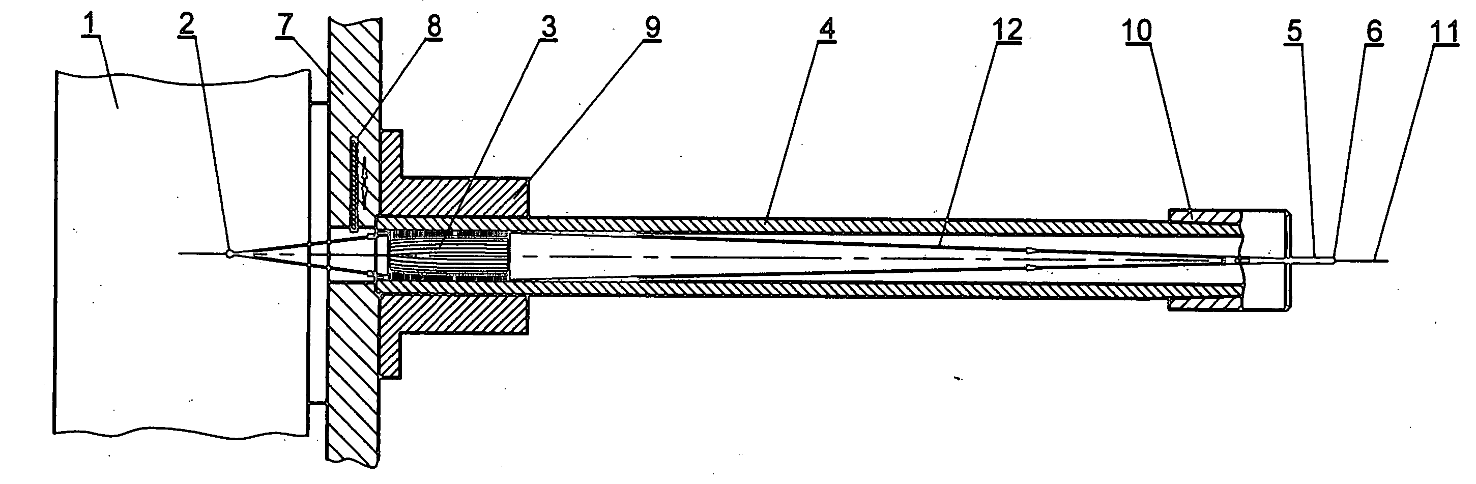
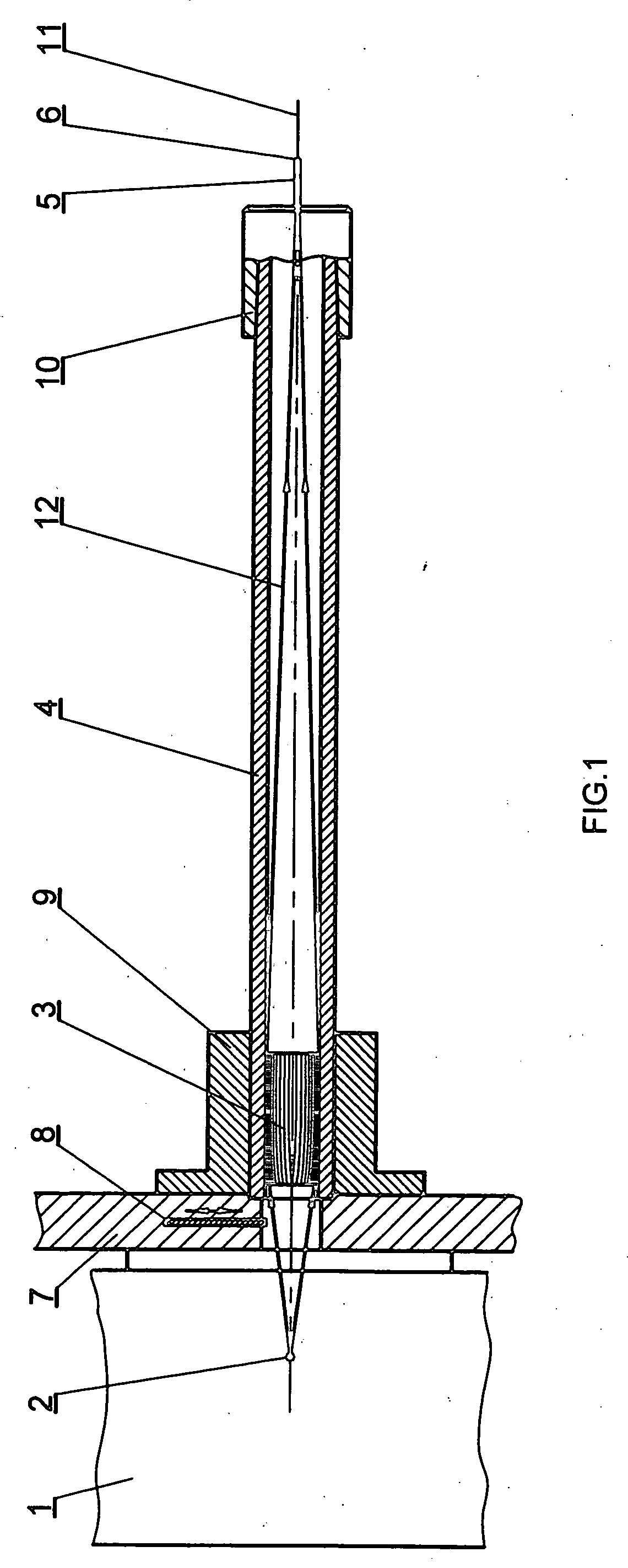
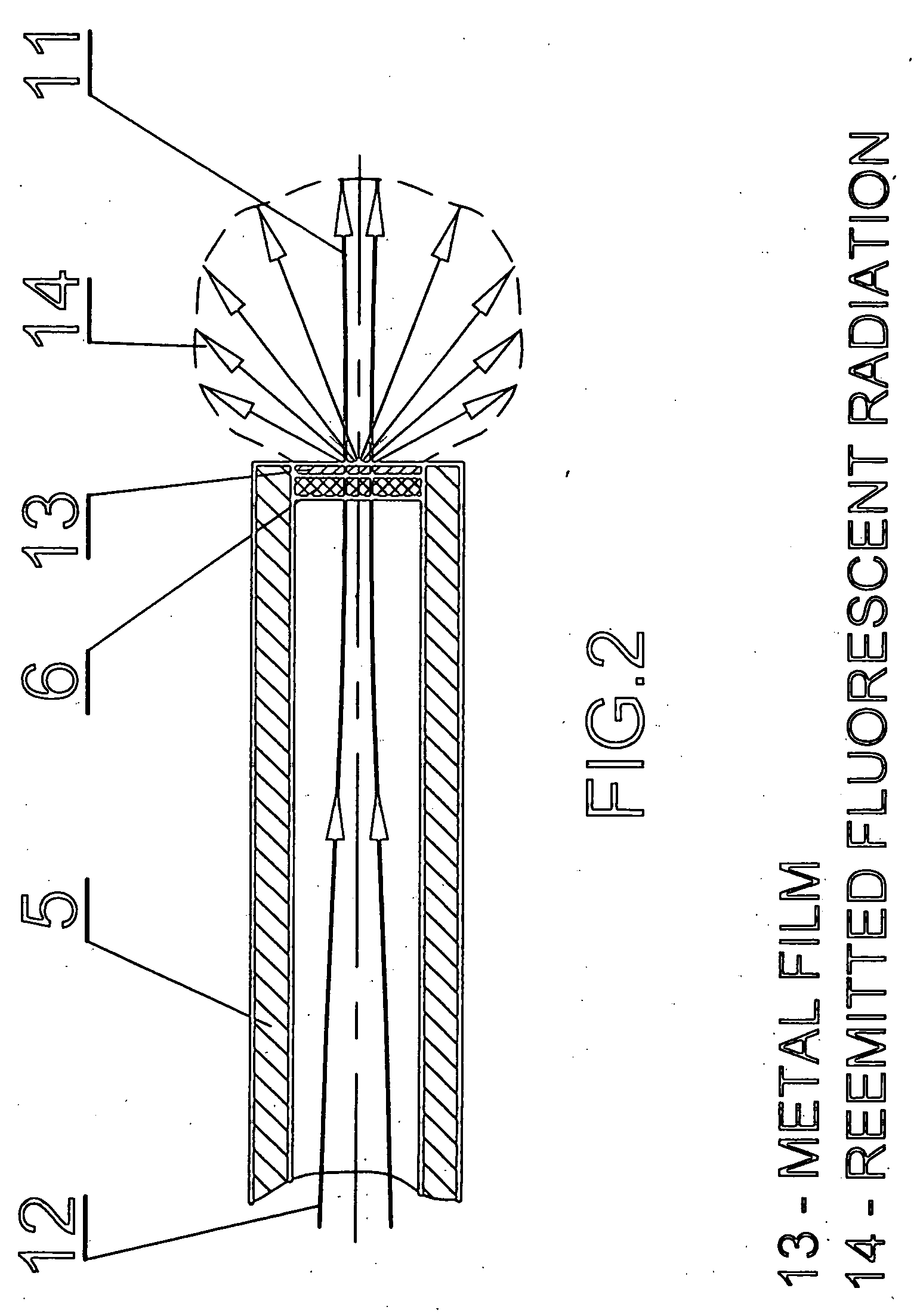
[0037]FIG. 1 shows one embodiment of an x-ray system containing an x-ray source 1 with a point focus 2, a capillary lens 3, an optical collimator 4 linked to the x-ray source 1 through a collimator holder 9, a needle holder 10 attached to the optical collimator 4, preferably through a Morse cone connection, or other connection means that provides consistent alignment and a secure, yet interchangeable interference fit and an implantable needle 5 with an output window 6 at its terminating end.

[0038] The x-ray system uses a focused x-ray beam...


PUM


Abstract

The invention is directed to an x-ray device and method for radiation treatment comprising an x-ray source 1, a collimator 4 incorporating conditional optics, such as a capillary lens 3 for directing and focusing the x-ray radiation, and implantable needles. One or more capillary semilenses 16, 17 are positioned along the optical axis of the x-ray beam allow to form a movable focus by changing the distance between the semilenses. The input end of the collimator 4 is optically and mechanically conjugated with the x-ray source 1. The output end of the collimator is optically and mechanically conjugated with an originating end of the needle 5. At its output end is a transparent window 6 on which can repose a layer 13 that substantially absorbs and re-emits radiation which passes through the window 6.

Description

CROSS-REFERENCE TO RELATED APPLICATIONS [0001] This application claims the benefit of U.S. Provisional Patent Application No. 60 / 638,016 that was filed on Dec. 21, 2004, the disclosure of which is incorporated herein by reference.BACKGROUND OF THE INVENTION [0002] 1. Field of the Invention [0003] The present invention relates to a portable, needle x-ray device for use in delivering high dose rates of x-ray radiation to a specified region of the body for treatment. [0004] 2. Background Art [0005] Conventional medical x-ray sources used for radiation treatment are large, fixed position machines. Such machines operate in 150 kV to 20 MeV region depending on the desired depth of radiation treatment. Since present radiation therapy machines apply x-ray radiation to target regions internal to a patient from a source external to the target, substantial damage can be done to healthy tissue surrounding the area of treatment. [0006] An alternative form of radiation therapy, called brachythera...

Claims


Application Information

Patent Timeline
Patent Type & Authority Applications(United States)
IPC IPC(8): H05G2/00G21G4/00H01J35/00A61N5/10
CPCA61B6/08A61B6/542A61N5/1001A61N5/1027G21K1/06H01J35/32
Inventor GUTMAN, GEORGESTRUMBAN, EMIL
Owner ADVANCED X RAY TECH
Why Eureka
Try Eureka