Self-stabilizing encapsulated imaging system

a self-stabilizing, imaging system technology, applied in the field of gastrointestinal organ imaging, can solve problems such as preventing meaningful interpretation of acquired images

Active Publication Date: 2006-08-10
UTI LLP
View PDF81 Cites 83 Cited by
  • Summary
  • Abstract
  • Description
  • Claims
  • Application Information

AI Technical Summary

Benefits of technology

[0010] In still another aspect of the present invention, and in accordance with its objects and purposes, the method for imaging gastrointestinal organs hereof includes the steps of: introducing a capsule adapted for obtaining images of the internal walls of structures through which it passes and for wirelessly transmitting these images, into the gastrointestinal tract of a patient; moving the capsule through the gastrointestinal tract to an organ to be imaged; stabilizing the capsule in the organ of interest such that the capsule does not tumble; activating the imaging function of the capsule in the organ to be imaged; transmitting the images obtained by the capsule to an image processor external to the patient; and moving the capsule out of the gastrointestinal tract.
[0011] Benefits and advantages of the present invention include, but are not limited to, the ability to obtain accurate and interpretable images of the interior of a targeted gastrointestinal organ.

Problems solved by technology

Application of this technology to other gastrointestinal organs, including the esophagus, the stomach, the duodenum, and the colon exposes the difficulty that, for organs having significantly larger lumens than the small intestine, the average diameter of which is about 2.5 cm, the imaging capsule may tumble during its passage through a larger-lumen organ since the capsule lacks a mechanism for laterally stabilizing itself, thereby precluding meaningful interpretation of the acquired images.

Method used


Image

  • Self-stabilizing encapsulated imaging system
  • Self-stabilizing encapsulated imaging system
  • Self-stabilizing encapsulated imaging system

Examples


Embodiment Construction

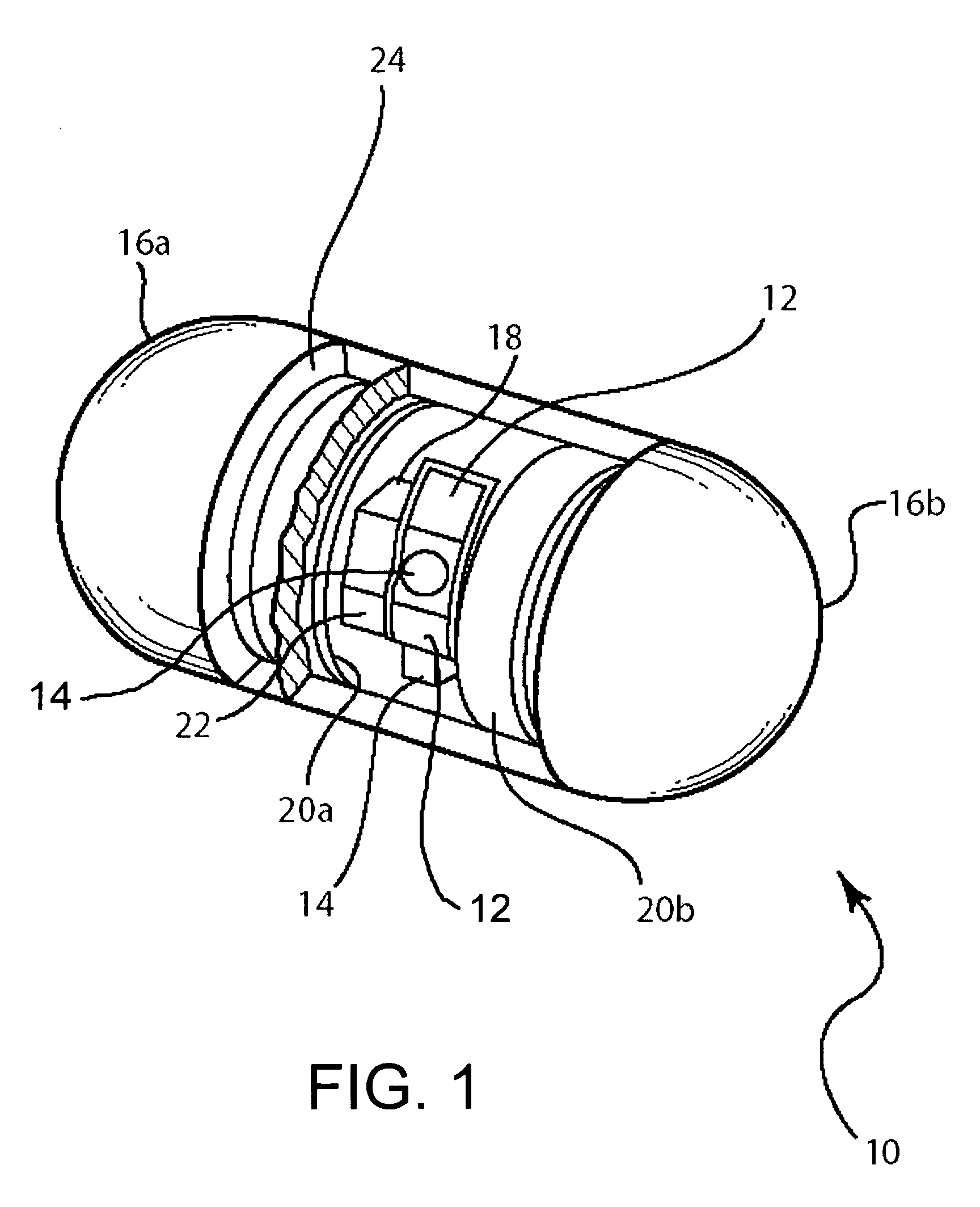
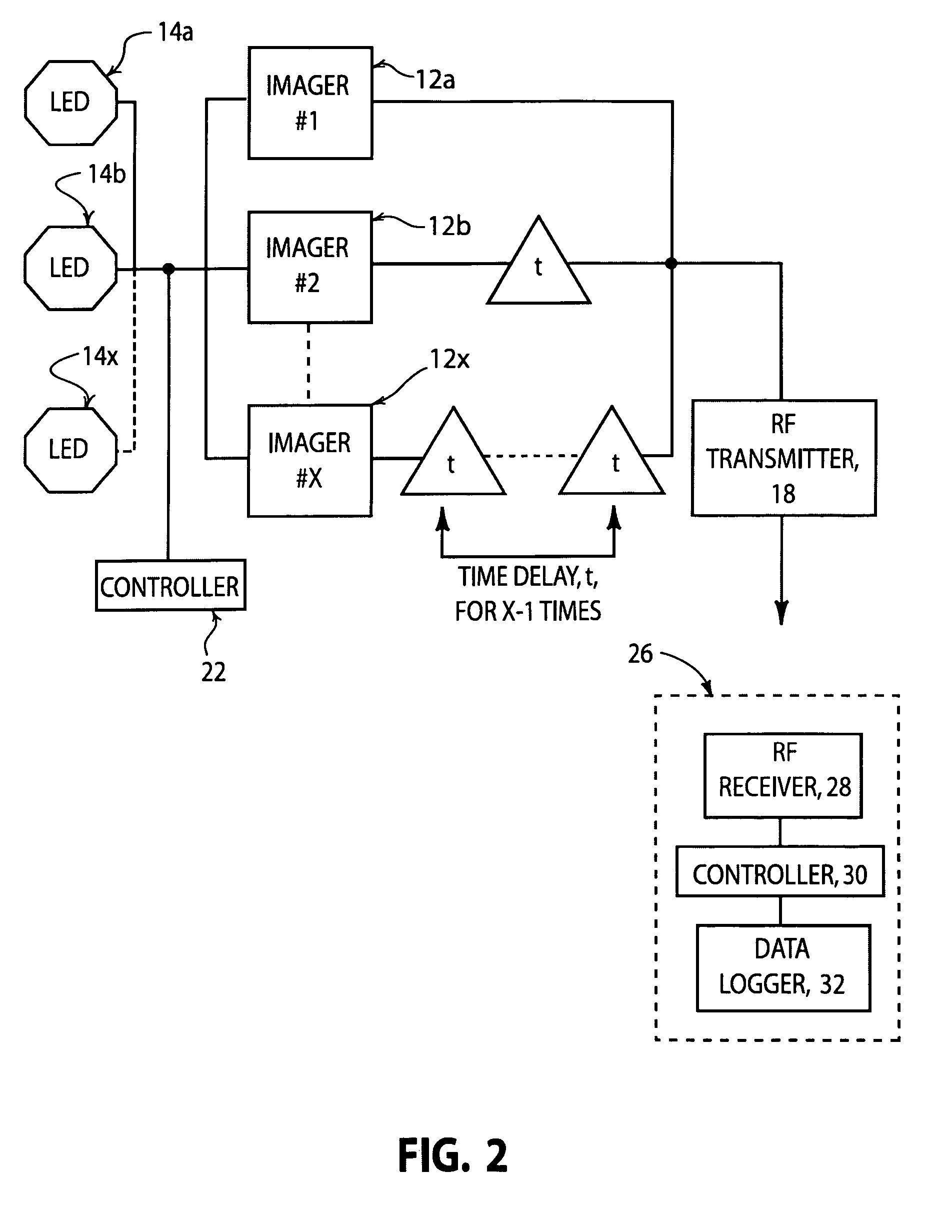
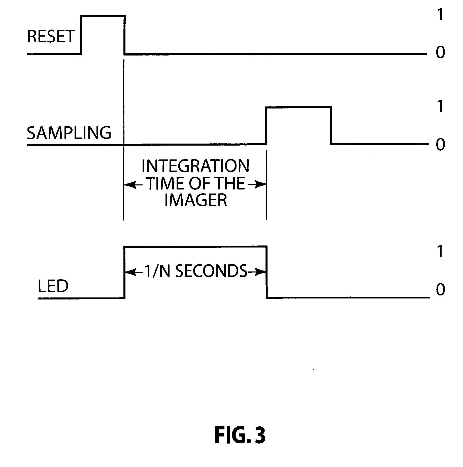
[0018] Briefly, the present invention includes an apparatus and method for imaging gastrointestinal organs from which meaningful images may be generated. The apparatus hereof comprises a wireless imaging capsule having an outer component that dissolves or breaks in the targeted organ, thereby permitting swelling of expandable materials disposed on each end of the capsule. As a result, the capsule is oriented and self-stabilizing against tumbling. At about the time of the expansion process, during this process or just afterwards, imaging components in the capsule are activated. These may include light emitting diodes (LEDs) for illuminating the interior of the organ of interest, and image sensors. The oriented and stabilized wireless capsule endoscope can then provide images from which details of the inner walls for organs having large lumens can be reconstructed. Although the colon has been chosen as an example of a large-lumen organ in the following description, application of the ...


PUM


Abstract

Wireless capsule endoscope technology has been used to image portions of the gastrointestinal (GI) tract, particularly the small bowel. However in other GI organs, especially those having larger-lumens, the capsule may become destabilized and tumble, precluding meaningful interpretation of the acquired images. The present invention describes a method and apparatus for permitting capsule imaging of organs having larger-lumens without tumbling, and includes an outer shell surrounding the capsule that targets the colon, as an example. Once the colon has been reached, the shell breaks or dissolves, and allows expansion of expandable materials attached to each end of the capsule, thereby stabilizing the capsule in the targeted organ, while permitting it to be moved by peristalsis and / or other means for locating the capsule. Imagers and light emitting diodes (LEDs) are activated during the expansion process, and enable overlapping of images. The capsule is moved through the colon, taking images at chosen frame rates with data being wirelessly transmitted by means of an RF transmitter, and is eventually expelled from the body.

Description

CROSS-REFERENCE TO RELATED APPLICATIONS [0001] This application claims the benefit under 35 USC 119(e) of U.S. provisional patent application No. 60 / 650,278 filed Feb. 4, 2005.FIELD OF THE INVENTION [0002] The present invention relates generally to imaging of the gastrointestinal organs and, more particularly, to the imaging of gastrointestinal organs using a self-orienting, capsule-based imaging system. BACKGROUND OF THE INVENTION [0003] Recent advances in low-power complementary metal-oxide silicon (CMOS) imagers, mixed signal application specific integrated circuit (ASIC) and white light emitting diodes (LEDs) has led to a swallowable video capsule for endoscopy [See, e.g., Arkady Clukhovsky, “Wireless Capsule Endoscopy” , Sensor Review, 23, no. 2, pp. 128-133 (2003).]. Capsule-based imaging of gastrointestinal organs was introduced in 1997 [See, e.g., Iddan et al., “In Vivo Video Camera System”, U.S. Pat. No. 5,604,531, Feb. 18, 1997.], and has since become a preferred imaging m...

Claims


Application Information

Patent Timeline
Patent Type & Authority Applications(United States)
IPC IPC(8): A61B1/00
CPCA61B1/0615A61B1/042A61B1/00006A61B1/00016A61B1/041A61B1/00177A61B1/00148
Inventor MINTCHEV, MARTIN P.YADID-PECHT, ORLY
Owner UTI LLP
Why Eureka
Try Eureka