Vaccines for proliferative ileitis and methods of making and using the same

a technology for ileitis and vaccines, applied in the field of vaccines for proliferative ileitis, can solve the problems of limited effective control measures for proliferative ileitis, porcine proliferative enteritis, and proliferative ileitis of porcine proliferative enteritis, and achieve the most potential use potential in diagnostic and antigen quantitation, and inhibit the development of cytopathic

Inactive Publication Date: 2006-08-31
THE ARIZONA BOARD OF REGENTS ON BEHALF OF THE UNIV OF ARIZONA
View PDF5 Cites 11 Cited by
  • Summary
  • Abstract
  • Description
  • Claims
  • Application Information

AI Technical Summary

Benefits of technology

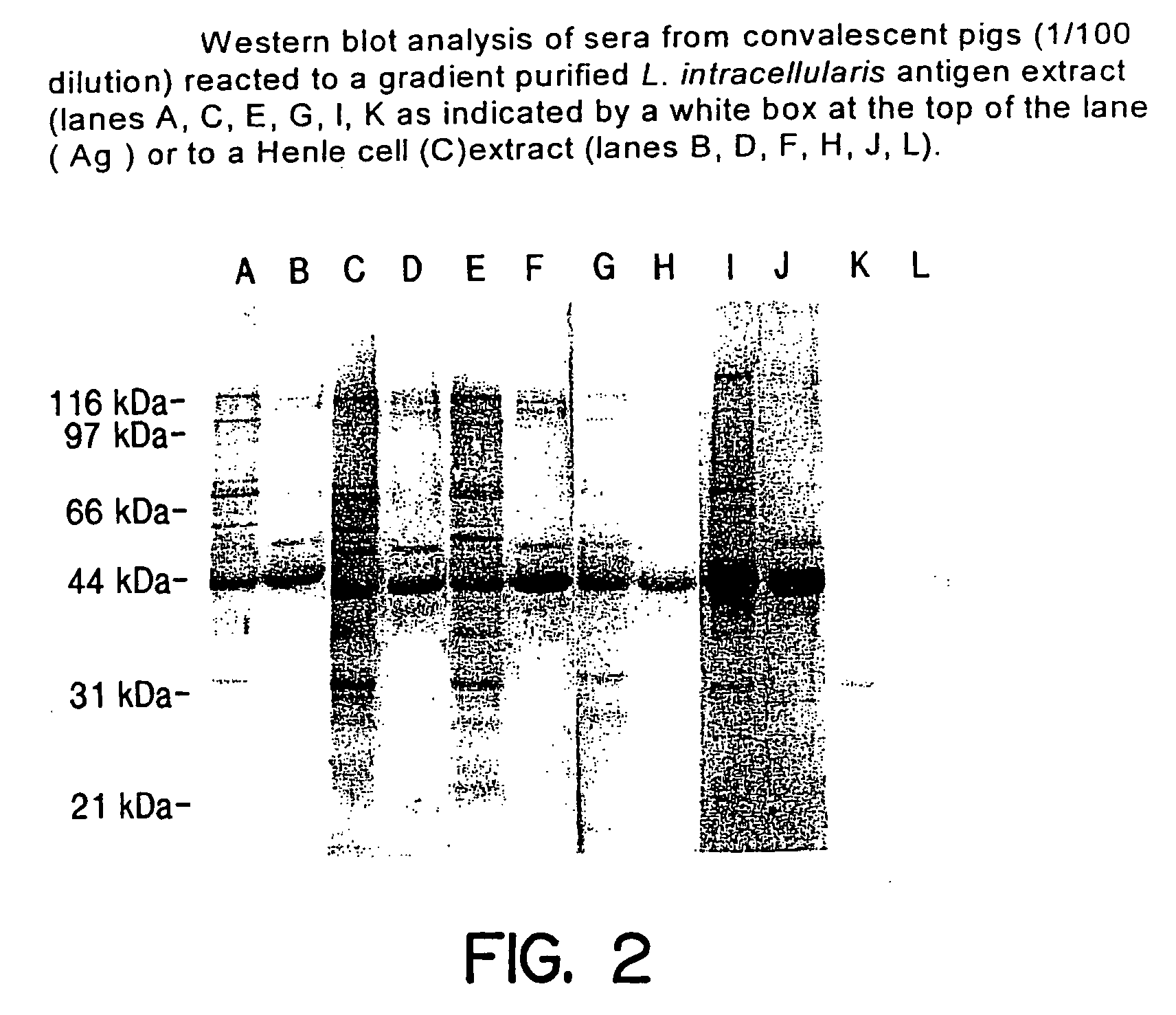
[0018] It is a final object of this invention to provide monoclonal antibodies (MoAbs) which can be used for diagnosis of proliferative ileitis or for development of antigen quantitation tests. Some of these MoAbs have been demonstrated to be neutralizing in that they inhibit the development of the cytopathic effect (CPE) when L. intracellularis is grown in Henle cells. By CPE is meant that the tissue culture cell monolayers are so adversely affected by the L. intracellularis that the cells die and slough off of the surface of the vessel onto which they are attached. Useful MoAbs include but are not limited to clones 2A2, 2B6, 5A2, 3A1, 2C1, 3D4, and 1C2, which correlate to antigens having molecular weights of 43-44, 60 kDa, 41 and 43-44 kDa, 41 kDa, 60 kDa, 71 kDa, and ≧115 kDa, respectively. Of these, MoAbs 2A2(detecting an antigen with a molecular weight between 43 and 44 kDa), 5A2 (detecting an antigen with molecular weights of 41, 43-44 kDa), 3A1 (detecting an antigen with a molecular weight of 41 kDa), 3D4 (detecting an antigen with a molecular weight of 71 kDa) and 1C2 (detecting an antigen with a molecular weight equal to or greater than 115 kDa) are neutralizing and have the most potential for use in diagnostic and antigen quantitation assays as well as for use in identifying target vaccine antigens including recombinant antigens. These studies indicate that an effective L. intracellularis vaccine must produce an immune response, possibly antibodies, to one or more of the antigens with molecular weights of 41 kDa, 43-44 kDa, 71 kDa and 115 kDa or greater.

Problems solved by technology

Porcine proliferative ileitis, sometimes referred to as porcine proliferative enteritis, is a major problem for the U.S. swine industry.
Effective proliferative ileitis control measures have been limited.
A basic trial-and-error therapeutic regimen, which includes the use of oral and parenteral broad-spectrum antibiotics, antihistamines, corticosteroids, nitroimidazole, and B vitamins, usually becomes quite costly and typically proves ineffective.

Method used


Image

  • Vaccines for proliferative ileitis and methods of making and using the same
  • Vaccines for proliferative ileitis and methods of making and using the same
  • Vaccines for proliferative ileitis and methods of making and using the same

Examples


example 1

[0050] Growth of L. intracellularis in tissue culture has been routinely accomplished. One method involves growing the organism in Henle 407 cells (ATCC No. CCL6) by infecting the cells with L. intracellularis bacterial seed organisms isolated from gut homogenates of pigs exhibiting clinical proliferative ileitis. Sections of intestine from infected pigs were removed, washed to remove mucus and suspended in Hanks Balanced Salts Solution (HBSS) containing hyaluronidase to detach epithelial cells from the lamina propria. The sections were then washed and the enterocytes were harvested by centrifugation. The enterocytes were washed a second time and then were exposed to gentamycin sulfate and amphotericin B for 24 hours at 4° C. to kill contaminating gut microflora. The treated cells were harvested, washed in HBSS, and lysed with 0.5% deoxycholate for 1 hour at 37° C. With constant agitation to release the intracellular organism. The lysates were passed through a sterile 0.65 u membran...

example 2

[0051]L. intracellularis also has been grown on rat intestinal epithelial cells (IEC), ATCC No. 1589. Since growth of L. intracellularis on these cells does not result in CPE, the monitoring of growth was accomplished either by use of standard PCR methods or by standard fluorescent antibody detection methods using a monoclonal antibody (MoAb). Quantitation via the PCR method was measured via use of a densitometer and competitive PCR procedures known to the art.

[0052] The process of growing L. intracellularis on IEC cells comprised the steps of 1) inoculation of the IEC cells with L. intracellularis organisms; 2) incubation of the infected IEC cell culture in a media capable of supporting growth of L. intracellularis at 37° C. in the presence of atmospheric conditions which allow growth of the organisms as well as growth of the IEC cells; 3) harvesting the L. intracellularis and 4) passaging the culture at approximately ten (10) day intervals to scale up the yield of organisms.

[005...

example 3

[0058]L. intracellularis IL-B (ATCC No. 55370) was grown in IEC cells using T-25 flasks, according to the procedure described previously (Example 2). Infected IEC cells were cultured for 5 to 7 days in gaspaks in an atmosphere of CO2:O2:N2 (8:8:84). Supernatant was removed and the cells were treated with 0.2% KCL for 5 min. and 0.1% KCL for 25 min. The KCL was removed and the cells were harvested by scraping. The harvested cells were passed through a 22 gauge needle to break down the cell structure. The cell lysate was subjected to low speed centrifugation for 10 min. and the semi-purified organisms remaining in the supernatant were harvested by high speed centrifugation. Antigen was pooled from 25 flasks and a portion of the antigen was subjected to a french press treatment for the production of soluble antigen. The remainder was aliquoted and stored at −70° C. This soluble antigen was formulated into a vaccine according to the following procedure. Vaccine antigen was formulated wi...


PUM

PropertyMeasurementUnit
molecular weightsaaaaaaaaaa

Abstract

A proliferative ileitis vaccine comprising tissue culture grown Lawsonia intracellularis and methods of making said vaccines. Proliferative ileitis vaccines described include those containing whole L. intracellularis, extracts of L. intracellularis, protective immunogenic subunits of L. intracellularis, recombinant immunogens of L. intracellularis and naked DNA of L. intracellularis. The vaccines of this invention may be inactivated or modified live and contain adjuvants and/or stabilizers. The vaccines of this invention may be in a liquid or lyophilized form. Also disclosed are monoclonal antibodies which neutralize the growth of L. intracellularis and which may be used for diagnosing proliferative ileitis as well as for quantitating antigen during vaccine production.

Description

BACKGROUND OF INVENTION [0001] 1. Field of the Invention [0002] The present invention relates generally to the preparation of vaccines and diagnostics for protecting pigs from proliferative ileitis caused by Lawsonia intracellularis. [0003] 2. Brief Description of the Prior Art [0004] Porcine proliferative ileitis, sometimes referred to as porcine proliferative enteritis, is a major problem for the U.S. swine industry. Proliferative ileitis is an intestinal disease complex of pigs characterized by crypt hyperplasia and by the presence of intracellular campylobacter-like organisms. Recognition of the disease has increased dramatically in the past ten years, with the incidence ranging as high as 20% and losses estimated at $50 million annually in the U.S. alone. Especially alarming is the apparent increase in incidence among the seed stock industry. The disease has been found worldwide and usually affects post-weaning pigs between six and twenty weeks of age. The clinical signs of pig...

Claims


Application Information

Patent Timeline
Patent Type & Authority Applications(United States)
IPC IPC(8): A61K39/02A61K48/00A61P1/00C12N15/09A61P1/04A61P31/04C07K16/12C12N1/20C12P21/08C12Q1/68
CPCA61K39/105C07K16/12A61P1/00A61P1/04A61P31/04
Inventor JONES, LYNN A.
Owner THE ARIZONA BOARD OF REGENTS ON BEHALF OF THE UNIV OF ARIZONA
Why Eureka
Try Eureka