
Method and apparatus for blood separations

a technology of blood separation and method, applied in the direction of centrifuge, centrifugal force sediment separation, separation process, etc., can solve the problems of low blood supply, widespread shortage of blood products, and less efficient manual methods of blood collection and separation than automated methods such as apheresis, so as to reduce direct collection and processing costs and minimize errors

Inactive Publication Date: 2006-10-12
TERUMO MEDICAL CORP
View PDF11 Cites 53 Cited by
  • Summary
  • Abstract
  • Description
  • Claims
  • Application Information

AI Technical Summary

Benefits of technology

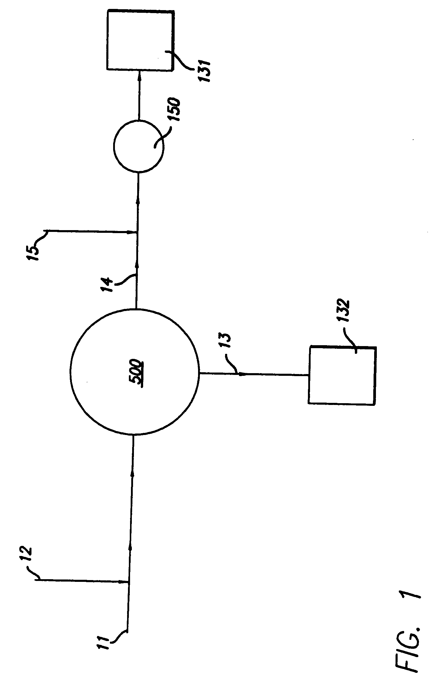
The present invention is a blood collection and processing system that reduces costs, automates procedures, and reduces errors. It can collect both single and double units of red blood cells, and can be used at remote or fixed sites. The system includes a disposable set with a manifold, continuous-flow centrifuge, and various components attached by tubing. The system also includes roller pump mechanisms, valves, sensors, and electronics to control the system's operation. The CFC disk in the system has an annular separation channel and a plasma shelf, and can transport fluids and blood products. The system can produce different types of blood products, such as leukoreduced RBCs and plasma, from whole blood collected from a donor. The technical effects of the invention include reducing costs, automating procedures, and reducing errors in blood collection and processing.

Problems solved by technology

In general, manual methods of collection and separation of blood are less efficient than automated methods such as apheresis.
Blood supply is low.
Nonetheless, the adoption of these programs, along with the increasing prevalence of aggressive medical procedures requiring blood components, has resulted in widespread shortages of blood products.
Additionally, there is a shrinking donor base.
However, many blood banks currently do not have the capacity or apheresis equipment required to perform double red cell collection.
Medicare and private insurers have limited reimbursements to hospitals for the purchase of blood units.
At many blood centers, the fully loaded cost to collect and process one unit of red blood cells exceeds its selling price since hospitals have enforced price pressures on blood centers.
Operating in compliance with regulations and practices when utilizing manual collection and processing procedures imposes an enormous quality assurance burden, under which more than one-half of blood centers in the United States still fail to operate.
Additionally, blood bank organizations have experienced significant price erosion for their blood products and have had to absorb costly, unfunded new safety and quality control procedures and tests mandated by the FDA.
Leukocytes are currently removed from red cells and platelets by manual filtration processes which are time consuming and labor intensive.
Although manual processes for blood collection and separation have some serious disadvantages, they are generally far less expensive than the automated alternatives, such as apheresis, as they do not require specialized staff, expensive equipment and disposables.
Additionally, the cumbersome (large and heavy) apheresis equipment does not lend itself to transportation to or use at mobile collection sites, where the majority of blood donations are collected.
In part for the foregoing reasons, although apheresis is used extensively for certain procedures, such as platelet collection where up to sixty-five percent of platelets collected in the United States are collected using plateletpheresis, apheresis has not achieved high penetration or displaced the current manual processes for blood collection and separation where one or more red cell products are obtained.
Similarly, double unit collection has not been implemented, in part, because current procedures for double unit collection are expensive and relatively complex.
Finally, for some procedures, such as leukocyte filtering, there are few, if any, alternatives to a time consuming and expensive manual process.

Method used


Image

  • Method and apparatus for blood separations
  • Method and apparatus for blood separations
  • Method and apparatus for blood separations

Examples


example 1

A Blood Collection and Separation Process

[0125] Before blood donation begins, a disposable set is removed from its sterile package and hung on the console. Solution bags (anticoagulant, red blood cell additive solution, and saline) are attached to the console. Solution bags could be pre-attached but are assumed in these processes to be attached at disposable set-up. The solution bags may have Luer-lock or spike attachments. Bacterial (0.2 micron) filters are used in the flow paths from these bags to maintain sterility. The bags are hung in designated locations on the console. The console calibrations and system software status are performed automatically before blood donation begins. Data collection is performed manually by the user with a bar code wand reader and automatically via the console.

[0126] The processes of the present invention are automatic. The automatic process begins after the phlebotomist (user) places the access needle in the donor's vein and after the non-anticoa...

example 2

One Unit of Leukoreduced RBCs and Plasma

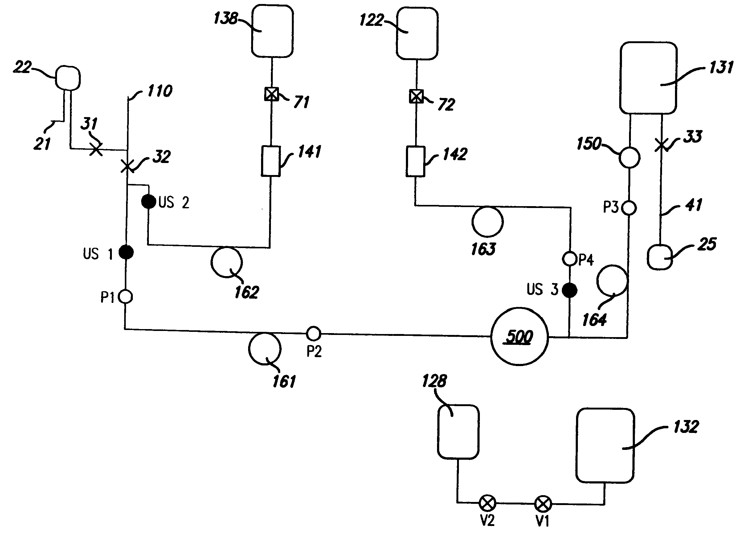
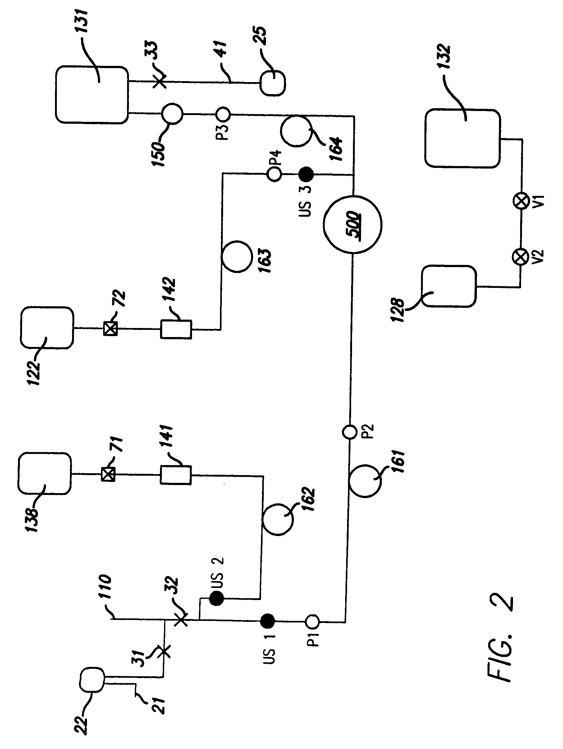
[0139] In one embodiment of the invention, one unit of whole blood is collected from a donor to produce one unit of leukoreduced RBCs in storage solution and plasma. This embodiment of the invention is depicted in FIG. 2. The system depicted in FIG. 2 includes a donor needle 110, sample pouch 22, sample site 21, manual clamps 31, 32 and 33, ultrasonic air sensors US1, US2 and US3, solenoid valves V1 and V2, pressure sensors P1, P2, P3 and P4, anticoagulant bag 138 and storage solution bag 122 attached by connectors 71 and 72, bacterial filters 141 and 142, anticoagulant pump 162, solution pump 163, blood pump 161, RBC pump 164, air bag 128, plasma bag 132, CFC 500, leukofilter 150, RBC bag 131, and air pouch 25 connected to line segment 41.

[0140] As seen in the schematic depicted in FIG. 2, this process automatically takes whole blood from the donor; adds anticoagulant from an anticoagulant bag 138; separates the blood into concentrated red ...

example 3

Whole Blood Separation into Leukoreduced RBCs and Plasma Products

[0147] A more detailed description of the user implementation of the process depicted in FIG. 2 (one unit of leukoreduced RBCs in storage solution and plasma are produced), is described here. The user plugs in the system, switches the system on, and closes the console door. The system warms up to the operating temperature range and then initializes in which the system boots up. The system then performs a self check in which it internally checks to see if components, as for example, pumps, valves, and sensors, are responding properly. The user unpacks the disposable set and waits until the user interface display 790 (FIG. 23) indicates “ready to accept disposable.” The user then opens the console door, installs the disposable set into the console, closes the console door, clamps the donor needle 110 line, clamps the sample pouch 22 line, clamps the line segment 41, hangs the pre-attached bags (anticoagulant bag 138, st...


PUM

PropertyMeasurementUnit
angleaaaaaaaaaa
volumeaaaaaaaaaa
volumeaaaaaaaaaa

Abstract

Described herein is a method and apparatus for collecting and separating whole blood into its components, including collecting an amount of leukoreduced red blood cells. The collection and separation system includes a console and a disposable set. The method may include processing the blood through the centrifuge, collecting the leukoreduced red blood cells, and collecting and returning plasma to the source. The disposable set may include a manifold, a CFC, and various components attached by tubing. These components may include one or more solution bags, blood product bags, bacterial filters, leukofilters, and a donor blood collection tube with access needle.

Description

RELATED APPLICATIONS [0001] This application is a continuation of U.S. patent application Ser. No. 11 / 102,215, filed on Apr. 8, 2005, which is incorporated herein by reference.BACKGROUND OF THE INVENTION [0002] 1. Field of the Invention [0003] The present invention generally relates to blood processing systems for the automated collection of blood and separation of blood into its component parts. More particularly, the present invention relates to a centrifuge which can separate blood into two or more components and may be used in such blood processing systems. [0004] 2. Description of Related Art [0005] The adult human body contains approximately 10 units (or approximately 5,000 mL) of whole blood consisting of both cellular and liquid portions. The cellular portion (about 45% by volume) comprises red blood cells, white blood cells and platelets. The liquid portion (about 55% by volume) is made up of plasma and soluble blood proteins. Each of these components can be directly transf...

Claims


Application Information

Patent Timeline
Patent Type & Authority Applications(United States)
IPC IPC(8): C02F1/38
CPCA61M1/0209A61M1/3693A61M2205/12A61M2205/505A61M1/3696A61M2209/084B04B5/0442A61M1/0236A61M2209/082A61M1/0218A61M1/0231A61M1/3633A61M1/362227A61M1/36226A61M1/362265A61M1/362223A61M1/36224A61M1/362261A61M1/362266A61M1/36225
Inventor ROBINSON, THOMAS CHARLESSAHINES, THOMAS P.D'ELIA, RICHARD R.ZAMORA, SALVADOR M.LEWIS, BRIAN D.FERNANDEZ, ROBERT K.BRIONES, VICTOR A.NIER, DAVID M.MCNAMARA, THOMAS NOLAND
Owner TERUMO MEDICAL CORP
Learn More