Apparatus for insertion of a medical device during a medical imaging process

a medical device and imaging technology, applied in the direction of magnetic variable regulation, instruments, catheters, etc., can solve the problems of prostate cancer, trus-guided biopsy fails to correctly detect the presence of prostate cancer in approximately 20% of cases, and targeted local therapy is not possible today, so as to minimize dynamic tissue deformation, minimize the effect of dynamic tissue deformation, and maximize the accuracy of needle placemen

Active Publication Date: 2006-10-26
THE JOHN HOPKINS UNIV SCHOOL OF MEDICINE
View PDF8 Cites 110 Cited by
  • Summary
  • Abstract
  • Description
  • Claims
  • Application Information

AI Technical Summary

Benefits of technology

[0017] The devices, systems, apparatuses, and methods of the present invention are particularly adaptable for use in image-guided prostate biopsy that employs high resolution MRI imaging inside a closed MRI scanner, while maintaining safe transrectal access. In addition, such devices, apparatuses, systems and methods embody a remotely controlled needle insertion technique, as compared to the conventional manual manipulation technique, thereby maximizing needle placement accuracy and also minimize dynamic tissue deformation during the procedure. The device, system, apparatus and methods of the present invention also can employ real-time MRI guidance while the system is compatible with high-field MRI scanners with no imaging artifacts. In addition, a device and / or apparatus of the present invention fits inside a closed magnet, assumes very little workspace for motion, minimizes organ motion and deformation in a non-invasive manner and uses at least three degree-of-freedom motion to reach a selected target.

Problems solved by technology

Prostate diseases represent a significant health problem in the United States.
At the same time, however, TRUS-guided biopsy fails to correctly detect the presence of prostate cancer in approximately 20% of cases [Norberg M, Egevad L, Holmberg L, Sparen P, Norlen B J, Busch C. The conventional sextant protocol for ultrasound-guided core biopsies of the prostate underestimates the presence of cancer.
For the same reason, targeted local therapy today also is not possible with the use of TRUS guidance.
Unfortunately, MR imaging alone, without concurrent biopsy, suffers from low diagnostic specificity.
In addition, there are other fundamental obstacles that must be addressed when using MRI imaging techniques in prostate biopsy and related localized therapy of the prostate.
Conventional high-field MRI scanners use whole-body magnets that surround the patient completely and do not allow access to the patients during imaging.
Thus, the workspace inside the bore of the whole-body magnet is so extremely limited, that conventional medical robots and mechanical linkages do not fit inside the whole-body magnet.
Due to these ultra-strong magnetic fields, ferromagnetic materials and electronic devices are not allowed to be in the magnet due to safety and / or imaging concerns, which excludes the use of traditional electromechanical robots and mechanical linkages.
This technique has limitations, however, because it involves the use of an open MRI scanner.
Perhaps most importantly, the incurred cost and complexity of open MRI imaging are substantial, especially when compared to transrectal ultrasound imaging.
While this approach seems to be acceptable when used in a research type of environment, it adds to the complexity and cost of the open MRI.
Therefore, the out comes of TRUS guided procedures show significant variability among practitioners.
Recently, a 6-DOF robot has been presented for transperineal needle placement into the prostate, but that kinematic concept is not applicable in transrectal procedures [G. Fichtinger, T. L DeWeese, A. Patriciu, A. Tanacs, D. Mazilu, J. H. Anderson, K. Masamune, R H. Taylor, D. Stoianovici: Robotically Assisted Prostate Biopsy And Therapy With Intra-Operative CT Guidance: Journal of Academic Radiology, Vol 9, No 1, pp.
This solution is not suitable for a closed magnet configuration.
In addition, the long arms of this robotic manipulator amplify the effects of flexure and sagging, which can render this system inaccurate for certain procedures.
Moreover, because the device is intended to be mounted permanently with respect to the MRI scanner, the robotic manipulator is not flexibly adaptable to different sides of the body.
This robot is demonstrated in accessing the breast, but it is not readily adaptable for abdominal and intracavity use.
There also has been published variations of an in-MRI robot for stereotactic brain surgery, but the actual embodiments of that system also are not applicable in transrectal biopsy [Masamune et. al., Development of an MRI-compatible needle insertion manipulator for stereotactic neurosurgery.

Method used


Image

  • Apparatus for insertion of a medical device during a medical imaging process
  • Apparatus for insertion of a medical device during a medical imaging process
  • Apparatus for insertion of a medical device during a medical imaging process

Examples


example

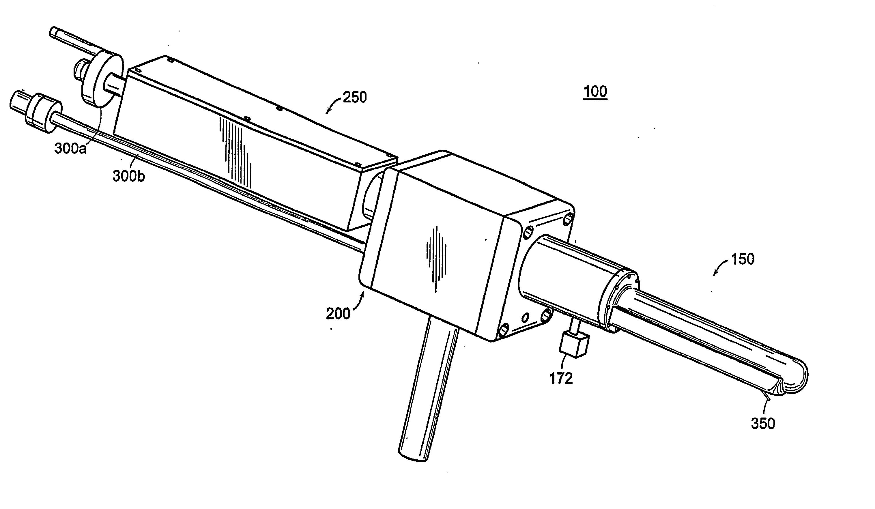
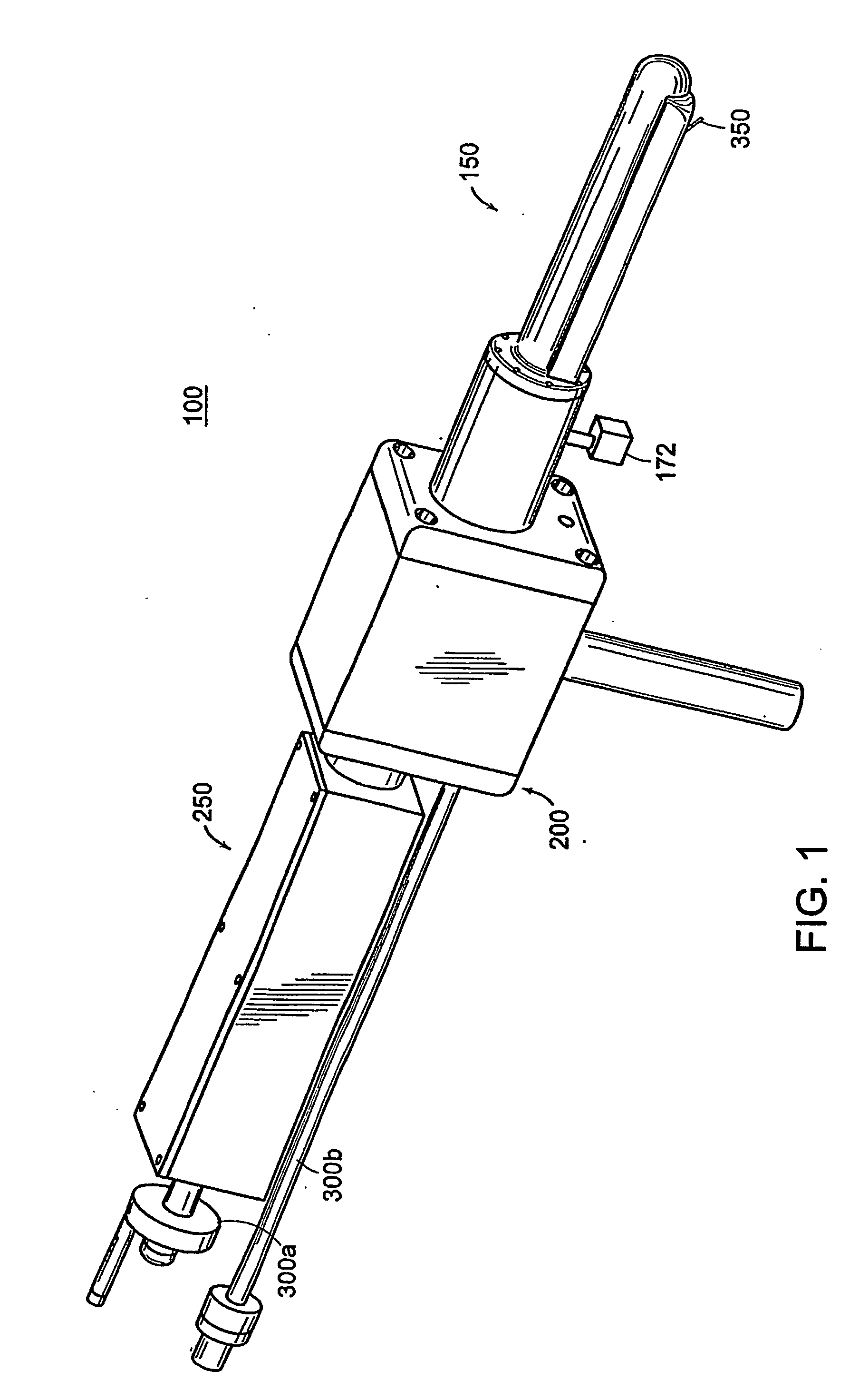
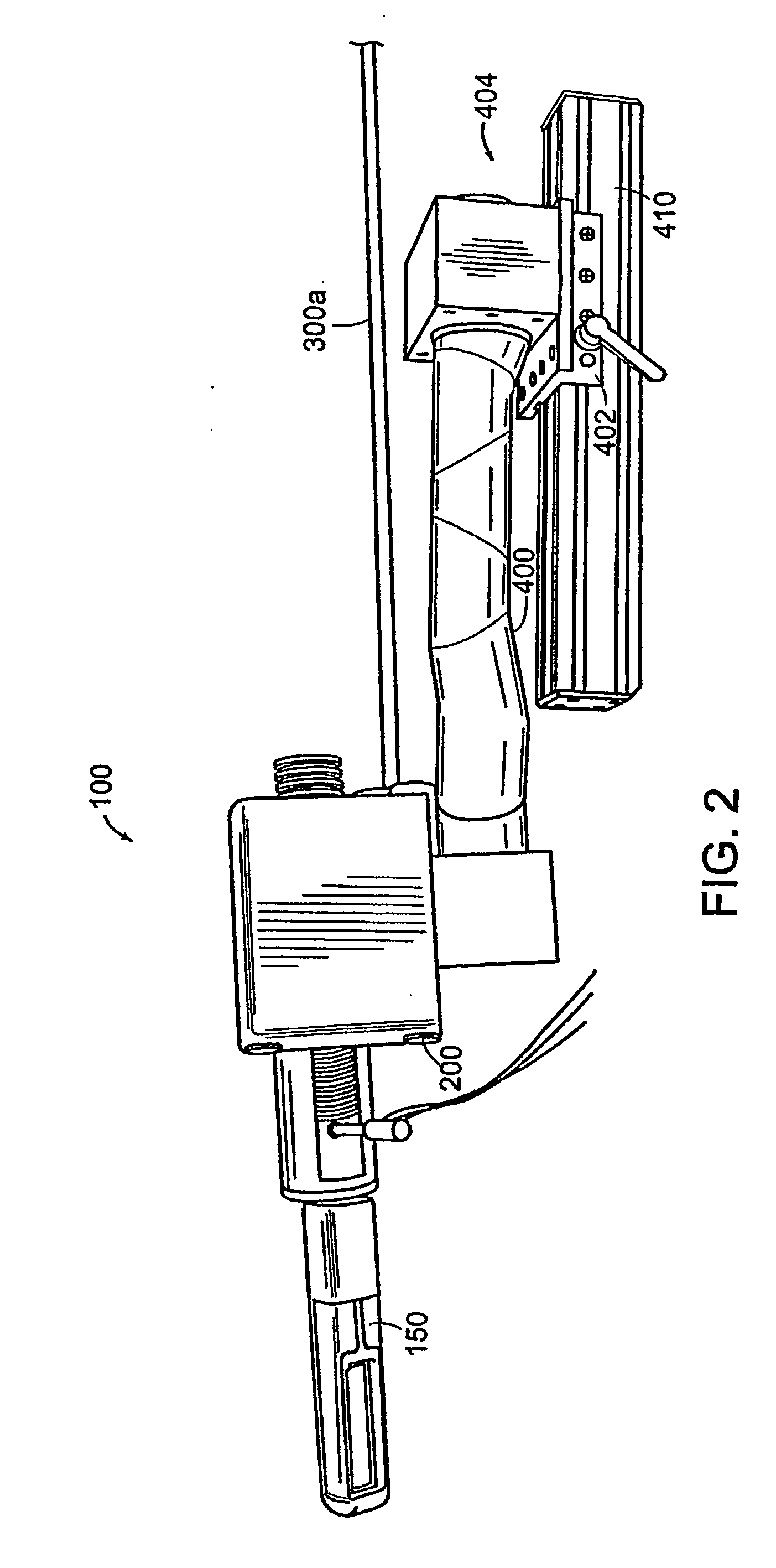
[0142] A mechanically actuated, transrectal needle guide is used to perform MR guided needle placements in the prostate. With a microcoil tracking method, the position and orientation of the biopsy needle guide in the MR imaging volume (60 msec) could be quickly and accurately located. Knowing the position of the biopsy needle allows for acquisition of realtime images of a plane including the needle and registration of the needle position with previously acquired, high-resolution images of the prostate. In four canine studies, the functionality and applications of a system was demonstrated.

[0143] A thin-walled, cylindrical plastic sheath (Delrin plastic, DuPont Inc., Wilmington, Del.) with a radius of 1.5 cm is inserted into the subject's rectum, forming a stable and stationary entry point through which the prostate can be accessed. Integral to the sheath is a single turn imaging loop (with a diameter of 2.5 cm) for local imaging of the prostate. The sheath has a window, located wi...


PUM


Abstract

The end-effector (150) includes a sheath (152) and a medical device or needle carrier (154) that is disposed within the interior compartment (166) of the sheath. Aperture (162) is located in a portion of the sheath proximal a distal end of the sheath that is inserted into a natural or artificial cavity. This device is guided by a real-time imager.

Description

[0001] This application claims the benefit of U.S. Provisional Application Ser. No. 60 / 374,376 filed Apr. 22, 2002, the teachings of which are incorporated herein by reference in their entirety.STATEMENT REGARDING FEDERALLY-SPONSORED RESEARCH [0002] The U.S. Government has provided funding under contract No. / grant No. EEC9731478 awarded by the National Science Foundation and thus the government may have certain rights to and / or in the invention.FIELD OF INVENTION [0003] The present invention generally relates to devices, apparatuses and methods for inserting a medical device such as a needle into a mammalian body while the body is within the imaging field of a medical imager, particularly devices, apparatuses and methods for inserting and guiding a needle to a target site within a body while the body is within the imaging field of a medical imager, and more particularly to devices, apparatuses and methods for inserting and remotely guiding a needle to a target site within a body sel...

Claims


Application Information

Patent Timeline
Patent Type & Authority Applications(United States)
IPC IPC(8): A61B5/05A61B10/02A61B5/055A61B5/07A61B8/12A61B10/00A61B17/00A61B17/34A61B19/00G01R33/28
CPCA61B5/055A61B8/445A61B6/12A61B8/0833A61B8/0841A61B8/12A61B10/0241A61B17/3403A61B17/3478A61B19/5244A61B2017/00274A61B2018/00547A61B2019/507A61B2019/5251A61B2019/5276A61B2019/528A61B2019/5291A61B2019/5454A61B2019/5458G01R33/287A61B5/06A61B5/065A61B34/20A61B2034/107A61B2034/2051A61B2090/365A61B2090/378A61B2090/3784A61B2090/3954A61B2090/3958
Inventor FICHTINGER, GABORATALAR, ERGINWHITCOMB, LOUISLSUSIL, ROBERTCTANACS, ATTILAKRIEGER, AXEL
Owner THE JOHN HOPKINS UNIV SCHOOL OF MEDICINE
Why Eureka
Try Eureka