System and method for imaging and treatment of tumorous tissue in breasts using computed tomography and radiotherapy

a breast cancer and computed tomography technology, applied in the field of breast cancer imaging and treatment of tumorous tissue using computed tomography and radiotherapy, can solve the problems of missing cancers, unnecessary surgical procedures, limited prediction value and specificity of x-ray mammography, etc., and achieve the effect of preventing the formation of vacuum

Inactive Publication Date: 2006-11-23
VARIAN MEDICAL SYSTEMS
View PDF40 Cites 186 Cited by
  • Summary
  • Abstract
  • Description
  • Claims
  • Application Information

AI Technical Summary

Benefits of technology

[0009] In some embodiments, the barrier is provided with a plurality of holes surrounding the opening area for preventing formation of vacuum in a region beyond the breast.
[0010] In some embodiments, the holder is an open ledge that supports the breast from below with no vacuum environment. This support ledge can be a simple flat surface oriented horizontally or at some small angle (e.g. 5 to 30 degrees) to the horizontal. In some embodiments, this open ledge has the curved shape of a half cup. In some embodiments, this ledge is a combination of flat and curved surfaces. In some embodiments the breast support is a flexible material that provides positioning and immobilization much like that of a brassiere.
[0011] The barrier can be in the shape of a cone, partial spheroid, many faceted pyramid or any arbitrary combination of flat and / or curved surfaces that support and / or protect the patient while allowing proper X-ray exposure of the breast and its axillary extension from the moving gantry. In some embodiments, the barrier can comprise an inner section and an outer section. The inner section is detachable from the outer section and provided with the opening adapted to allow a patient's breast and arm passing therethrough. In some embodiments, the inner section is rotatable with respect to the outer section such that the location of the opening in the barrier is changeable.

Problems solved by technology

The predictive value and specificity of x-ray mammography remain limited however, due to projecting a three-dimensional object into a two-dimensional image and due to poor contrast detectability.
Incorrectly diagnosing malignant tissue as healthy can result in missing cancers in their early stages while incorrectly diagnosing healthy tissue as malignant can lead to unnecessary surgical procedures.
In indeterminate cases, biopsy is often necessary, despite the disadvantages of high cost and the stress imposed on patients.
One limitation of this type of system is that it only images the breast itself without providing image information on the important soft tissue between the breast and the axilla (arm pit) where primary breast lesions often spread to infect lymph nodes in this region.
Another limitation is that it cannot make immediate use of widely available CT gantry systems and external beam radiotherapy systems, which primarily have axes of rotation that are horizontal.
One limitation of this type of system is that it scans not only the target breast and surrounding soft tissue, but also simultaneously exposes the heart, lungs and other body parts to unnecessary radiation.

Method used


Image

  • System and method for imaging and treatment of tumorous tissue in breasts using computed tomography and radiotherapy
  • System and method for imaging and treatment of tumorous tissue in breasts using computed tomography and radiotherapy
  • System and method for imaging and treatment of tumorous tissue in breasts using computed tomography and radiotherapy

Examples


Embodiment Construction

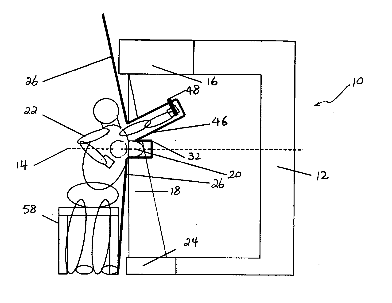
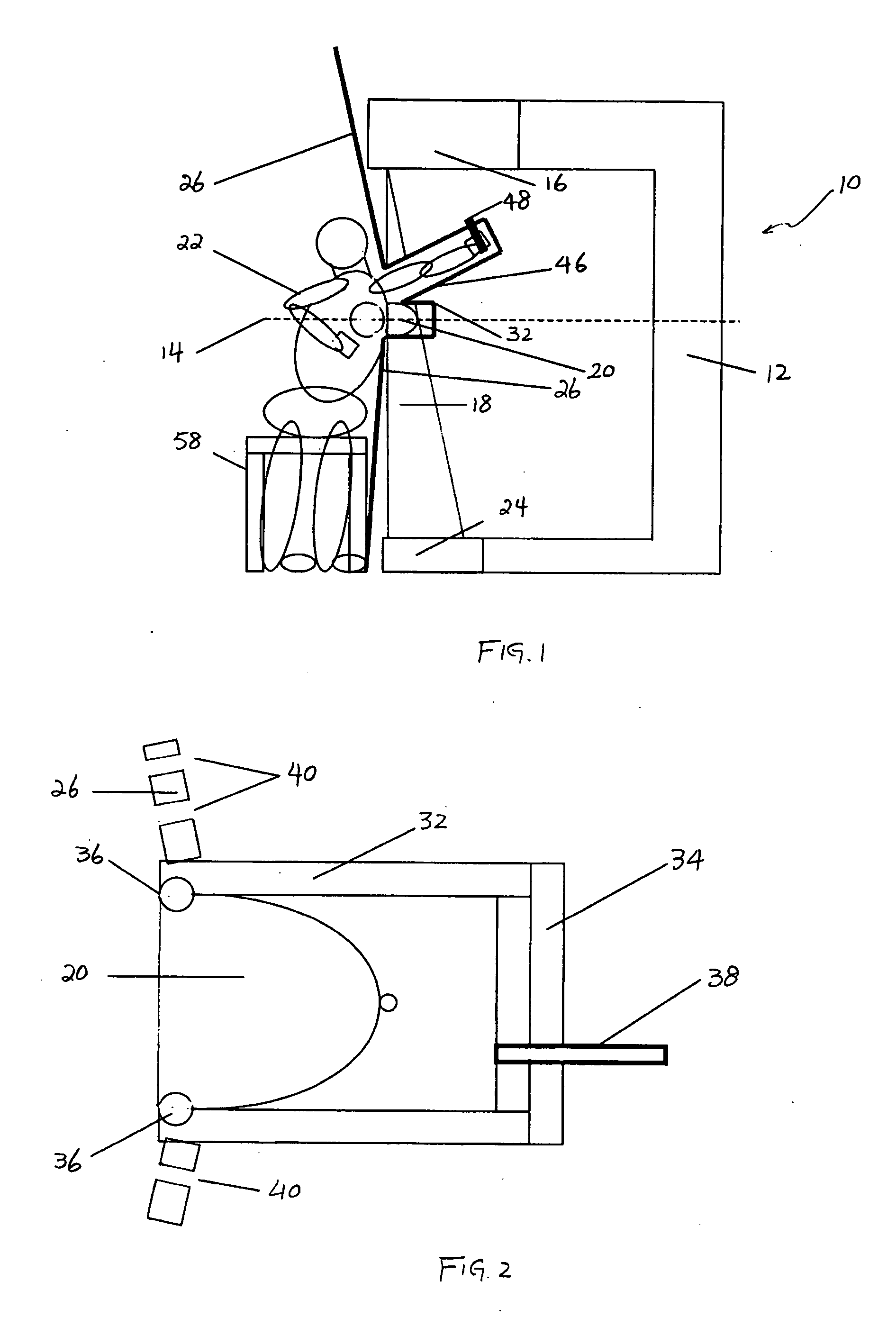
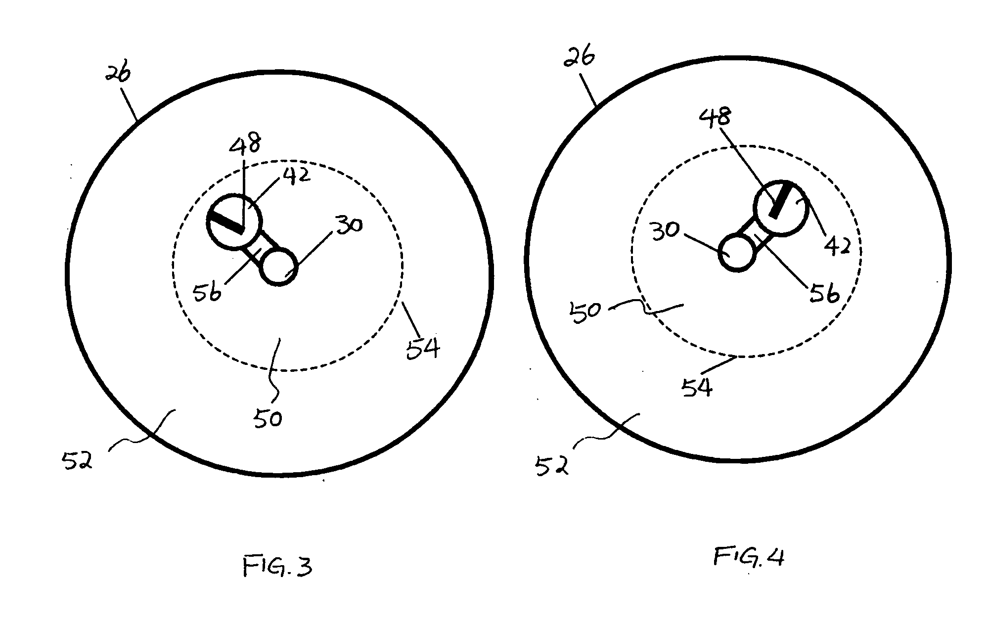
[0043] Referring to FIGS. 1 to 18, where like elements are designated by like references,-various exemplary embodiments of the radiation system and method of the present invention will now be described. In general, the radiation system includes a gantry comprising a radiation source for generating a radiation beam to irradiate a portion of a body, a detector spaced from the radiation source, and a structure positioned between the body and the gantry. In the present specification, the invention is described with embodiments where a human breast is irradiated, for example, for the purpose of forming an image thereof. It will be appreciated that the claimed invention may be used on animals as well as humans, and may be used on different body parts. The structure is described in the illustrative embodiments as a protective barrier that prevents at least a portion of the radiation from reaching other portions of the body, but can perform different or additional functions. For example, th...


PUM


Abstract

The present invention provides a system 10 for irradiating a breast 20 of a patient 22. The system 10 comprises a gantry 12 rotatable about a horizontal axis 14 and comprising a radiation source 16 for generating a radiation beam 18 and a detector 24 spaced from the radiation source 16, and a barrier 26 disposed between the patient 22 and the gantry 12. The barrier 26 is provided with an opening 30 adapted to allow a breast 20 passing therethrough to be exposed to the radiation beam 18. In some embodiments, the barrier 26 is provided with an opening 30 adapted to allow both the breast 20 and the tissue leading from the breast to axilla and the muscle tissue of the adjacent chest wall passing therethrough to be exposed to the radiation beam 18.

Description

FIELD OF THE INVENTION [0001] This invention relates generally to a system and method of providing radiation imaging and / or treatment and in particular, to a system and method for imaging and treatment of tumorous tissue in breasts using computed tomography (CT) and radiotherapy. BACKGROUND [0002] Radiological evaluation of breasts is important not only for early detection of disease in breasts, but also for staging, treatment targeting and monitoring responses to treatment. Conventional x-ray mammography has been shown a cost-effective tool for early detection of breast cancer. In conventional x-ray mammography, a breast is positioned on a platform and compressed with plastic plates. The breast is compressed in order to even out the breast thickness so that the correct dose of X-rays can be delivered for clear images without over exposure in thinner regions and under exposure in thicker regions and also to spread out the tissue to reduce the likelihood of obscuring benign and malig...

Claims


Application Information

Patent Timeline
Patent Type & Authority Applications(United States)
IPC IPC(8): A61B6/04
CPCA61B6/032A61B6/0414A61B6/107A61B6/405A61B6/4441A61N2005/1097A61B6/482A61B6/502A61B6/541A61N5/10A61N5/1081A61B6/466
Inventor PARTAIN, LARRY D.HUMBER, DAVID H.
Owner VARIAN MEDICAL SYSTEMS
Why Eureka
Try Eureka