Kits, apparatus and methods for magnetically coating medical devices with living cells

a technology of living cells and medical devices, applied in the field of medical devices, can solve the problems of poor blood compatibility of these surfaces, need to provide surfaces, and risk of acute thrombosis and chronic instability, and achieve the effects of convenient coating of medical devices, easy and quick magnetization, and convenient attachmen

Active Publication Date: 2006-12-21
MAYO FOUND FOR MEDICAL EDUCATION & RES
View PDF41 Cites 48 Cited by
  • Summary
  • Abstract
  • Description
  • Claims
  • Application Information

AI Technical Summary

Benefits of technology

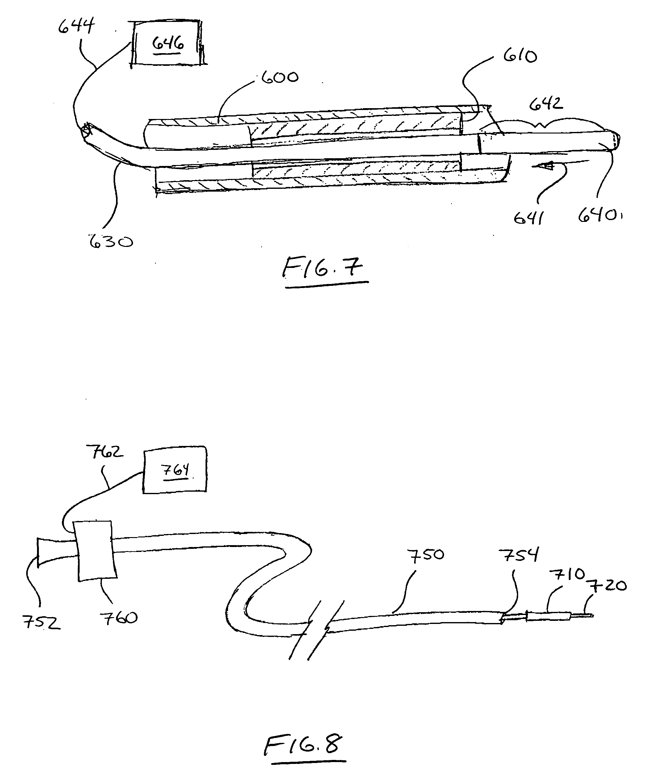
[0011] One potential advantage of the present invention is the ability to rapidly attract and attach large numbers of magnetic cells to the magnetic contact surfaces of a medical device. This is possible because the device can be located in a carrier liquid containing high concentrations of magnetic cells before or after implantation. The carrier liquid with magnetic cells may be in contact with the medical device in vitro or in vivo. In either case, it may be preferred that the carrier liquid have a concentration of magnetic cells that is high enough to facilitate coating of the medical device within an acceptable period of time, e.g., several hours or less.
[0012] If the magnetic coating is performed in vivo, it may be preferred that the carrier liquid with magnetic cells be delivered to a defined volume in which the medical device is located. If the medical device is implanted within, e.g., a blood vessel, the defined volume may be created by using a catheter to seal the vessel on one or both sides of the implanted medical device, followed by delivery of the carrier liquid to the location of the implanted medical device.
[0013] Another potential advantage of the present invention is that the medical devices may be located in any suitable location within the body and coated with magnetic cells after implantation, i.e., the medical devices and methods may be used in blood vessels, but the potential locations are not limited to blood vessels. For example, the medical devices may be located and magnetically coated within the gastrointestinal tract, salivary glands, bile ducts, pancreatic ducts, renal system (kidney, ureters, bladder, urethra, etc.), airways in lungs, conduits in the brain and spinal cord, surgically created conduits or lumens in an organ, congenital abnormalities in any organ, etc.
[0014] Another potential advantage of the present invention is that medical devices constructed of metals and / or metal alloys may be easily and quickly magnetized by exposure to a magnetic field (if the metals or metal alloys do not inherently exhibit magnetic fields). In some embodiments, the apparatus and methods of the present invention provide the ability to magnetize a medical device as part of the implantation process or to magnetize the medical device in vivo. In some instances, it may be possible to use known medical devices such as, e.g., stents, heart valves, etc. in connection with the present invention.
[0015] Yet another potential advantage of the present invention is the ability to demagnetize an implanted medical device after magnetically coating the medical device by, e.g., locating a degaussing element proximate the medical device in vivo after a selected period of time, e.g., 24 hours to 48 hours. By magnetically coating the medical device, the advantages associated with magnetic coating as discussed herein may be obtained and any potential disadvantages associated with leaving a magnetized medical device may be alleviated by demagnetizing the medical device after the magnetically attracted biologic cells have formed alternative attachments to the medical device.
[0016] Still another potential advantage of some embodiments of the present invention is that cells can be magnetically attracted to and retained on the medical devices without requiring additional modifications for cellular attachments such as receptor-ligand binding, antigen-antibody interactions, or other coatings (such as, e.g., fibronectin) to obtain adequate cell adhesion. Examples of some such binding techniques that may be avoided by use of the present invention include, but are not limited to, e.g., ligand binding, fibronectin, antibodies, proteins, etc.

Problems solved by technology

A recurring issue in the field of medical devices, especially implantable medical devices, is the need to provide surfaces that are compatible with the environment in which they are contained.
Problems associated with implanted medical devices that include surfaces in contact with a patient's bloodstream may include, for example, the risk of acute thrombosis and chronic instability—such as calcification—of the implant surface.
Poor blood compatibility of these surfaces is almost always a predominant reason for the limitations of these implants, such as the loss of heart valve functionality over time or poor patency rates in small diameter conduits due to acute thrombosis or intimal hyperplasia.
Attempts to modify the surfaces of synthetic grafts to overcome the patency problems associated with thrombosis or intimal hyperplasia have generally shown poor long-term outcomes, as these surfaces are unable to maintain a sustained anti-thrombogenic bioactivity as discussed in, e.g., U.S. Patent Application Publication No.
Problems arise, however, in the retention of suitable cells on the surfaces of the devices.
Unfortunately, however, in vivo recruitment techniques suffer from a number of disadvantages.
One significant disadvantage is the inability to determine whether the magnetic forces exhibited by the surfaces are sufficient to attract target cells before implantation.
A failure to properly magnetize a device including magnetic material that is not permanently magnetic may result in less than optimal in vivo recruitment.
Another potential disadvantage is the time required to recruit significant numbers of target cells to the desired surfaces.
Furthermore, even after significant periods of time, the target cells may be attached at low density over the surfaces, resulting in less than optimal performance.
If the surfaces are located in areas of limited or no blood flow, then recruitment may be further hampered or prevented.

Method used


Image

  • Kits, apparatus and methods for magnetically coating medical devices with living cells
  • Kits, apparatus and methods for magnetically coating medical devices with living cells
  • Kits, apparatus and methods for magnetically coating medical devices with living cells

Examples


Embodiment Construction

[0049] In the following detailed description of illustrative embodiments, reference is made to the accompanying drawings that form a part hereof, and in which are shown, by way of illustration, specific embodiments in which the invention may be practiced. It is to be understood that other embodiments may be utilized and structural changes may be made without departing from the scope of the present invention. Furthermore, like reference numbers denote like features in the different figures.

[0050] It should be noted that as used herein and in the appended claims, the singular forms “a”, “and”, and “the” include plural referents unless the context clearly dictates otherwise. Thus, for example, reference to “a magnetic cell” includes a plurality of such cells and reference to “the magnetic contact surface” includes reference to one or more magnetic contact surfaces and equivalents thereof known to those skilled in the art.

Medical Devices with Magnetic Contact Surfaces

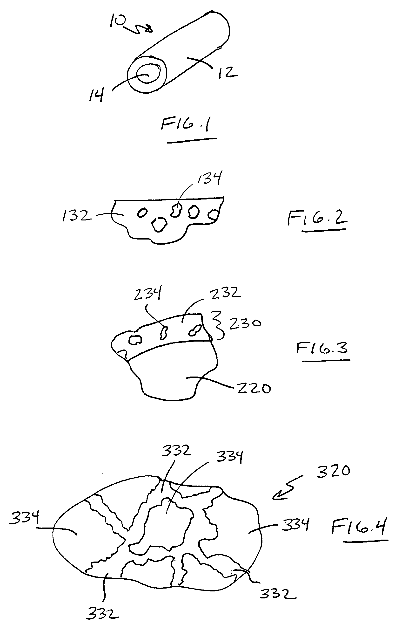
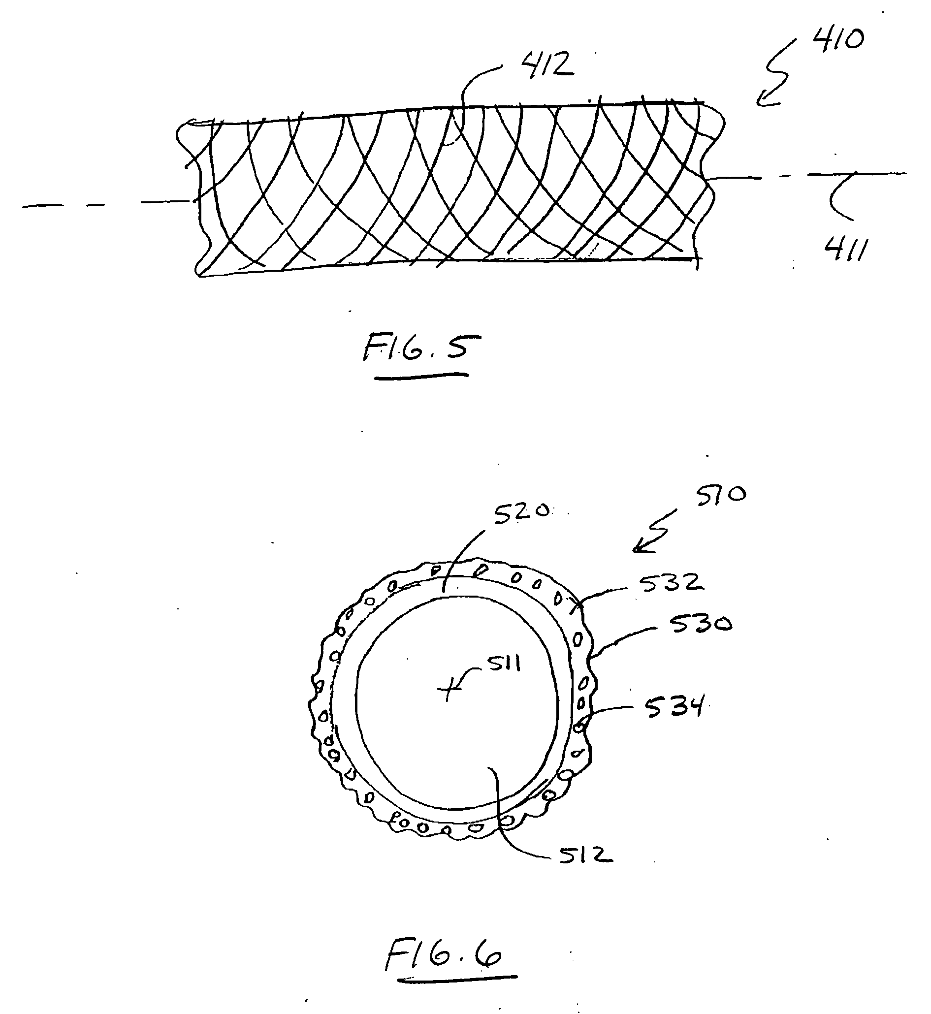
[0051]FIG. 1 de...


PUM

PropertyMeasurementUnit
timeaaaaaaaaaa
inner diametersaaaaaaaaaa
diameteraaaaaaaaaa

Abstract

Medical devices with surfaces on which viable biologic cells are magnetically attracted and retained are disclosed along with methods of magnetic coating. The medical devices can be located in a carrier liquid containing high concentrations of magnetic cells before or after implantation. The carrier liquid with magnetic cells may be contact with the medical device in vitro or in vivo. In either case, the carrier liquid may have a concentration of magnetic cells that is high enough to facilitate coating of the medical device within an acceptable period of time, e.g., several hours or less. Magnetization of medical devices before, during, and/or after implantation and apparatus for performing the same are disclosed. Degaussing of magnetic medical devices is also disclosed.

Description

RELATED APPLICATIONS [0001] This application claims the benefit of U.S. Provisional Application No. 60 / 691,090, filed on 16 Jun. 2005, titled MEDICAL DEVICES WITH DISTRIBUTED MAGNETIC FIELDS AND METHODS OF MANUFACTURING THE SAME, which is hereby incorporated by reference in its entirety. [0002] This application is also a continuation-in-part of PCT / US2004 / 040242, filed on 2 Dec. 2004, titled KITS, APPARATUS AND METHODS FOR MAGNETICALLY COATING MEDICAL DEVICES WITH LIVING CELLS, which claims the benefit of U.S. Provisional Application No. 60 / 526,522, filed on 3 Dec. 2003, titled COATING MAGNETIC MEDICAL DEVICES WITH LIVING CELLS, both of which are hereby incorporated by reference in their entireties.[0003] A recurring issue in the field of medical devices, especially implantable medical devices, is the need to provide surfaces that are compatible with the environment in which they are contained. [0004] Problems associated with implanted medical devices that include surfaces in contac...

Claims


Application Information

Patent Timeline
Patent Type & Authority Applications(United States)
IPC IPC(8): A61N1/39
CPCA61F2/04A61F2/06A61F2/07A61F2/24A61F2/82A61F2/90A61N1/05A61N1/375A61N2/06A61N2001/0585A61F2210/009A61F2210/0004A61F2/958A61L27/28A61L27/54A61L27/56A61L27/58
Inventor SANDHU, GURPREET S.SIMARI, ROBERT D.SANDHU, NICOLE P.GULATI, RAJIV
Owner MAYO FOUND FOR MEDICAL EDUCATION & RES
Why Eureka
Try Eureka