
Devices and methods for enrichment and alteration of circulating tumor cells and other particles
a technology of circulating tumor cells and particles, applied in the field of medical diagnostics and microfluidics, can solve the problems of lysis of tumor cells, unaligned obstacles may be distributed, and the expense of a potentially increased loss to and dilution of waste fractions, so as to reduce the hydrodynamic size of particles, increase deformation, and reduce volume
- Summary
- Abstract
- Description
- Claims
- Application Information
AI Technical Summary
Benefits of technology
Problems solved by technology
Method used
Image

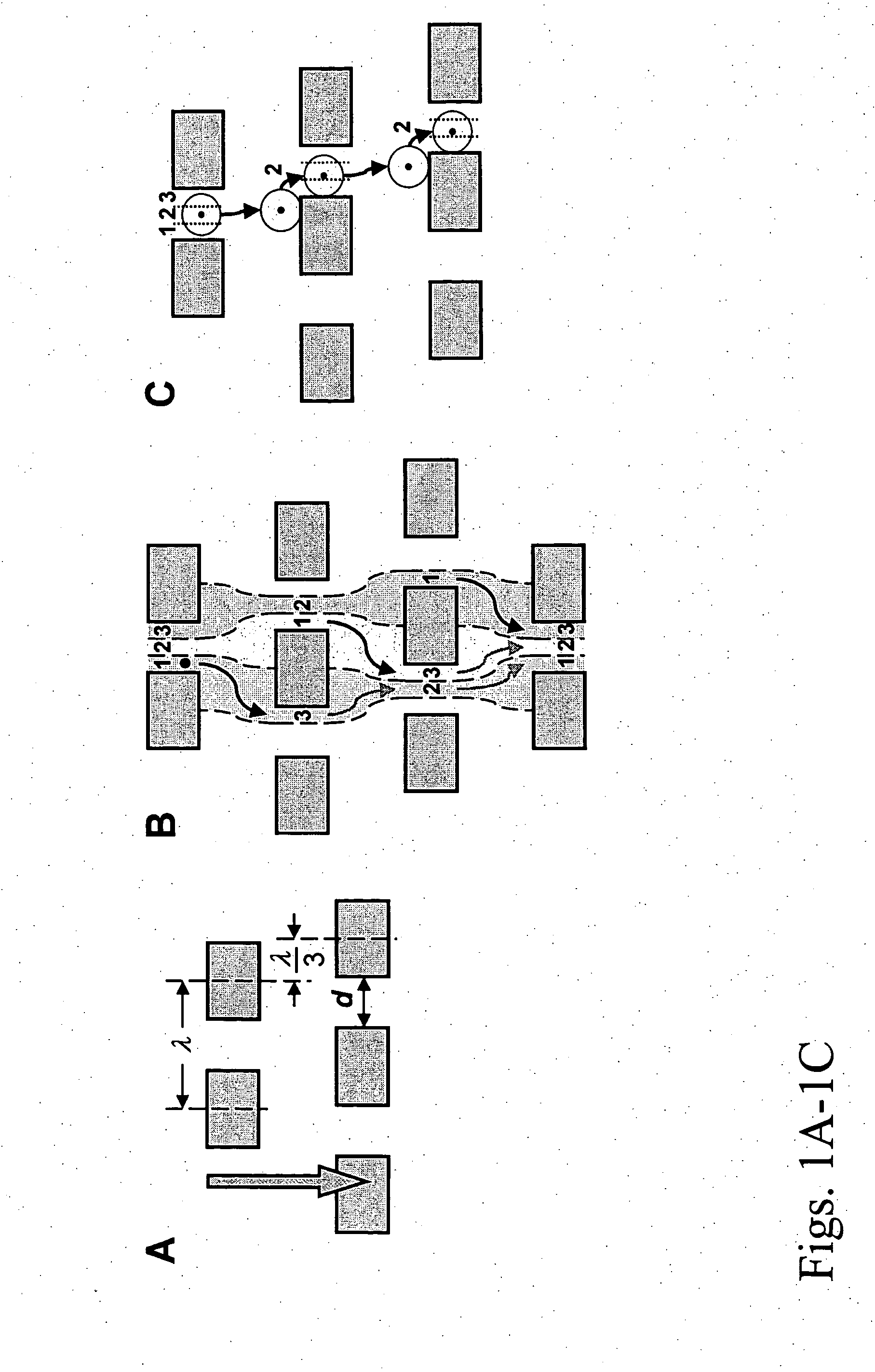
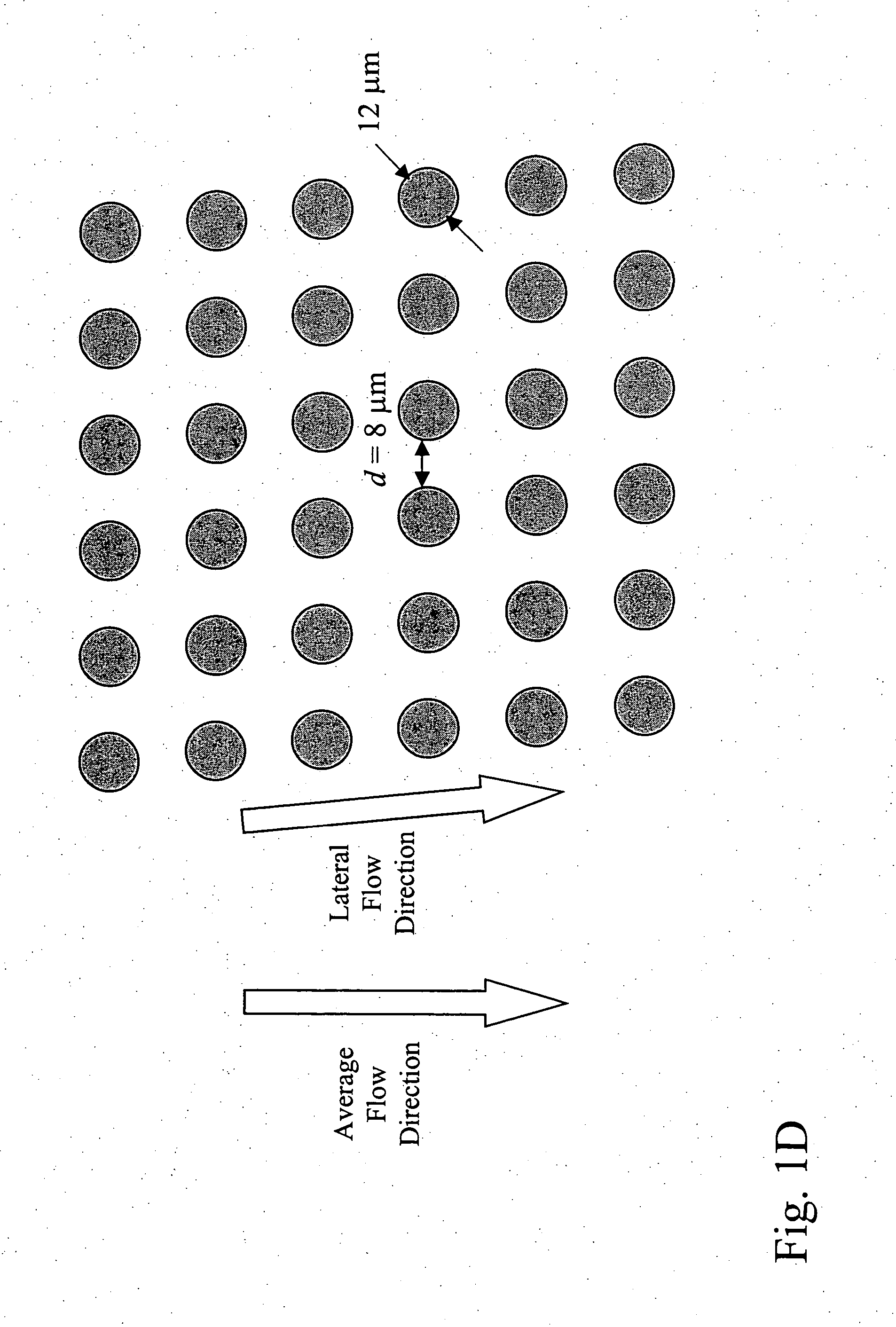
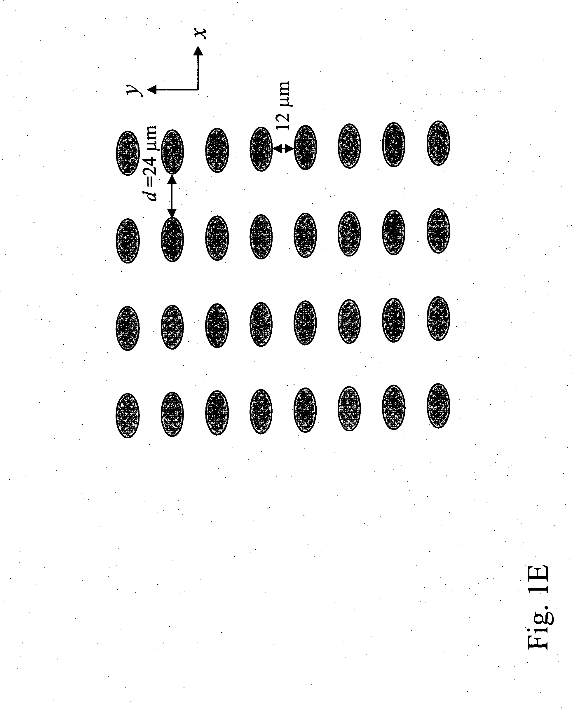
Examples
example 1
[0252] Microfluidic devices of the invention were designed by computer-aided design (CAD) and microfabricated by photolithography. A two-step process was developed in which a blood sample is first debulked to remove the large population of small cells, and then the rare target epithelial cells target cells are recovered by immunoaffinity capture. The devices were defined by photolithography and etched into a silicon substrate based on the CAD-generated design. The cell enrichment module, which is approximately the size of a standard microscope slide, contains 14 parallel sample processing sections and associated sample handling channels that connect to common sample and buffer inlets and product and waste outlets. Each section contains an array of microfabricated obstacles that is optimized to enrich the target cell type by hydrodynamic size via displacement of the larger cells into the product stream. In this example, the microchip was designed to separate red blood cells (RBCs) an...
example 2
Device Embodiments
[0255] A design for preferred device embodiments of the invention is shown in FIG. 57A, and parameters corresponding to three preferred device embodiments associated with this design are shown in FIG. 57B. These embodiments are particularly useful for enrich epithelial cells from blood.
example 3
Determining Counts for Non-Epithelial Cell Types
[0256] Using the methods of the invention, one may make a diagnosis based on counting cell types other than CTCs or other epithelial cells. A diagnosis of the absence, presence, or progression of cancer may be based on the number of cells in a cellular sample that are larger than a particular cutoff size. For example, cells with a hydrodynamic size of 14 microns or larger may be selected. This cutoff size would eliminate most leukocytes. The nature of these cells may then be determined by downstream molecular or cytological analysis.
[0257] Cell types other than epithelial cells that would be useful to analyze include endothelial cells, endothelial progenitor cells, endometrial cells, or trophoblasts indicative of a disease state. Furthermore, determining separate counts for epithelial cells, e.g., cancer cells, and other cell types, e.g., endothelial cells, followed by a determination of the ratios between the number of epithelial ce...
PUM
| Property | Measurement | Unit |
|---|---|---|
| Length | aaaaa | aaaaa |
| Mass | aaaaa | aaaaa |
| Mass | aaaaa | aaaaa |
Abstract
Description
Claims
Application Information