Cytotoxicity mediation of cells evidencing surface expression of CD44

a cytotoxicity and surface expression technology, applied in the field of cancer diagnosis and treatment, can solve the problems of insufficient evidence, inconsistent findings, and increased likelihood of tumor invasiveness and induction of angiogenesis through the ecm

Inactive Publication Date: 2007-03-08
F HOFFMANN LA ROCHE INC
View PDF21 Cites 3 Cited by
  • Summary
  • Abstract
  • Description
  • Claims
  • Application Information

AI Technical Summary

Benefits of technology

[0042] In the preventative in vivo model of human breast cancer, H460-16-2 treatment was significantly (p<0.0001) more effective in suppressing tumor growth during the treatment period than an isotype control antibody, which was identical to H460-16-2 in structure and size but incapable of binding MB-231 cells. At the end of the treatment phase, mice given H460-16-2 had tumors that grew to only 1.3 percent of the control group. During the post treatment follow-up period, the treatment effects of H460-16-2 were sustained and the mean tumor volume in the treated groups continued to be significantly smaller than controls until the end of the measurement phase. Using survival as a measure of antibody efficacy, it was estimated that the risk of dying in the H460-16-2 treatment group was about 71 percent of the antibody buffer control group (p=0.028) at 70 days post-treatment. These data demonstrated that H40-16-2 treatment conferred a survival benefit compared to the control-treated groups. H460-16-2 treatment appeared safe, as it did not induce any signs of toxicity, including reduced body weight and clinical distress. Thus, H460-16-2 treatment was efficacious as it both delayed tumor growth and enhanced survival compared to the control-treated groups in a well-established model of human breast cancer.
[0043] In addition, H460-16-2 demonstrated anti-tumor activity against MB-231 cells in an established in vivo tumor model (as outlined in Ser. No. 10 / 603,000). Treatment with H460-16-2 was compared to the standard chemotherapeutic drug, Cisplatin, and it was shown that the Cisplatin and H460-16-2 treatment groups had significantly (p<0.001) smaller mean tumor volumes compared with groups treated with either antibody dilution buffer or the isotype control antibody. H460-16-2 treatment mediated tumor suppression that was approximately two-thirds that of cisplatin chemotherapy but without the significant (19.2 percent) weight loss (p<0.003) and clinical distress, including 2 treatment-associated deaths, observed with Cisplatin treatment. The anti-tumor activity of H460-16-2 and its minimal toxicity make it an attractive anti-cancer therapeutic agent. In the post-treatment period, H460-16-2 showed a significant survival benefit (p<0.02) as the risk of dying in the H460-16-2 group was about half of that in the isotype control antibody group at >70 days after treatment. The observed survival benefit continued past 120 days post-treatment where 100 percent of the isotype control and cisplatin treated mice had died compared to 67 percent of the H460-16-2 treatment group. H460-16-2 maintained tumor suppression by delaying tumor growth by 26 percent compared to the isotype control antibody group. At 31 days post treatment, H460-16-2 limited tumor size by reducing tumor growth by 48 percent compared to the isotype control group, which is comparable to the 49 percent reduction observed at the end of the treatment. In the established tumor model of breast cancer, these results indicated the potential of H460-16-2 to maintain tumor suppression beyond the treatment phase and demonstrated the ability of the antibody to reduce the tumor burden and enhance survival in a mammal.
[0053] In all, this invention teaches the use of the H460-16-2 antigen as a target for a therapeutic agent, that when administered can reduce the tumor burden of a cancer expressing the antigen in a mammal (thus delaying disease progression), and can also lead to a prolonged survival of the treated mammal. This invention also teaches the use of a CDMAB (H460-16-2), and its derivatives, to target its antigen to reduce the tumor burden of a cancer expressing the antigen in a mammal, and to prolong the survival of a mammal bearing tumors that express this antigen. In addition, this invention teaches that after binding to its antigen, H460-16-2 can interfere with a cancer cell's ability to interact with hyaluronic acid and can also cause a cancer cell to undergo apoptosis. Furthermore, this invention also teaches the use of detecting the H460-16-2 antigen in cancerous cells that can be useful for the diagnosis, prediction of therapy, and prognosis of mammals bearing tumors that express this antigen.
[0054] If a patient is refractory to the initial course of therapy or metastases develop, the process of generating specific antibodies to the tumor can be repeated for re-treatment. Furthermore, the anti-cancer antibodies can be conjugated to red blood cells obtained from that patient and re-infused for treatment of metastases. There have been few effective treatments for metastatic cancer and metastases usually portend a poor outcome resulting in death. However, metastatic cancers are usually well vascularized and the delivery of anti-cancer antibodies by red blood cells can have the effect of concentrating the antibodies at the site of the tumor. Even prior to metastases, most cancer cells are dependent on the host's blood supply for their survival and anti-cancer antibody conjugated to red blood cells can be effective against in situ tumors as well. Alternatively, the antibodies may be conjugated to other hematogenous cells, e.g. lymphocytes, macrophages, monocytes, natural killer cells, etc.

Problems solved by technology

Also, the intracellular degradation of HA by lysosomal hylauronidases after internalization of the CD44-HA complex can potentially increase the likelihood of tumor invasiveness and induction of angiogenesis through the ECM.
However, inconsistent findings were observed in a majority of tumor types and this is probably due to a combination of reagents, technique, pathological scoring and cell type differences between researchers.
Taken together, there is no evidence that antibodies such as these are suitable for use as cancer therapeutics since they either are not directed against cancer (e.g. activate lymphocytes), induce cell proliferation, or when used with cytotoxic agents inhibited drug-induced death of cancer cells.
Survival of the treated animals was concomitantly increased.
The antibody was only effective if administered before lymph node colonization, and was postulated to interfere with cell proliferation in the lymph node.
None of these studies demonstrated human utility for this antibody.
One notable feature of this antibody was that it recognized all isoforms of CD44, which suggests limited possibilities for therapeutic use.
Further limiting the application of BIWA 1 is the immunogenicity of the murine antibody (11 of 12 patients developed human anti-mouse antibodies (HAMA)), heterogenous accumulation throughout the tumor and formation of antibody-soluble CD44 complexes.
Although deemed to be acceptable in both safety and tolerablility for the efficacy achieved, these studies have higher rates of adverse events compared to other non-radioisotope conjugated biological therapies in clinical studies.
Although there were some effects on tumor size these effects did not fulfill the criteria for objective responses to treatment.
Thus, it can be seen that although Mab U36 is a highly specific antibody the disadvantage of requiring a radioimmunoconjugate to achieve anti-cancer effects limits its usefulness because of the toxicity associated with the therapy in relation to the clinical effects achieved.
These results, though they are encouraging and support the development of anti-CD44 monoclonal antibodies as potential cancer therapeutics, demonstrate limited effectiveness, safety, or applicability to human cancers.
More specifically the inventors point out that this antibody does not recognize human cancers.

Method used


Image

  • Cytotoxicity mediation of cells evidencing surface expression of CD44
  • Cytotoxicity mediation of cells evidencing surface expression of CD44
  • Cytotoxicity mediation of cells evidencing surface expression of CD44

Examples


example 2

Normal Human Tissue Staining

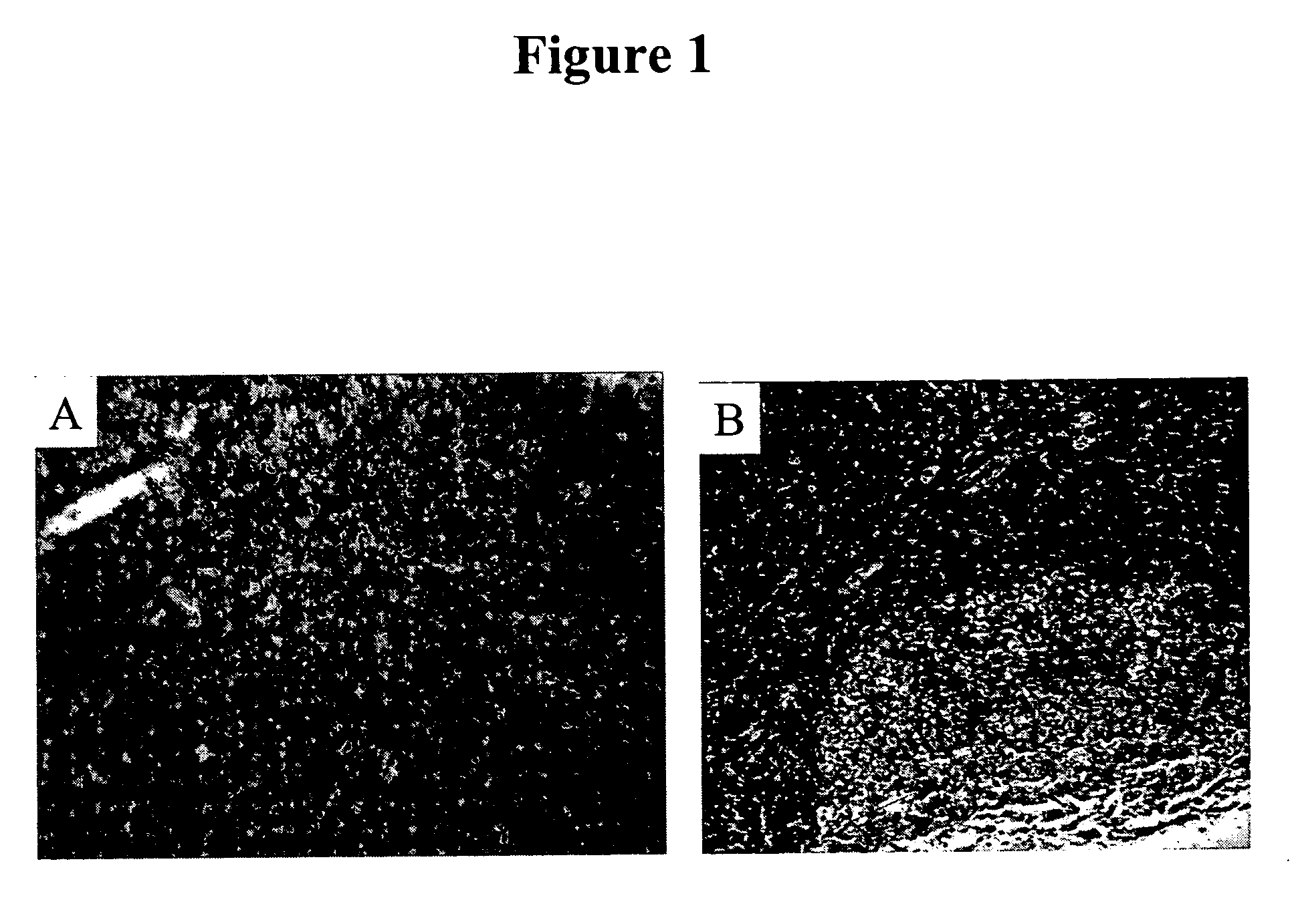
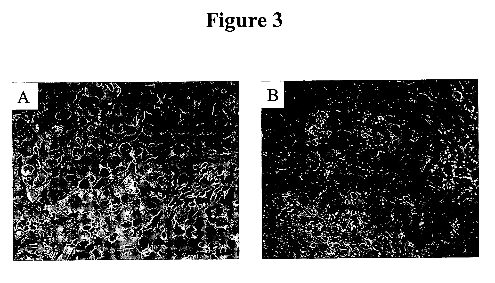
[0078] IHC studies were previously conducted to characterize H460-16-2 antigen distribution in humans (Ser. No. 10 / 603,000) and in comparison to L178 (Ser. No. 10 / 647,818). The current studies compare H460-16-2 to another antibody directed against CD44 (BU75) since the H460-16-2 antigen may be a cancer variant of CD44 as determined previously by biochemical methods. Binding of antibodies to 59 normal human tissues was performed using a human normal organ tissue array (Imgenex, San Diego, Calif.). All primary antibodies (H460-16-2; BU75 anti-CD44 (BIOCAN Scientific Inc., Mississauga, ON); and mouse IgG1 negative control (Dako, Toronto, ON)) were diluted in antibody dilution buffer (Dako, Toronto, ON) to a concentration of 5 μg / ml (found to be the optimal concentration in previous optimization steps). The negative control antibody has been shown to be negative to all mammalian tissues by the manufacturer. The procedure for IHC is as follows.

[0079] Tissue...

example 3

Human Breast Tumor Tissue Staining

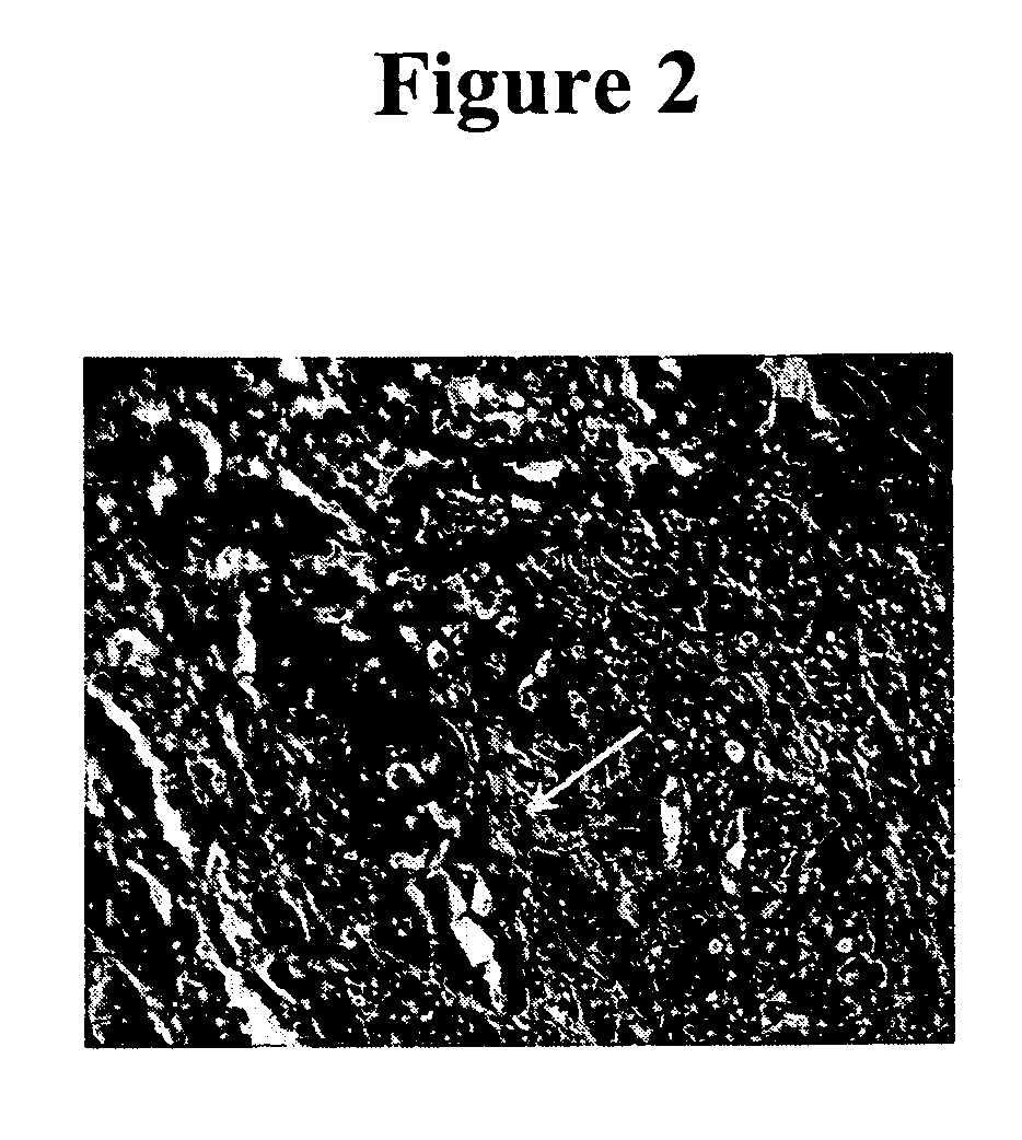
[0082] Previous IHC studies were undertaken to determine the cancer association of the H460-16-2 antigen with human breast cancers and whether the H460-16-2 antibody was likely to recognize human cancers (Ser. No. 10 / 603,000) and how it compared to anti-CD44 staining with L178 (Ser. No. 10 / 647,818). Currently, a comparison was made for BU75 anti-CD44 staining, c-erbB-2 anti-Her2 and an antibody directed towards Aspergillus niger glucose oxidase, an enzyme which is neither present nor inducible in mammalian tissues (negative control). A breast cancer tissue array derived from 50 breast cancer patients and 10 samples derived from non-neoplastic breast tissue in breast cancer patients was used (Imgenex Corporation, San Diego, Calif.). The following information was provided for each patient: age, sex, American Joint Committee on Cancer (AJCC) tumor stage, lymph node, estrogen receptor (ER) and projesterone receptor (PR) status. The procedure for IHC f...

example 4

In Vivo PC-3 Established Chemotherapy Combination Tumor Experiments

[0089] With reference to FIGS. 5 and 6, 6 to 8 week old male SCID mice were implanted with 1 million PC-3 human prostate cancer cells in 100 microlitres saline injected subcutaneously in the scruff of the neck. Tumor growth was measured with calipers every week. When the majority of the cohort reached a tumor volume of 80 mm3 (range 50-130 mm3) at 21 days post-implantation, 8 mice were randomly assigned into each of 4 treatment groups. H460-16-2 antibody, the chemotherapeutic drug Cisplatin, the combination of H460-16-2 and Cisplatin or buffer control was administered intraperitoneally with 15 or 6 mg / kg of antibody or Cisplatin respectively at a volume of 300 microliters after dilution from the stock concentration with a diluent that contained 2.7 mM KCl, 1 mM KH2PO4, 137 mM NaCl and 20 mM Na2HPO4. H460-16-2 or buffer control was then administered 4 times per week for the first week followed by 3 times per week fo...


PUM

PropertyMeasurementUnit
concentrationaaaaaaaaaa

Abstract

This invention relates to the diagnosis and treatment of cancerous diseases, particularly to the mediation of cytotoxicity of tumor cells; and most particularly to the use of cancerous disease modifying antibodies (CDMAB) or derivatives thereof, optionally in combination with one or more chemotherapeutic agents, as a means for initiating the cytotoxic response. The invention further relates to binding assays which utilize the CDMABs of the instant invention.

Description

REFERENCE TO RELATED APPLICATIONS [0001] This application is a continuation of application Ser. No. 10 / 810,165 filed Mar. 26, 2004, which is a continuation-in-part of application Ser. No. 10 / 647,818 filed Aug. 22, 2003, which is a continuation-in-part of application Ser. No. 10 / 603,000, filed Jun. 23, 2003, which is a continuation-in-part of application Ser. No. 09 / 727,361, filed Nov. 29, 2000, which is a continuation-in-part of application Ser. No. 09 / 415,278, filed Oct. 8, 1999, now U.S. Pat. No. 6,180,357 B1, the contents of each of which are herein incorporated by reference.FIELD OF THE INVENTION [0002] This invention relates to the diagnosis and treatment of cancerous diseases, particularly to the mediation of cytotoxicity of tumor cells; and most particularly to the use of cancerous disease modifying antibodies (CDMAB), optionally in combination with one or more chemotherapeutic agents, as a means for initiating the cytotoxic response. The invention further relates to binding ...

Claims


Application Information

Patent Timeline
Patent Type & Authority Applications(United States)
IPC IPC(8): A61K51/00A61K39/395A61K47/48A61K49/00A61P35/00C07K16/00C07K16/28C07K16/30G01N33/50G01N33/566G01N33/567G01N33/569G01N33/574
CPCC07K16/3015G01N33/57449A61K39/39558A61K45/06A61K47/48561A61K49/0058A61K51/1027A61K2039/505B82Y5/00C07K16/00C07K16/2884C07K16/30G01N33/57484C07K2316/95G01N33/5011G01N33/5014G01N33/5082G01N33/566G01N33/56972G01N33/574G01N33/57415G01N33/57419G01N33/57423A61K2300/00A61K47/6849A61P35/00C07K2317/73
Inventor YOUNG, DAVID S. F.HAHN, SUSAN E.FINDLAY, HELEN P.
Owner F HOFFMANN LA ROCHE INC
Why Eureka
Try Eureka