

Method for three-dimensional cartilage tissue engineering using bone marrow cells in tissue engineering bone marrow cells in simulated microgravity environment
a three-dimensional cartilage and microgravity environment technology, applied in the direction of skeletal/connective tissue cells, biocide, prosthesis, etc., can solve the problems of cell damage, cell damage, and inability to give satisfactory therapeutic effects, etc., to achieve the effect of regenerating and/or repairing cartilage defects of patients
- Summary
- Abstract
- Description
- Claims
- Application Information
AI Technical Summary
Benefits of technology
Problems solved by technology
Method used
Image

Examples
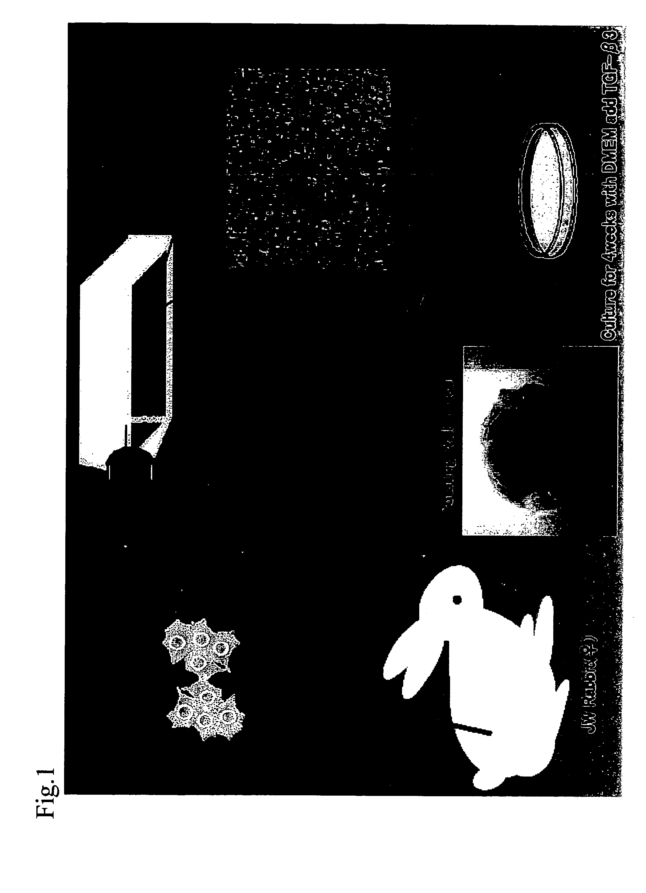
example 1
Carrtilage Tissue Engineering from Mesenchymal Stem Cells Derived from Rabbit Bone Marrow
1. Culture of Mesenchymal Stem Cells Derived from Rabbit Bone Marrow
(1) Preparation of Mesenchymal Stem Cells Derived from Rabbit Bone Marrow
[0042] Mesenchymal stem cells derived from rabbit bone marrow were extracted from the femur of a 2-week-old JW rabbit (female) in accordance with the method of Maniatopoulos et al. (Maniatopoulos, C., Sodek, J., and Melcher, A. H., 1988, Cell Tissue Res., 254, pp. 317-330). The sampled cells were cultured in DMEM containing 10% FBS (Sigma) and Antibiotic-Antimycotic (GIBCO BRL) for 3 weeks, and they were allowed to grow.
(2) Culture of Mesenchymal Stem Cells Derived from Rabbit Bone Marrow
[0043] The mesenchymal stem cells derived from rabbit bone marrow thus prepared were suspended in 10 ml of DMEM culture medium(Sigma) containing 10−7 M dexamethasone (Sigma), 10 ng / ml TGF-β3 (Sigma), 50 μ g / ml ascorbic acid (Wako), ITS +Premix (BD), 40 μg / ml L-proli...
example 2
Assay of Strength of RWV-Xultured Tissue
[0058] Mechanical strength of the RWV-cultured tissue was measured using the EIKO TA-XT2i (Eko Instruments). The RWV-cultured tissue prepared by the procedure of Example 1 was cut into 2-mm square pieces and then compressed at a rate of 0.1 mm / sec. The stress-strain curve was determined from the compression load (Pa) and the distance (mm), and the strength was calculated based thereon.
[0059]FIG. 8 shows the results of a comparison of the compression strength of the cartilage tissue 4 weeks after the initiation of culture and that of the articular cartilage tissue of a rabbit.
example 3
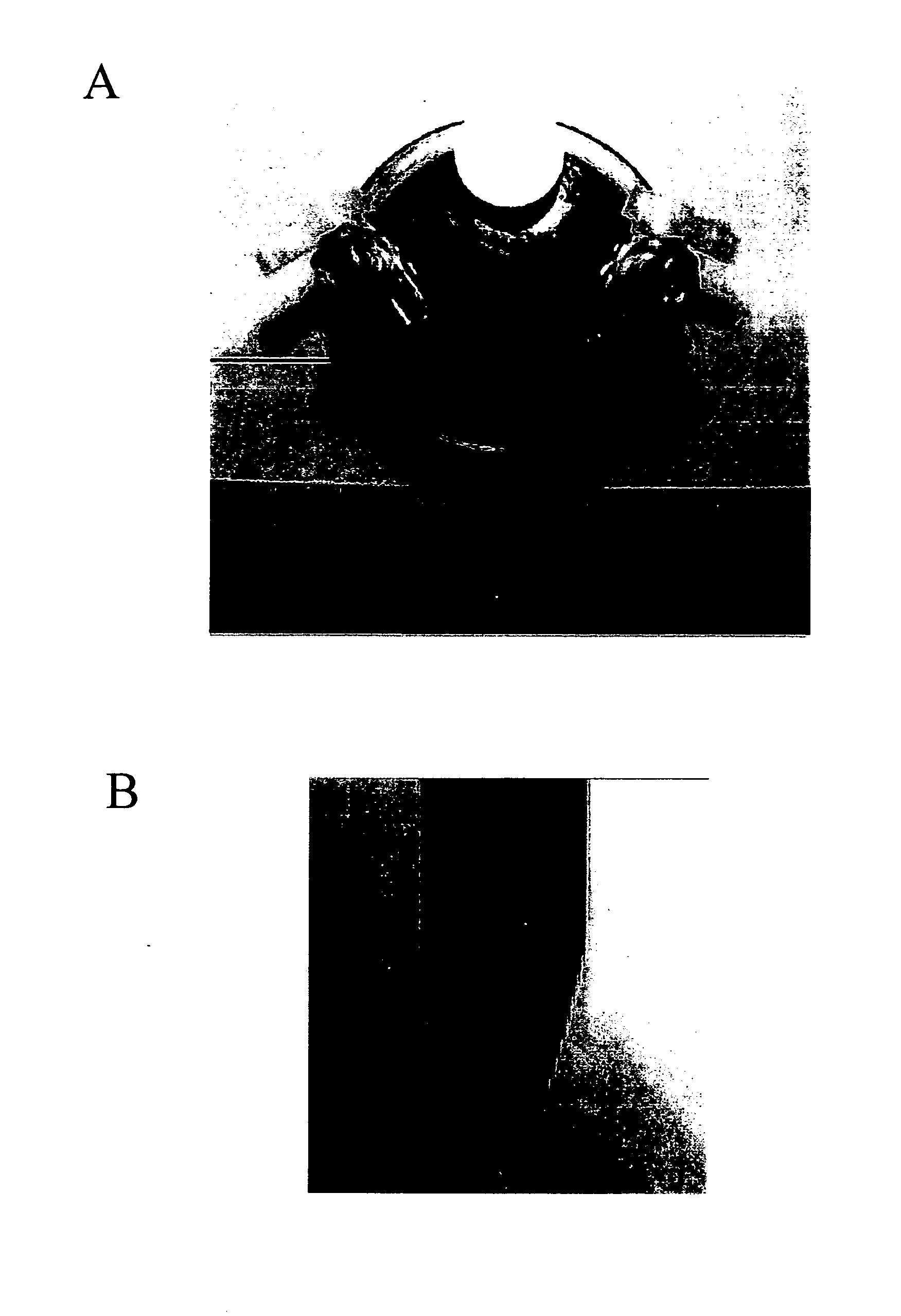
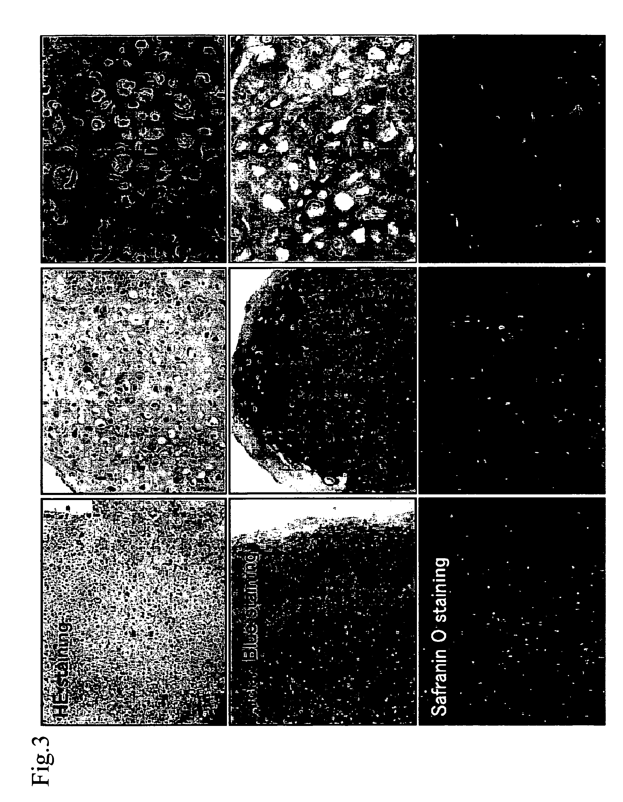
Experimentation Concerning Transplantation of RWV-Cultured Tissue Into Osteochondral Defect in Rabbit Knee Joint
1. Transplantation into Osteochondral Defect in Rabbit Knee Joint
[0060] The RWV-cultured tissue prepared by the procedure of Example 1 (cultured in vitro for 2 weeks) was transplanted into an osteochondral defect in a rabbit knee joint, and the hardness at the site of transplantation and the results of histological observation thereof were evaluated.
[0061] The rabbit was intravenously anesthetized with 0.6 mg / kg of Somnopentyl. The weight-bearing area of the left femoral condyle (the left knee joint) was designated as the site of surgical operation. A vertical skin incision was made on the lateral side of the patella, and the articular capsule was incised through a medial parapatellar approach. The patella was dislocated via lateral reflection, and an osteochondral defect 4 mm in depth was provided in the femoral trochlea using a drill with a diameter of 5 mm (the bott...
PUM
| Property | Measurement | Unit |
|---|---|---|
| concentration | aaaaa | aaaaa |
| concentration | aaaaa | aaaaa |
| pH | aaaaa | aaaaa |
Abstract
Description
Claims
Application Information