Amniotic fluid cell-free fetal DNA fragment size pattern for prenatal diagnosis

Inactive Publication Date: 2007-05-31
NEW ENGLAND MEDICAL CENT HOSPITALS
View PDF0 Cites 21 Cited by
  • Summary
  • Abstract
  • Description
  • Claims
  • Application Information

AI Technical Summary

Benefits of technology

[0011] The present invention provides an improved system for analyzing a fetus' genetic information. In particular, the present invention allows for the rapid prenatal screening of certain chromosomal abnormalities. More specifically, the present invention encompasses the recognition by the Applicants that the fragment size pattern of cell-free fetal DNA isolated from amniotic fluid is

Problems solved by technology

Furthermore, genetic disorders and congenital anomalies contribute substantially to long-term disability; they are associated with enormous medical-care costs (A. Czeizel et al., Mutat. Res. 1984, 128: 73-103; Centers of Disease Control, Morb. Mortal. Weekly Rep. 1989, 38: 264-267; S. Kaplan, J. Am. Coll. Cardiol.
Examination of the karyotypes determined by these banding methods can reveal the presence of numerical and structural chromosomal abnormalities over the whole genome.
In addition to tissue sampling and selective staining, conventional banding methods also require cell culturing, which can take between 10 and 15 days depending on the tissue source, and preparation of high quality metaphase spreads, which is tedious, time-consuming and labor-intensive (B. Eiben et al., Am. J. Hum. Genet.
Furthermore, conventional chromosome analysis methods have limited sensitivity, and their standard 450-550 band level of resolution does not allow detection of small or subtle chromosomal aberrations, such as, for example, those associated with microdeletion/microduplication syndromes.
However, in contrast to conventional banding analysis, certain molecular cytogenetic methods

Method used


Image

  • Amniotic fluid cell-free fetal DNA fragment size pattern for prenatal diagnosis
  • Amniotic fluid cell-free fetal DNA fragment size pattern for prenatal diagnosis
  • Amniotic fluid cell-free fetal DNA fragment size pattern for prenatal diagnosis

Examples


example 1

Amniotic Fluid Fetal DNA Isolation and Preliminary Tests

[0128] Frozen amniotic fluid supernatant specimens (38) were obtained from the Tufts-New England Medical Center (Tufts-NEMC) Cytogenetics Laboratory (D. W. Bianchi et al., Clin. Chem. 2001, 47: 1867-1869). All samples were collected for routine indications, such as advanced maternal age, abnormal maternal serum screening results, or detection of a fetal sonographic abnormality. The standard protocol in the Cytogenetics Laboratory is to centrifuge the amniotic fluid sample upon receipt, place the cell pellet into tissue culture, assay an aliquot of the fluid for alpha-fetoprotein and acetyl cholinesterase levels, and store the remainder at −20° C. as a back-up in case of assay failure. After six months, the frozen amniotic fluid supernatant samples are normally discarded.

[0129] The frozen fluid samples obtained from the Cytogenetics Laboratory were initially thawed at 37° C. and then mixed with a vortex for 15 seconds. An aliq...

example 2

Improvements in Amniotic Fluid Fetal DNA Isolation Method

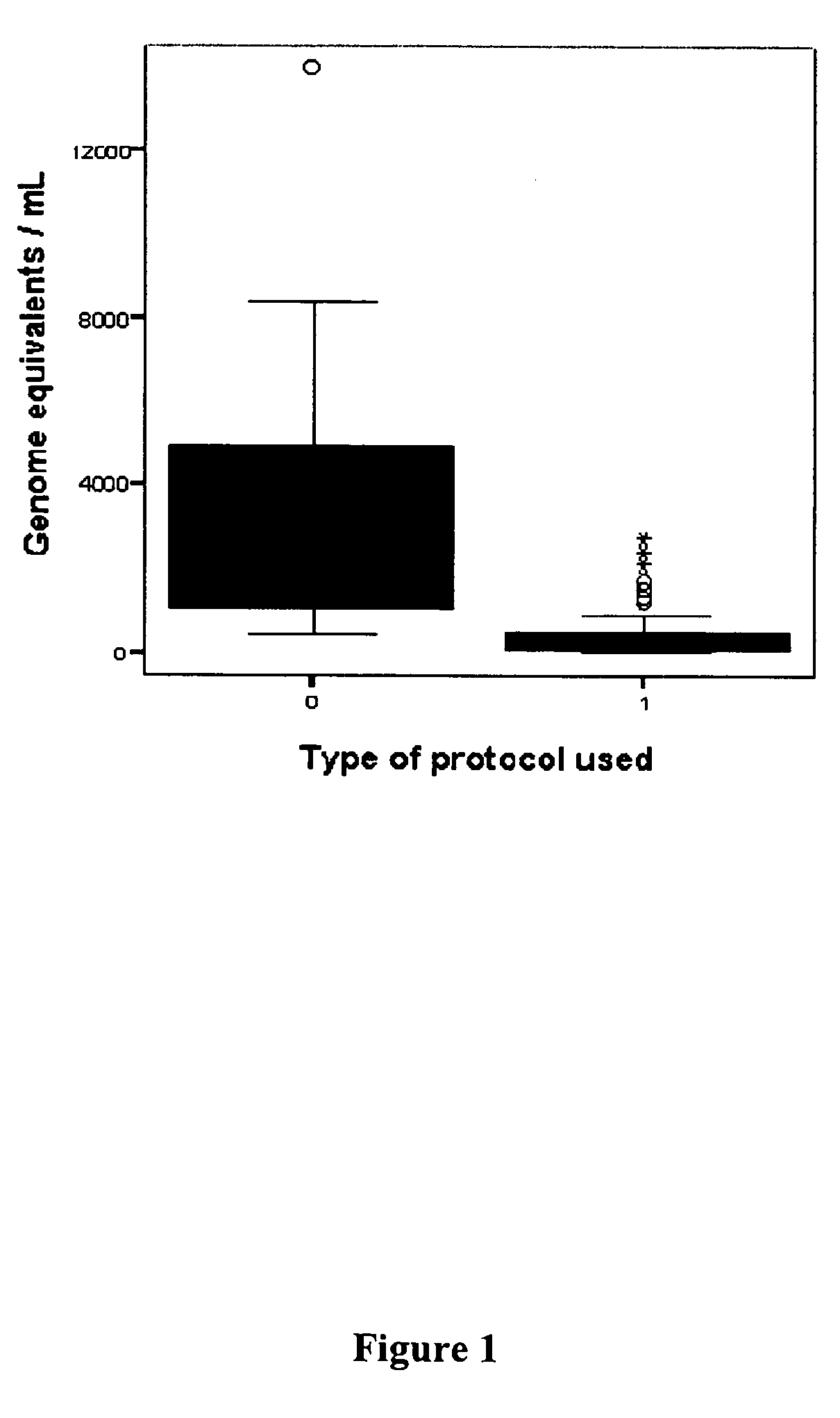
[0134] Using the “Blood and Body Fluid” vacuum protocol, only a minor proportion of amniotic fluid supernatant samples could be further analyzed (e.g., with genomic microarrays, in which a minimum of 100 ng of DNA is necessary) (P. B. Larrabee et al., Am. J. Hum. Genet., 2004, 75: 485-491).

[0135] As reported above, the original method was based on known protocols for the extraction of cell-free fetal DNA from maternal plasma / serum, as specific guidelines for the extraction of DNA from amniotic fluid did not exist. Therefore, further investigation was needed to optimize cell-free fetal DNA extraction from amniotic fluid supernatant to more fully exploit this promising source of genetic material.

[0136] Approval for this study was obtained from the institutional review Board of Tufts-New England Medical Center (Boston, Mass.) and Women and Infants Hospital (Providence, R.I.). For protocol optimization, five large volume sample...

example 3

Investigation of Amniotic Fluid Fetal DNA Fragmentation Pattern

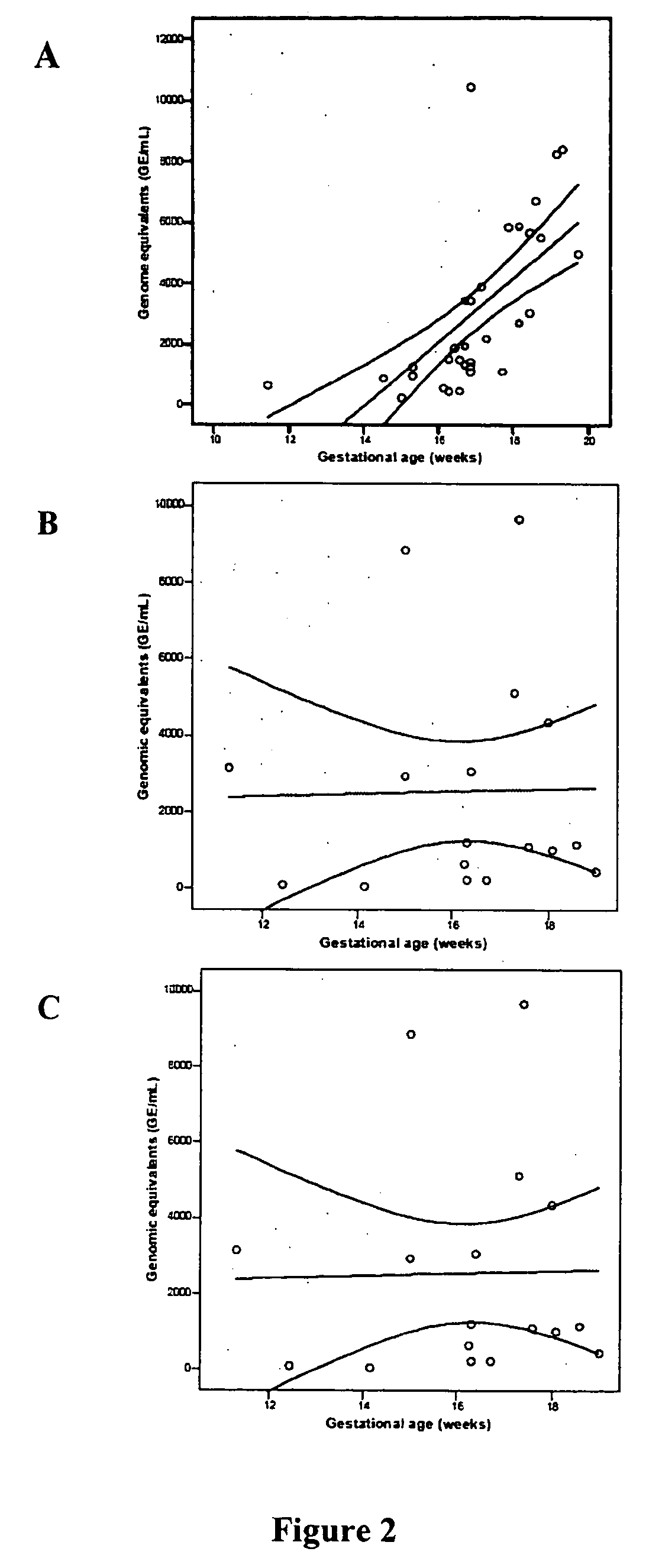
[0145] To date, no study has addressed the biochemical properties of cell-free fetal DNA in amniotic fluid. This is in contrast to maternal plasma, in which it has been shown that circulating fetal DNA sequences are smaller than maternal-derived ones, on the order of less than 300 base pairs (Y. Li et al., Clin. Chem., 2004, 50: 1002-1011; K. C. A. Chan et al., Clin. 2004, 50: 88-92). This distinct property has been used to increase the yield of fetal DNA extracted from maternal samples to permit non-invasive prenatal diagnosis of β-thalassemia (Y Li et al., JAMA, 2005, 292: 843-849).

[0146] The Applicants have hypothesized that cell-free fetal DNA in amniotic fluid would have different biophysical properties that cell-free fetal DNA in maternal plasma. Since second trimester amniotic fluid is composed predominantly of fetal urine, the Applicants speculated that passage of cell-free fetal DNA through the fetal kidneys m...


PUM

PropertyMeasurementUnit
Timeaaaaaaaaaa
Volumeaaaaaaaaaa
Volumeaaaaaaaaaa

Abstract

The present invention relates to improved methods of prenatal diagnosis, screening, monitoring and/or testing. The inventive methods include analysis of the fragment size distribution of cell-free fetal DNA isolated from amniotic fluid. The inventive methods allow for rapid screening of fetal characteristics such as chromosomal abnormalities and for prenatal diagnosis of a variety of diseases and conditions. Since the new methods do not require cell culture, they can be performed more rapidly than conventional fetal karyotypes.

Description

RELATED APPLICATIONS [0001] This application claims priority from Provisional Patent Application No. 60 / 713,540, filed Sep. 1, 2005 and entitled “Amniotic Fluid Cell-Free Fetal DNA Fragment Size Pattern for Prenatal Diagnosis”. The Provisional Application is incorporated herein by reference in its entirety. The present application is also related to U.S. Application Ser. No. 10 / 577,341 filed Apr. 28, 2006, which is a U.S. National Phase Application under 35 U.S.C § 371 of International Application PCT / US04 / 035929 (published PCT application No. WO 2005 / 044086) filed Oct. 29, 2004, which itself claims priority from Provisional Application No. 60 / 515,735 filed Oct. 30, 2003. Each of these applications is incorporated herein by reference in its entirety.GOVERNMENT INTERESTS [0002] The work described herein was funded by the National Institutes of Health (Grant No. NIH HD42053). The United States government may have certain rights in the invention.BACKGROUND OF THE INVENTION [0003] Genet...

Claims


Application Information

Patent Timeline
IPC IPC(8): C12Q1/68G06F19/00
CPCC12Q1/6883C12Q2565/125C12Q2600/156
Inventor BIANCHI, DIANA W.JOHNSON, KIRBY L.LAPAIRE, OLAV
Owner NEW ENGLAND MEDICAL CENT HOSPITALS
Why Eureka
Try Eureka