Engineered blood vessels

a technology of blood vessels and blood vessels, applied in the direction of prosthesis, skeletal/connective tissue cells, biocide, etc., can solve the problems of lack of ec lining on the lumen of the graft, lack of burst strength of the construct, and insufficient veins for bypass surgery

Active Publication Date: 2007-06-07
RGT UNIV OF MINNESOTA
View PDF21 Cites 57 Cited by
  • Summary
  • Abstract
  • Description
  • Claims
  • Application Information

AI Technical Summary

Benefits of technology

[0016] In yet another embodiment, the present invention relates to compositions in vitro comprising ECs and SMCs incorporated in a matrix circumferentially positioned around a tubular support, wherein one or more mitogenic and attractant factors (in combination) or one or more mitoattractant factors capable of permeating the support are present within the support.
[0017] In yet another embodiment, the present invention relates to a composition in vitro comprisin...

Problems solved by technology

Many patients who are in need of bypass surgery do not possess sufficient veins to act as replacements for their diseased arteries.
However, as with other acellular studies, this study suffered from a lack of non-thrombogenic EC lining on the lumen of the graft.
However, the constructs lacked burst strength.
One published in vivo study using the self-assembly approach was limited by use of xenogeneic cells...

Method used


Image

  • Engineered blood vessels
  • Engineered blood vessels
  • Engineered blood vessels

Examples


example 1

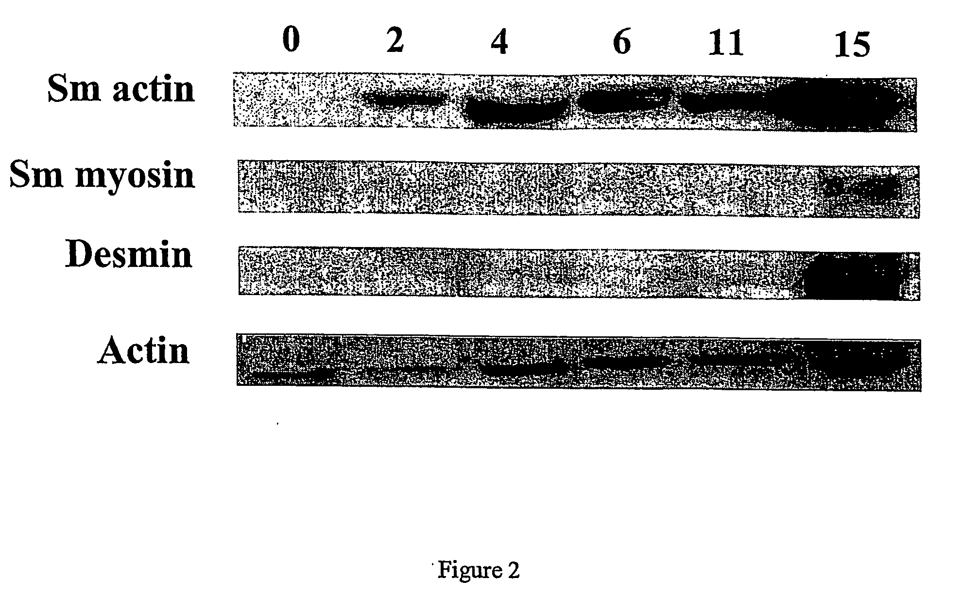
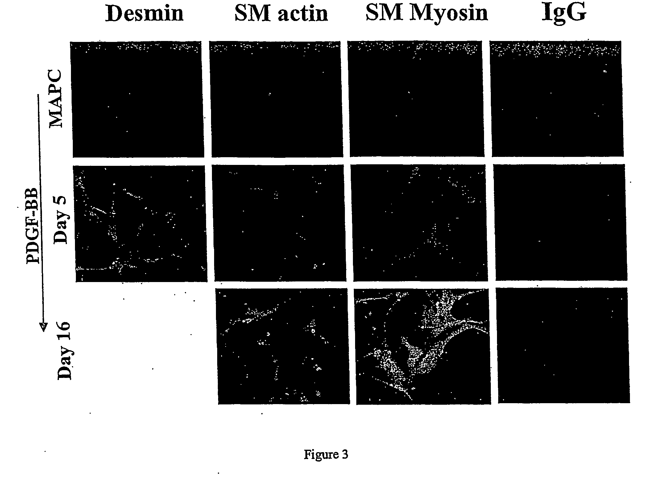
Culture And Differentiation Conditions For MAPCs

MAPC Isolation and Culture Conditions

[0144] Bone marrow (BM) was obtained from 55 healthy volunteers donors (age 2-50 years) after informed consent using guidelines from the University of Minnesota Committee on the use of Human Subject in Research. MAPCs were generated as previously described (Furcht et aL). Briefly, BM mononuclear cells (BMMNC) were depleted of CD45+ and glycophorin-A+ cells using micromagnetic beads (Miltenyii Biotec, Sunnyvale, Calif.). CD45− / GlyA− cells (5×10³ cells) diluted in 200 μL expansion medium comprising 58% low-glucose Dulbecco's minimal essential medium, low-glucose formulation (DMEM-LG) (Gibco-BRL, Grand Island, N.Y.), 40% MCDB-201 (Sigma Chemical Co, St Louis, Mo.), supplemented with 1× insulin-transferrin-selenium (ITS), 1× linoleic-acid bovine serum albumin (LA-BSA), 10−8M Dexamethasone, 10−4M ascorbic acid 2-phosphate (all from Sigma), 100 U penicillin and 1,000 U streptomycin (Gibco) and 10% FCS ...

example 2

Formation Of a Blood Vessel By Incorporation Of Cells In a Matrix

[0151] In this Example, a blood vessel is engineered by incorporation of SMCs and ECs in a fibrin gel tube with VEGF stimulus in the lumen.

[0152] Three solutions are required to make a fibrin gel: fibrinogen solution, cell suspension, and thrombin solution. The fibrinogen solution is composed of 5 mg / mL of fibrinogen in 20 mM HEPES buffered saline while the thrombin solution is 150 units of thrombin in 1% water by volume and 9% saline by volume in serum free culture medium with a 0.004 M CaCl2 catalyst. The cell suspension is at six times the final concentration; therefore, the suspension is at 1.5×10⁶ cell / mL to have a final concentration of 0.25×10⁶ cells / mL. The fibrinogen solution, the cell suspension, and the thrombin solution are mixed at a ratio of 4:1:1 by volume and gel in less than one hour at 37° C.

[0153] The mold to form a tubular fibrin gel for co-culturing cells is composed of a tubular porous polyethy...

example 3

Characterization Of Layered Circumferential Vascular Structure

[0160] After culturing the constructs from Example 2 on the agarose-coated porous polyethylene tube for 7 days, ECs were mainly located close to the lumenal surface (FIG. 8), however the cells were still somewhat disorganized, as seen in FIG. 9. After 3 weeks of culture, the cells began to align and form prototypical layers, as detected by vWF staining, LDL uptake, and DAPI staining (FIG. 10). ECs were seen close to the lumenal surface. FIG. 11A show a cross-section of the entire construct at lower magnification immunostained for α-smooth muscle-actin (red), collagen type IV (green), and DAPI (blue). SMCs populated the interior of the construct and ECs were absent from the outer surface, which was surrounded by medium that was not supplemented with VEGF, indicating that ECs can be selectively localized to surfaces after incorporation by presenting a concentration gradient of VEGF, presumably by a chemomitoattractant resp...


PUM


Abstract

The present invention relates to engineered blood vessels and methods of making such vessels using matrices comprising endothelial and smooth muscle cells, or cells capable of differentiating into endothelial and smooth muscle cell lineages (e.g., stem cells, or the progenitors thereof).

Description

RELATED APPLICATIONS / PATENTS & INCORPORATION BY REFERENCE [0001] This application claims priority to U.S. Application Ser. No. 60 / 484,563, filed Jul. 1, 2003, and U.S. Application Ser. No. 60 / 484,595 filed Jul. 2, 2003. [0002] Each of the applications and patents cited in this text, as well as each document or reference cited in each of the applications and patents (including during the prosecution of each issued patent; “application cited documents”), and each of the PCT and foreign applications or patents corresponding to and / or claiming priority from any of these applications and patents, and each of the documents cited or referenced in each of the application cited documents, are hereby expressly incorporated herein by reference. More generally, documents or references are cited in this text, either in a Reference List before the numbered paragraphs, or in the text itself; and, each of these documents or references (“herein-cited references”), as well as each document or referen...

Claims


Application Information

Patent Timeline
IPC IPC(8): A61K35/34C12N5/08C12NC12N5/071
CPCA61L27/3808A61L27/3826A61L27/383A61L27/3843A61L27/3891A61L27/507C12N5/0691C12N2500/25C12N2501/11C12N2501/135C12N2501/15C12N2501/165C12N2501/39C12N2506/02C12N2506/03C12N2506/11C12N2533/30C12N2533/54C12N2533/56C12N2506/1353A61L27/3834
Inventor TRANQUILLO, ROBERT T.ROSS, JEFFREYREYES, MORAYMA
Owner RGT UNIV OF MINNESOTA
Why Eureka
Try Eureka