Method for drug delivery to ocular tissue using microneedle

a technology of ocular tissue and microneedles, which is applied in the field of ophthalmic therapies, can solve the problems of significant side effects, difficult to deliver effective doses of drugs to the back of the eye, and difficult to deliver drugs to the ey

Active Publication Date: 2007-11-08
GEORGIA TECH RES CORP +1
View PDF26 Cites 187 Cited by
  • Summary
  • Abstract
  • Description
  • Claims
  • Application Information

AI Technical Summary

Benefits of technology

[0021] In one embodiment, the drug depot provides extended or sustained release of a drug. In one embodiment, the drug formulation comprises microparticles or nanoparticles which provide controlled release of the drug.

Problems solved by technology

The delivery of drug to the eye is extremely difficult, particularly delivery of macromolecules and delivery to the back of the eye.
It is difficult to deliver effective doses of drug to the back of the eye using conventional delivery methods such as topical application, which has poor efficacy, and systemic administration, which often causes significant side effects.
Direct injection into the eye, using conventional needles and syringes is often effective, but requires professional training and raises concerns about safety.

Method used


Image

  • Method for drug delivery to ocular tissue using microneedle
  • Method for drug delivery to ocular tissue using microneedle
  • Method for drug delivery to ocular tissue using microneedle

Examples


example 1

Drug-Coated Solid Microneedles

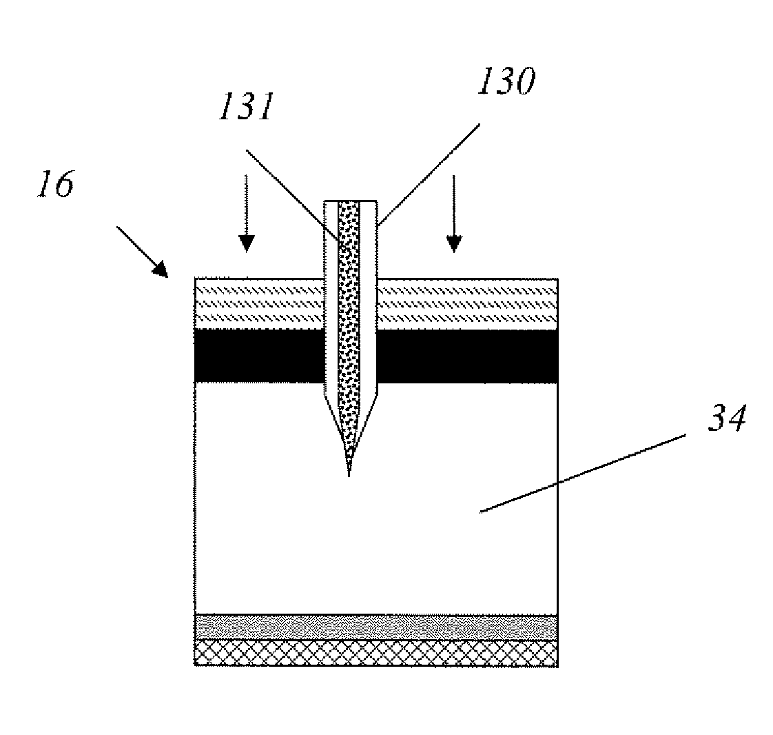
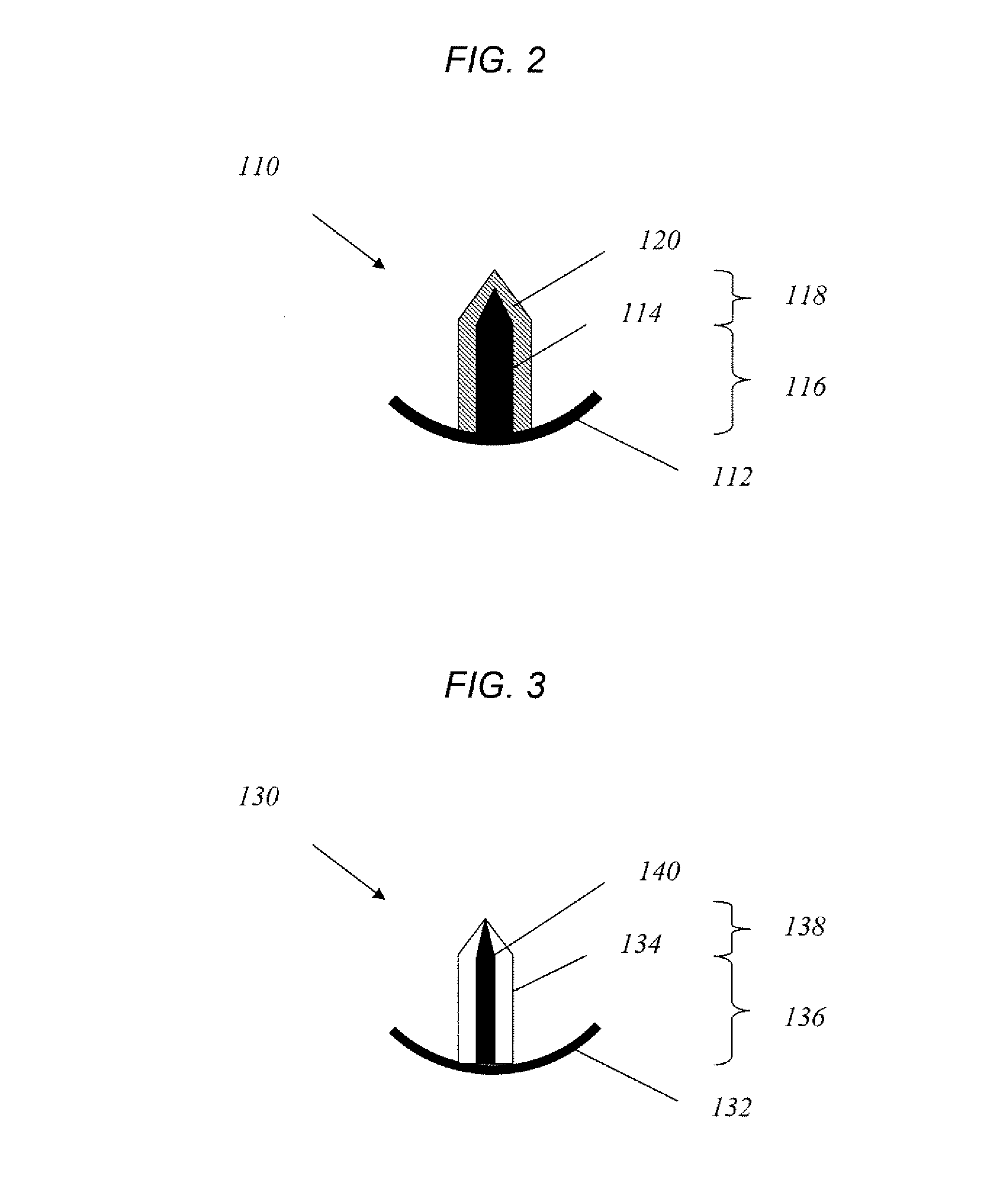
[0079] Single solid microneedles were fabricated using an infrared laser to cut microneedles from 75 micron thick stainless steel sheets. To deburr and sharpen the microneedle edges, electropolishing was done in a 1:3:6 v / v mixture of water:phosphoric acid:glycerine at 70° C. The final microneedle dimensions were 500 μm in length, 50×200 μm in cross section at the base, and 55° in tip angle. For in vivo rabbit experiments, the microneedles were modified to 500 μm in length, 50×100 μm in cross section at the base, and 45° in tip angle, to avoid penetration through the thinner rabbit corneal tissue. To facilitate handling microneedles using forceps during insertion into tissue, an extended metal substrate was attached to the needle base, which measured 1 cm in length, 4 mm in width and 50 μm in thickness, was included in the needle design.

[0080] The microneedles were coated at room temperature using a dip coating method and an aqueous coating solution. ...

example 2

In Vitro Drug Delivery to Human Sclera

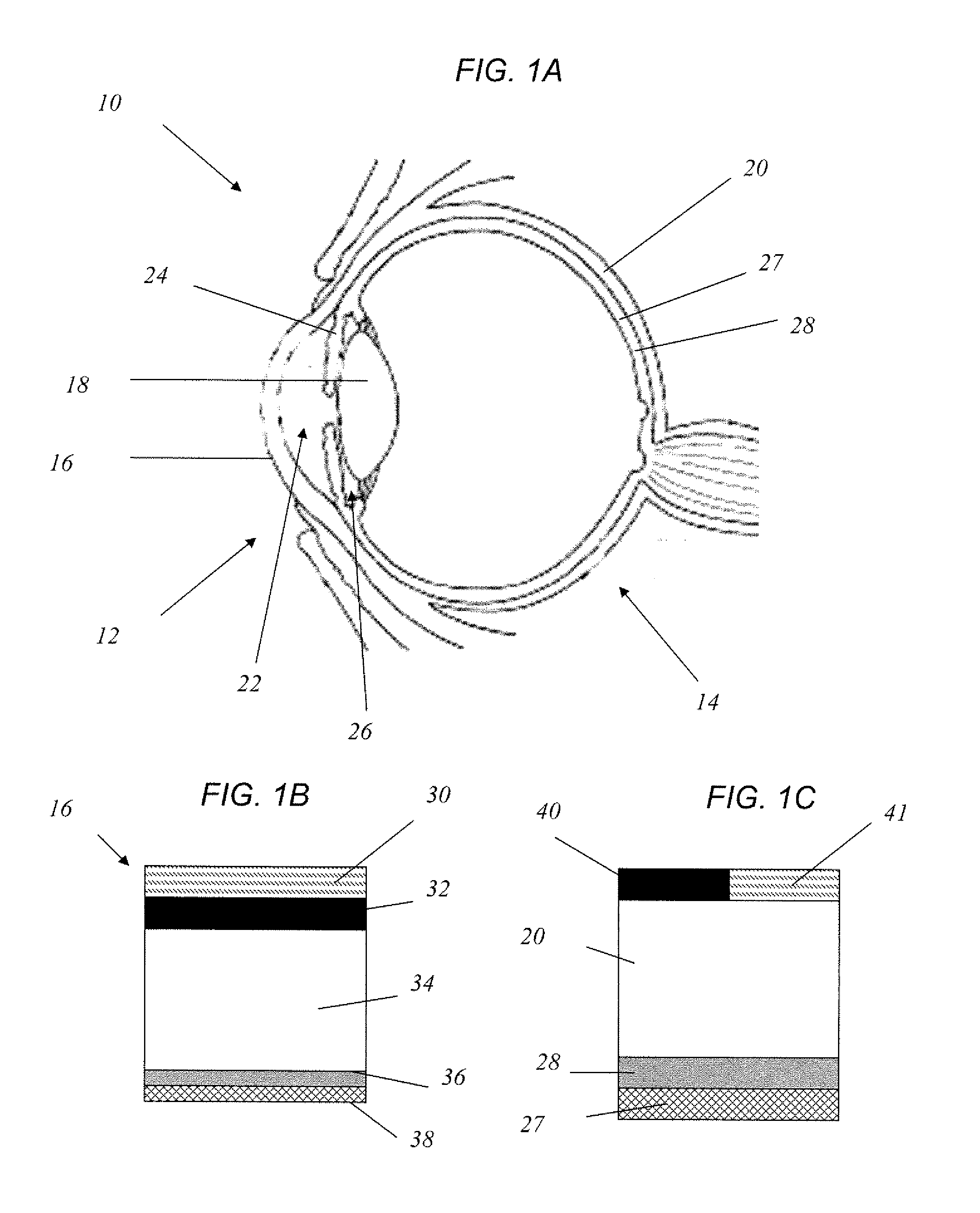
[0081] Human sclera was obtained from the Georgia Eye Bank with the approval of the Georgia Tech IRB. Pieces of the scleral tissue were cut to a size of 0.7×0.7 cm using surgical scissors and rinsed with deionized water. Adherent tissues associated with the retina, choroid, and episclera were removed from the scleral tissue with a cotton swab and the scleral tissue was then placed on top of a hemispherical surface (0.6 cm in radius), which simulated the curvature of the ocular surface.

[0082] Uncoated single microneedles from Example 1 were manually inserted approximately half way into the sclera tissue and then removed. The insertion site was stained with a blue tissue dye and imaged by brightfield and fluorescence microscopy to assess the site and extent of delivery.

[0083] The microneedles were found to be sufficiently strong and sharp to penetrate into the sclera without bending or breaking (data not shown). Within two minutes, the micronee...

example 3

In Vivo Drug Delivery to Rabbit Cornea

[0087] New Zealand white rabbits were anesthetized using intramuscular injection of ketamine and xylazine. Then, the fluorescein-coated microneedles from Example 1 were inserted into the upper region of the cornea and left within the tissue for two minutes. After removal of the microneedle, fluorescein concentration in the anterior segment of the eye was measured by spectrofluorometry for ≦24 h (n=4).

[0088] To facilitate imaging and fluorometric analysis, fluorescein was delivered to the cornea, rather than the sclera, and monitored fluorescein concentration in the anterior of the rabbit eye for 24 hours after insertion of a microneedle coated with 280 ng fluorescein. As shown in FIGS. 8A-B, fluorescein concentration was measured over time as a function of the distance along the visual axis in the anterior segment of the eye from the cornea to lens. Prior to microneedle insertion, only small amounts of background fluorescence were detected in ...


PUM


Abstract

Methods and devices are provided for administering a drug to a patient's eye. The methods include (a) inserting a hollow microneedle into the sclera or corneal stroma without penetrating across the sclera or corneal stroma; and (b) infusing a fluid drug formulation through the microneedle and into the sclera or cornea. It further may include partially retracting the microneedle before infusion to enhance delivery. Alternatively, the methods may include (a) inserting a solid microneedle into the sclera or corneal stroma without penetrating across the sclera or corneal stroma, wherein the solid microneedle comprises a first quantity of a drug formulation and inserting causes the solid microneedle to form a pocket in the sclera or corneal stroma; and (b) releasing the drug formulation into the pocket to form a drug depot, whereby a drug is released from the depot. The methods and devices may include an array of multiple microneedles.

Description

CROSS REFERENCE TO RELATED APPLICATIONS [0001] This application claims benefit of U.S. Provisional Application No. 60 / 746,237, filed May 2, 2006. The application is incorporated herein by reference.STATEMENT REGARDING FEDERALLY SPONSORED RESEARCH OR DEVELOPMENT [0002] This invention was made with U.S. government support under Contract No. 8 RO1 EB00260-03 and Contract No. R24EY017045-01, which were awarded by the National Institute of Health. The U.S. government has certain rights in the invention.BACKGROUND OF THE INVENTION [0003] This invention is generally in the field of ophthalmic therapies, and more particularly to the use of microneedles for local drug delivery to and / or diagnostic sensing in ocular tissues. [0004] The delivery of drug to the eye is extremely difficult, particularly delivery of macromolecules and delivery to the back of the eye. Many inflammatory and proliferative diseases in the posterior region of the eye require long term pharmacological treatment. Example...

Claims


Application Information

Patent Timeline
Patent Type & Authority Applications(United States)
IPC IPC(8): A61M5/32
CPCA61M2037/0061A61F9/0017
Inventor PRAUSNITZ, MARK R.JIANG, NINGHAOEDELHAUSER, HENRY F.
Owner GEORGIA TECH RES CORP
Why Eureka
Try Eureka