Characterization of moving objects in a stationary background

a stationary background and moving object technology, applied in the field of chromophores, can solve the problems of significant irreversible damage already occurring, insufficient conventional imaging methods, and often impaired oxygen supply,

Inactive Publication Date: 2008-01-24
YEDA RES & DEV CO LTD
View PDF3 Cites 67 Cited by
  • Summary
  • Abstract
  • Description
  • Claims
  • Application Information

AI Technical Summary

Benefits of technology

[0049] When more than one path map exists for the same subject and region, the computing and control system is preferably constructed to enable interaction with all corresponding maps together, and in particular for displaying differences among them with emphasis, so that the operator can easily discern both the disappearance of paths along which motion was previously detected, and the appearance of new paths.

Problems solved by technology

In such cases, conventional imaging methods are not always adequate.
Oxygen supply, however, is often impaired as a result of several acute and / or chronic diseases, such as those involving local changes in blood vessels caused by mechanical obstruction or inflammatory processes.
Such changes can result, for instance, as an outcome of arteriosclerosis or diabetes, which can cause damage to the tissue at the systemic level and / or can cause well defined pathologies in specific organs, including the heart, brain, eyes, and others.
But these structural changes are indicative of significant irreversible damage which has already occurred.
It is, however, invasive, since it requires a blood sample from the point of interest and thus, in many cases, cannot be used.
Also, the measurement takes time and cannot be performed continuously.
Pulse oximetry, however, cannot be applied to blood vessels or blood vessel irrigated areas where, due to the viscous properties of the blood and the elastic properties of the blood vessel system, the heartbeat signal has decayed below the detectability threshold.
In vivo measurements, on the other hand, are more difficult.
The main difficulty with in vivo spectrometry methods is posed by the presence of pigments other than oxy- and deoxy-hemoglobin.
Thus, a spectral decomposition of the absolute reflection spectrum is often highly problematic, especially, for instance, in a location such as the retina, where many pigments are involved.
Another common disadvantage of all of the above techniques for in vivo oxygen saturation measurement is their intrinsically low spatial resolution, generally allowing the assessment only of systemic blood oxygenation values.
None of these techniques allows in vivo visualization of the oxygen saturation in distinct vessels, in particular not at the level of the capillary network and not in a comparative way across the different vascular compartments.
Those measurements were, however, confined to large vessels next to the optical disk in a swine animal model.
Neovascularization in the eye is a structural change that indicates the development of an ocular disease state, which carries a high risk of causing permanent and irreversible damage to the eyesight of a patient.
These factors indicate that a patient should be monitored closely for further signs of disease, but by themselves are not enough to begin treatments which themselves may have serious consequences for an individual's sight.
Causes that increase the concentration of angiogenesis factors (such as certain tumors), or that decrease the concentration of vasoinhibitory factors (such as vitrectomy or lensectomy) in the eye may also lead to an increased risk of neovascular disease.
New vessels are weaker than normal vessels, and prone to hemorrhages that can block sight and reduce blood supply.
Hemorrhaging may in turn promote retinal detachment, that leads directly to loss of sight.
This technique, though it usually saves the long-term vision of the patient, is partially destructive to existing visual acuity, and is attended by the risk of complications.
At the same time, early PRP treatment exposes a number of patients to disadvantage and risk, even though they would not in fact have developed PDR.
The most sensitive of the two examination techniques, fluorescein angiography, detects peripupillary or retinal leakage from newly grown vessels; however, it is an invasive technique that carries a risk of complications.
Furthermore, it is often not available to the primary care physicians on whom many patients at risk rely.
However, this visualization is not as sensitive as fluorescein angiography, and again, requires a physician trained to evaluate the findings.
Existing techniques for evaluating neovascularization suffer from the drawbacks of invasiveness, or of insensitivity, and require specially trained medical personnel and / or hospital facilities.

Method used


Image

  • Characterization of moving objects in a stationary background
  • Characterization of moving objects in a stationary background
  • Characterization of moving objects in a stationary background

Examples


Embodiment Construction

[0112] The invention will now be described in connection with certain preferred embodiments with reference to the following illustrative figures so that it may be more fully understood.

[0113] With specific reference now to the figures in detail, it is stressed that the particulars shown are by way of example and for purposes of illustrative discussion of the preferred embodiments of the present invention only, and are presented to provide what is believed to be the most useful and readily understood description of the principles, conceptual aspects and relevant details of the invention. The description, taken with the drawings, should make it apparent to those skilled in the art how the several forms of the invention may be embodied in practice.

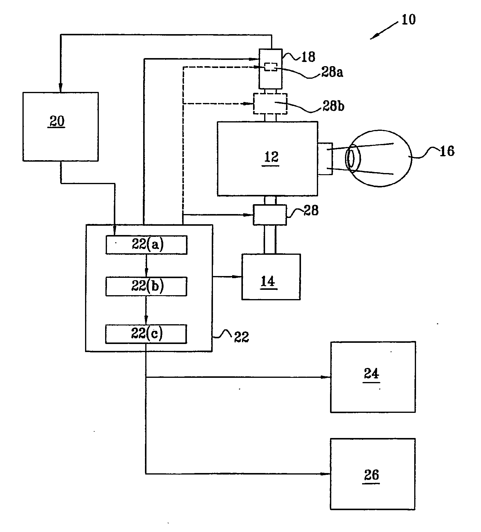
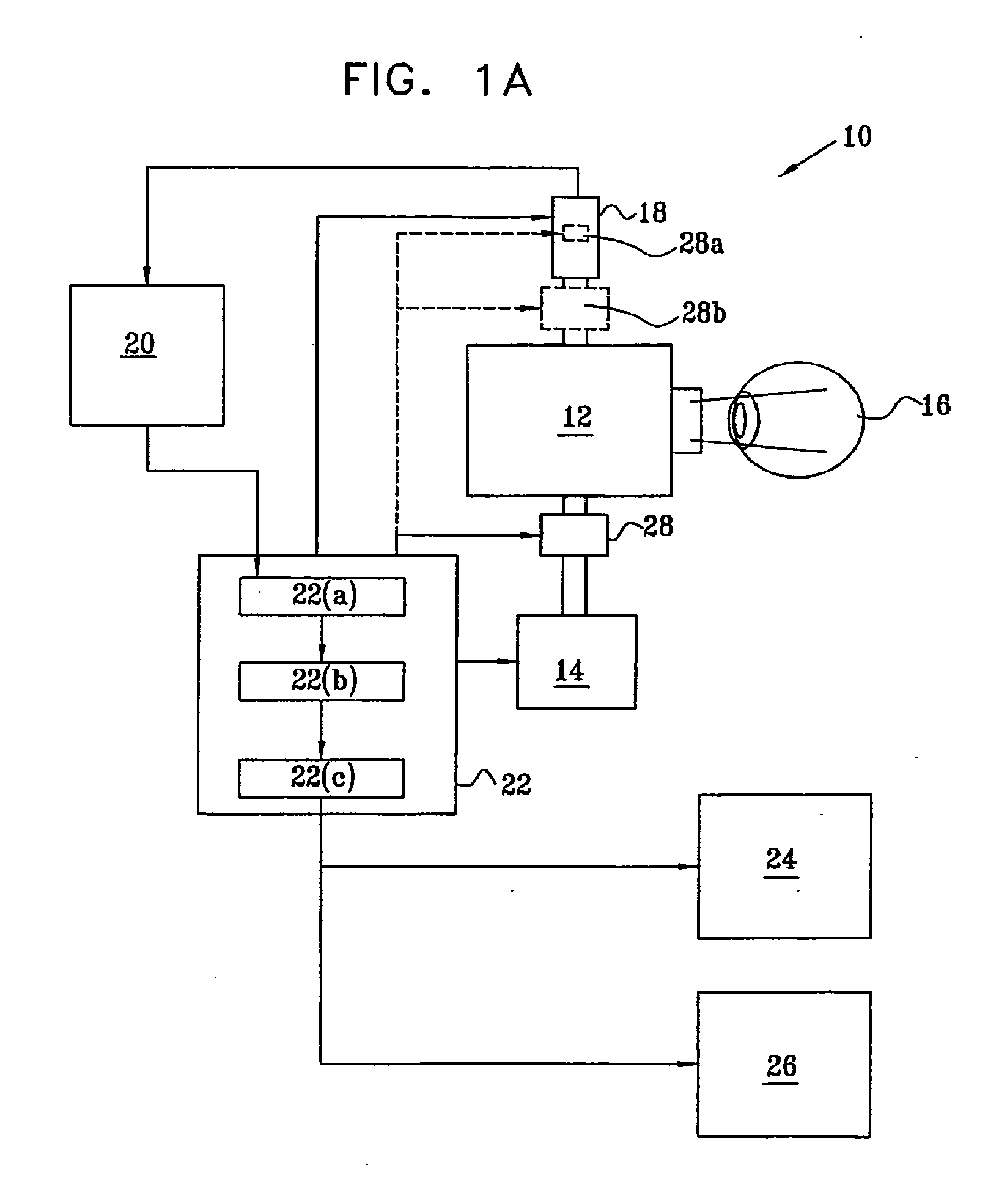
[0114] Reference is now made to FIG. 1A which is a schematic block diagram illustrating a system, constructed and operative according to a preferred embodiment of the present invention, for determining the oxygen saturation in the blood ves...


PUM


Abstract

A method and system for determination and mapping the quantity of chromophores having a distinct spectrum attached to moving objects in an spectrally rich environment that may include multiple chromophores attached to stationary objects. An area of inters is imaged at different times and different wavelengths, and the spectral properties of the or more chromophores attached to the moving objects are separated from the stationary spectral properties of the background, followed by spectral analysis of the moving objects to determine their quantity. Application to the retinal vasculature is illustrated, showing the imaging, analyzing and quantifying of the oxygen saturation of retinal blood, resolved for the different vascular compartments—capillaries, arterioles, venules, arteries, and veins. Changes in the structure of the vascular environment are also determined, whether growth of new vessels or the elimination of existing ones, by the generation of path maps based on analysis of differential images taken at a single wavelength of the moving components in the blood flow.

Description

FIELD OF THE INVENTION [0001] The present invention relates to the field of detecting chromophores attached to moving objects in a generally stationary spectral background, by separating the known distinct spectra of the moving objects from the overall background spectra, especially as applied to the non-invasive measurement of oxygen saturation in blood vessels by spectrally decomposing the separated spectrum of the moving red blood cells even in blood vessels which do not show significant pulsation, and to the characterization of the paths of blood flow. BACKGROUND OF THE INVENTION [0002] There are many applications, industrial, scientific and medical, in which it is necessary to determine the quantitative levels of particular components or details of a moving system, wherein the component or detail to be measured is situated in a background environment which may be visually difficult to differentiate from the component or detail to be measured. In such cases, conventional imaging...

Claims


Application Information

Patent Timeline
Patent Type & Authority Applications(United States)
IPC IPC(8): A61B5/00
CPCA61B3/1233A61B5/1459A61B5/14555A61B5/0261
Inventor GRINVALD, AMIRAMNELSON, DARINVANZETTA, IVO
Owner YEDA RES & DEV CO LTD
Why Eureka
Try Eureka