Method for preparing biological scaffold materials

a biological scaffold and material technology, applied in the field of biological scaffold materials, can solve the problems of limited life of biological scaffolds, inability to use biodegradable vascular grafts in the artery system, and other adverse effects, so as to improve the biodynamic properties, reduce the immunogenicity of the scaffold, and preserve the mechanical strength of the starting native tissue.

Inactive Publication Date: 2008-01-31
NAT CEREBRAL & CARDIOVASCULAR CENT
View PDF1 Cites 67 Cited by
  • Summary
  • Abstract
  • Description
  • Claims
  • Application Information

AI Technical Summary

Benefits of technology

[0013]Partial crosslinking and fixation of proteins serve to preserve the mechanical strength of the starting native tissue to a large extent. Heating of lyophilized tissue under vacuum induces dehydration-condensation reactions between amino groups and carboxyl and/or hydroxyl groups present in the protein. However, the degree of crosslinkin

Problems solved by technology

However, these materials are susceptible to calcification in vivo and, therefore, their life is limited to less than about 15 years in case of heart valves.
In addition to the limited life due to calcification, they have another disadvantage when used as vascular grafts which require to have sufficient elasticity and tensile strength.
However, constriction and other a

Method used


Image

  • Method for preparing biological scaffold materials
  • Method for preparing biological scaffold materials
  • Method for preparing biological scaffold materials

Examples


example 1

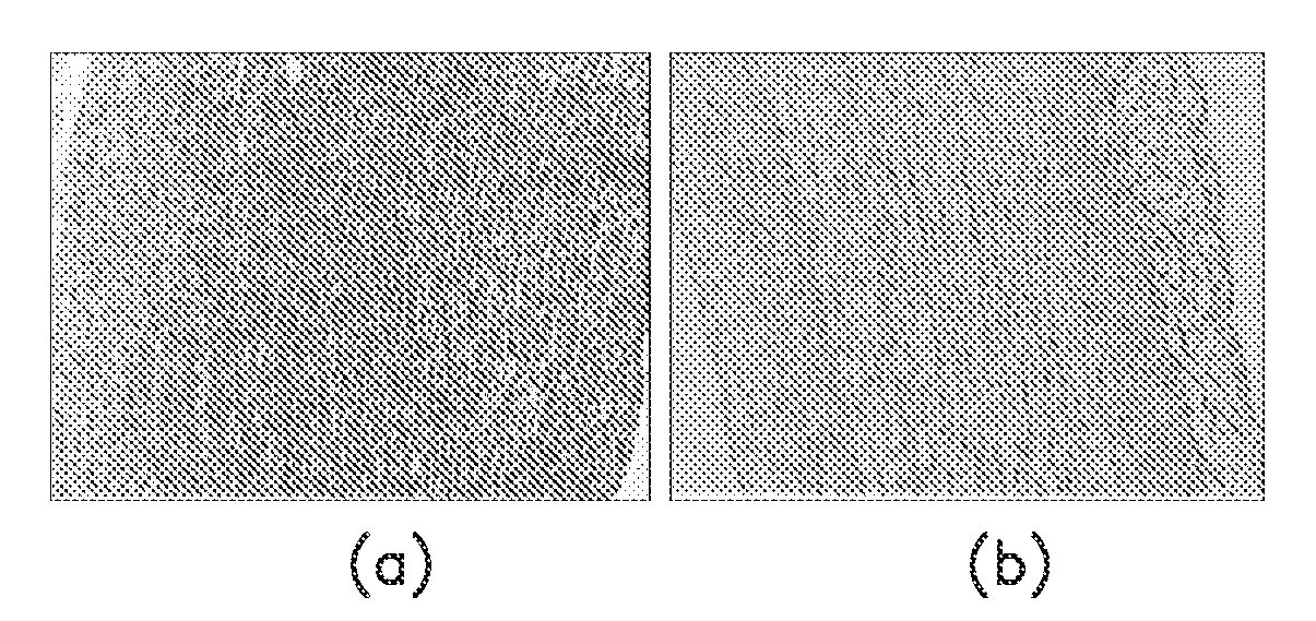
[0026]Porcine aorta purchased from Japan Farm Co., Ltd. was cut into tubular segments, lyophilized, and heated at 100° C., 120° C., 160° C., or 180° C. for a constant period of time of 24 hours in a vacuum chamber. The weight of the tissue segment was measured before and after the heat treatment to determine percent weight loss at varying temperatures. The results are shown in the graph of FIG. 1. The best result was obtained for the tissue heated at 120° C. for 24 hours when treating with elastase as described below.

[0027]Separately, an enzyme solution was prepared from a commercially available elastase preparation (3.95 U / mg, Funakoshi) by dissolving the enzyme in 0.0 M tris buffer (pH 8.0) to a concentration of 150 mg / L (0.57 U / ml), followed by the addition of 0.01M CaCl2 and 0.02% sodium azide.

[0028]The tissue treated at 120° C. for 24 hours was placed in the above elastase solution, incubated at 37° C. for 72 hours, and rinsed with physiological saline.

example 2

[0029]Porcine aorta segments as used in Example 1 were decellularized by placing in an ultrahigh pressure apparatus (Dr. SHEF available from Kobe Steel Ltd.) together with sterile water and applied a hydrostatic pressure of 10,000 atms for 10 minutes. The decellularized tissue was thoroughly washed with sterile water.

[0030]The procedure of Example 1 was repeated with the decellularized tissue under exactly the same conditions to prepare a scaffold material.

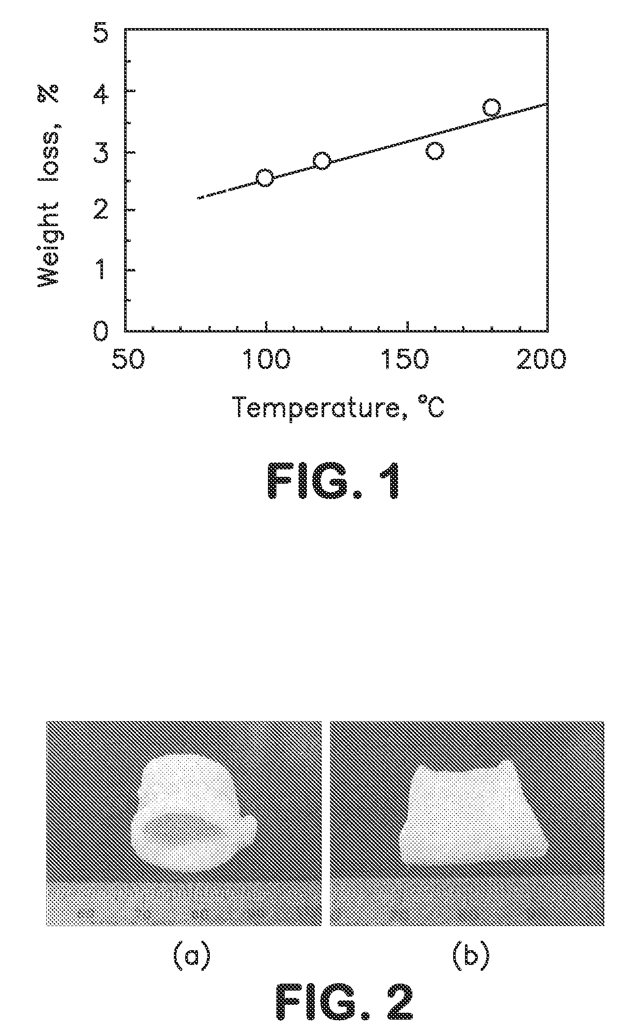
[0031]In FIG. 2, (a) is a photograph of native porcine aorta segment and (b) is a similar photograph of porcine aorta segment treated in Example 1. As seen, the tubular shape of native aorta was maintained after the treatment with heat and then with elastase.

[0032]The scaffold material prepared in Example 1 was tested for strength properties along the circumferential direction in comparison with native porcine aorta and native porcine pulmonary artery. FIG. 3 shows the stress vs. strain curve (a) of the scaffold of Example 1, in c...


PUM


Abstract

A method for preparing a scaffold material for use in the prosthesis therapy is disclosed. The method comprises (a) lyophilizing a segment of native soft tissue of mammalian origin, heating the lyophilized tissue at a temperature of 100-200° C., and incubating the tissue with elastase to selectively remove elastin leaving the extracellular components mainly comprised of collagen.

Description

FIELD OF THE INVENTION[0001]The present invention relates to a method for preparing biological scaffold materials for use in the prosthesis of impaired organs such as heart valves or vascular vessels.[0002]Prosthetic surgery is aimed to repair impaired organs by replacing with artificial devices. To this end scaffold materials are implanted and allowed to reconstruct the organ. Currently porcine or bovine tissues have been used as the scaffold material after treating with glutaraldehyde. However, these materials are susceptible to calcification in vivo and, therefore, their life is limited to less than about 15 years in case of heart valves. Glutaraldehyde-treated porcine or bovine tissues are used also as vascular grafts. In addition to the limited life due to calcification, they have another disadvantage when used as vascular grafts which require to have sufficient elasticity and tensile strength. The treatment with glutaraldehyde greatly decreases the elasticity and tensile stren...

Claims


Application Information

Patent Timeline
IPC IPC(8): A61F2/02A61L27/00
CPCA61K35/12A61L27/3604A61L2430/40A61L27/3645A61L27/3683A61L27/3641
Inventor FUJISATO, TOSHIYATERADA, DOHIKOSAWADA, KAZUYANAKATANI, TAKESHI
Owner NAT CEREBRAL & CARDIOVASCULAR CENT
Why Eureka
Try Eureka