Knockout Non-Human Animal

a non-human animal and knockout technology, applied in the field of knockout non-human animals, can solve the problem of failure of each organ via an immunoglobulin e-mediated inflammatory mechanism

Inactive Publication Date: 2008-02-07
SUMITOMO CHEM CO LTD +2
View PDF0 Cites 4 Cited by
  • Summary
  • Abstract
  • Description
  • Claims
  • Application Information

AI Technical Summary

Benefits of technology

[0007] The present inventors disrupted the PHF3 gene on a chromosome of a mouse ES cell, mixed the resulting ES cell with the blastocyst of a parent animal, allowed embryonic differentiation in the parent animal to continue until a baby was born, and thereby a chimeric primitive mouse was produced as an offspring. Then, the present inventors bred the chimeric primitive mouse to produce a progeny (hereinafter, the chimeric primitive mouse and a progeny thereof are referred to as the present knockout mouse in some cases). In the present knockout mouse, scab formation and cell infiltration into the skin dermis layer were high-frequently observed, and the total immunoglobulin E level in blood was extremely high. Based on these findings, the present inventors found that said animal can be a model mouse representing disease states similar to the development of atopic dermatitis, and as a result, completed the present invention.

Problems solved by technology

Allergic diseases including atopic dermatitis, asthma, allergic rhinitis, allergic conjunctivitis and the like cause hypersensitive responses to an environmental antigen and the like to which normal people do not respond, and lead to failure of each organ via an immunoglobulin E mediated inflammatory mechanism.

Method used


Image

  • Knockout Non-Human Animal
  • Knockout Non-Human Animal
  • Knockout Non-Human Animal

Examples


example 1

Preparation of PHF3 Gene Knockout ES Cell

[0235] PHF3 gene knockout ES cells were prepared by a gene trap method (International Publication WO01 / 005987) using a trap vector pU-17.

[0236] It was confirmed that a disrupted or recombined gene is only the PHF3 gene, according to the following method.

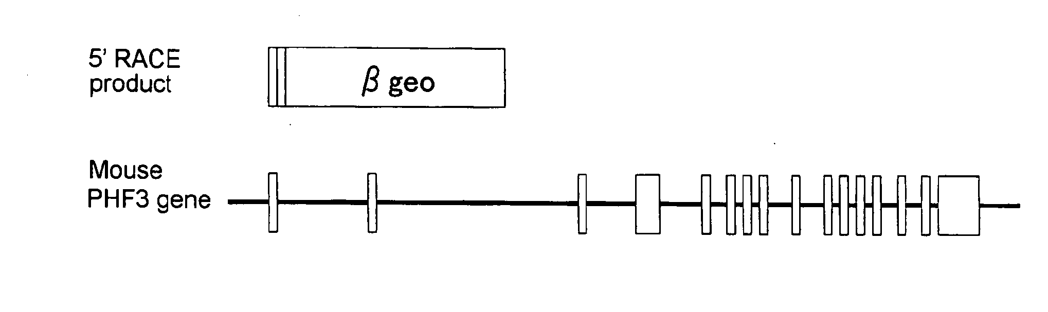
[0237] In the ES cell used, it was confirmed by PCR analysis that only a copy of the vector was introduced, and it was also confirmed by Southern blotting analysis that the full length of the vector was introduced. Further, when a partial sequence of the trapped gene (a cDNA sequence upstream of an insertion position of the trap vector) was determined by a 5′ RACE method, it was consistent with the nucleotide sequences of the first exon and the second exon of the PHF3 gene (GenBank Acc. No. XM—129836) (see FIG. 1). The mouse PHF3 gene is composed of 16 exons, and encodes a protein consisting of 2025 amino acids. A translation initiation codon of the gene is present in the second exon. In a ...

example 2

Production of PHF3 Gene Knockout Mouse

[0238] A chimeric mouse comprising cells in which the PHF3 gene was knocked out was produced according to the following method. The ES cell prepared in Example 1 in which the PHF3 gene was disrupted or recombined and an ICR line 8-cell stage embryo were aggregated by an aggregation method (Nagy A., 1993, Oxford University Press, Oxford, pp 147-179). On the following day, the chimeric embryo which had been developed into a morula or a blastocyst was transplanted into the uterus of a pseudo-pregnancy-induced ICR line female mouse (pseudo-pregnancy induction day 3) (Shinichi Aizawa: Gene targeting: Preparation of Mutated Mouse using ES Cell, Yodosha Co., Ltd, 1995). After 17.5 days from transplantation, the female mouse was subjected to caesarotomy to take out a fetal, and thereby a chimeric mouse was obtained.

[0239] The chimeric mouse was reared by a foster parent for 4 weeks (Shinichi Aizawa: Gene targeting: Preparation of Mutated Mouse using E...

example 3

Pathological Test of PHF3 Gene Homo-Knockout Mouse

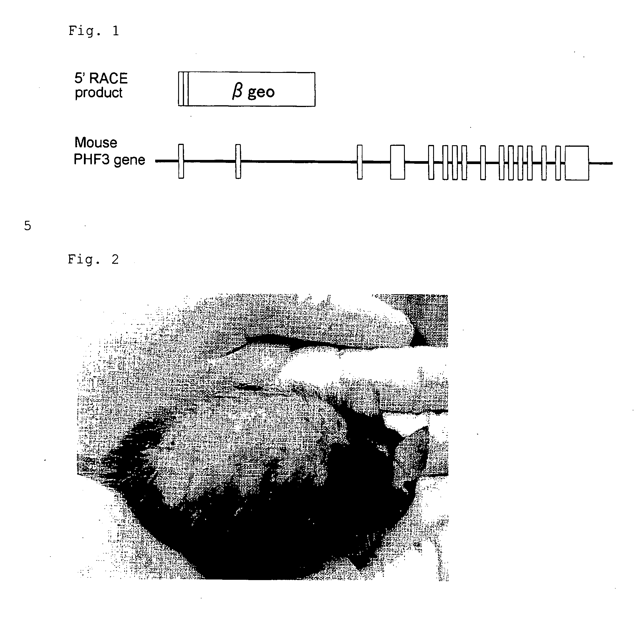
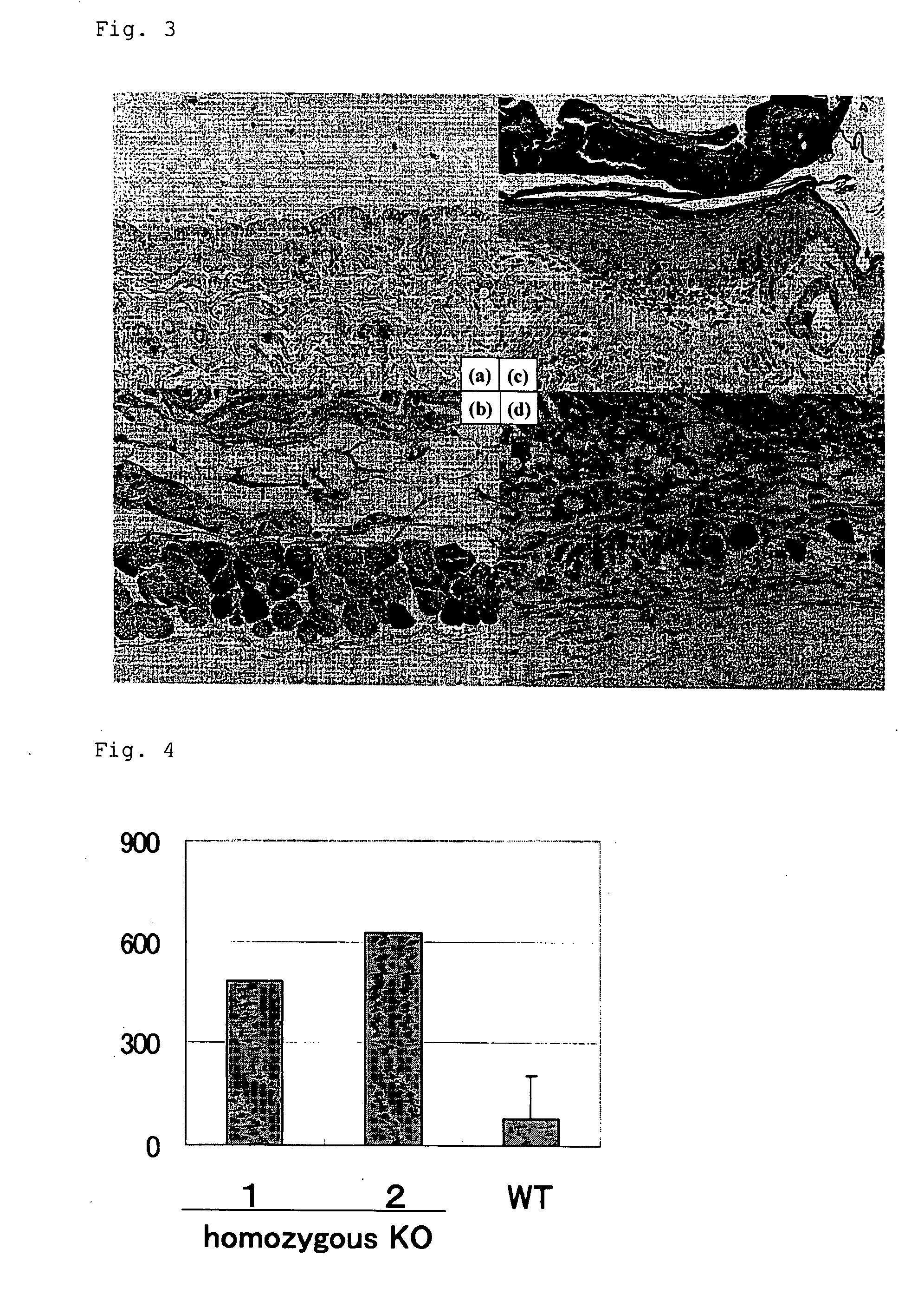
[0242] The atopic dermatitis symptoms found in the PHF3 gene homo-knockout mouse produced in Example 2 were histopathologically tested according to the following method. After the 8 week-old PHF3 gene homo-knockout mouse was led to death by exsanguination under ether anesthesia, a part of a skin tissue (abdominal) was taken from the mouse and fixed in a 10% neutral buffered formalin solution at room temperature for 1 week. The fixed skin tissue was alcohol-dehydrated, then embedded in paraffin, and thinly sliced at a thickness of 2 to 10 um using a microslicer. The resulting thin section was placed on a slide glass. The thin section was de-paraffined (7 min×3) with xylene, washed with 100% ethanol (30 sec×1, 2 min×2) and then distilled water, and then stained with hematoxylin / eosin (hereinafter, referred to as HE staining). The HE staining was performed by immersing the section in a hematoxylin solution (a solution of hematoxylin 2 ...


PUM

PropertyMeasurementUnit
temperatureaaaaaaaaaa
weightaaaaaaaaaa
weightaaaaaaaaaa

Abstract

The present invention relates to a knockout non-human animal whose genome comprises a disruption in at least one allele of the PHF3 gene, useful in developing drugs for diseases with the onset of atopic dermatitis or the like; a totipotent cell such as an ES cell which is essential to production of the above non-human animal; use thereof; and so on.

Description

TECHNICAL FIELD [0001] The present invention relates to a knockout non-human animal in which the PHF3 gene is disrupted and the like. BACKGROUND ART [0002] Allergic diseases including atopic dermatitis, asthma, allergic rhinitis, allergic conjunctivitis and the like cause hypersensitive responses to an environmental antigen and the like to which normal people do not respond, and lead to failure of each organ via an immunoglobulin E mediated inflammatory mechanism. [0003] There are a variety of genome regions involved in the development of allergic diseases, and there are many disease-causative genes corresponding to the genome regions (Halapi et al., Curr Opin Pulm Med., 10:22-30, 2004; Lee et al., Nat Genet., 26:470-473, 2000; Cookson et al., Lancet, 1:1292-1295, 1989; Bradley et al., Hum. Mol. Genet., 11;1539-1548, 2002). In some of genome regions involved in allergic diseases, a disease-causative gene and its physiological mechanism have been known. For example, it has been known...

Claims


Application Information

Patent Timeline
Patent Type & Authority Applications(United States)
IPC IPC(8): A61K38/00A01K67/00A61K48/00C12N15/00C12Q1/68G01N33/00C12N5/06C07K16/18A01K67/027
CPCA01K67/0276A01K2217/075A01K2227/105C12N2517/02C07K14/4702C12N15/8509A01K2267/0325
Inventor NAITO, YOSHIKAZUOEDA, KENJISUKATA, TOKUOINOUE, YOSHIFUMI
Owner SUMITOMO CHEM CO LTD
Why Eureka
Try Eureka