
Detection method for human pappilomavirus (HPV) and its application in cervical cancer

a detection method and technology for human pappilomavirus, applied in the field of detection method for human pappilomavirus and its application in cervical cancer, can solve the problems of high risk of progression toward invasive cancer, regress spontaneously, and usually self-limited

Inactive Publication Date: 2008-02-21
HEER MEDICAL TECH DEV CO LTD
View PDF51 Cites 14 Cited by
  • Summary
  • Abstract
  • Description
  • Claims
  • Application Information

AI Technical Summary

Benefits of technology

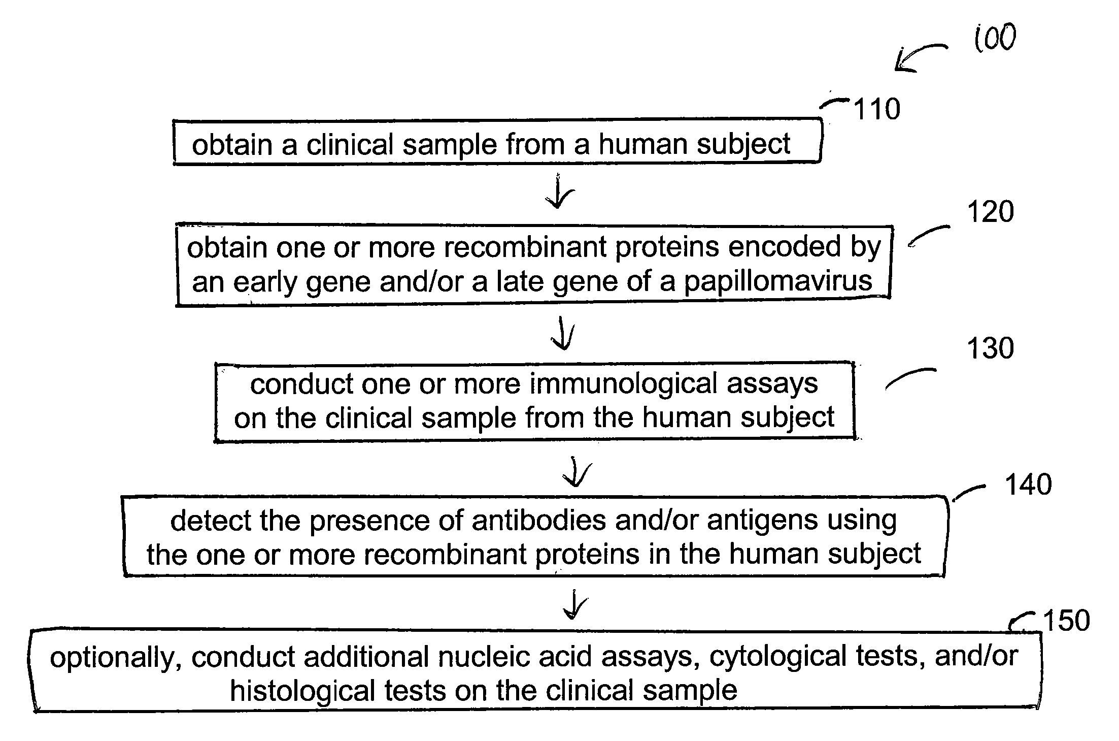
[0009] Embodiments of the invention generally relate to various methods, detection assays, kits, polypeptides, recombinant proteins, antibodies, and nucleic acids useful for detecting general HPV infection as well as high risk HPV infection. In one embodiment, a method of screening a human subject of papillomavirus infection includes obtaining a clinical sample from the human subject, obtaining a first recombinant protein encoded by an early gene of a papillomavirus, and obtaining a second recombinant protein encoded by a late gene of the papillomavirus. The method further includes conducting one or more immunological assays on the clinical sample from the human subject, detecting the presence of an antibody to the first recombinant protein in the human subject using the first recombinant protein, and detecting the presence of an antibody to the second recombinant protein in the human subject using the second recombinant protein.
[0010] In another embodiment, a method of screening a human subject infected with a human papillomavirus includes obtaining a clinical sample from the human subject, conducting a nucleic acid hybridization assay on the clinical sample, detecting the presence of a papillomavirus genome in the clinical sample from the human subject, and conducting one or more immunological assays on the clinical sample. The one or more immunological assays can be performed independently or concurrently of the nucleic acid hybridization assay using the same or different clinical sample but from the same human subject. The one or more immunological assays are performed to detect the presence of an antibody to an early papillomavirus vi

Problems solved by technology

The presence of these lesions, preferentially observed in women aged 35-40 yrs, are at high risk of progression toward invasive cancer.
However, the development of cervical cancer is a multiple step process that cannot be explained simply by infection with specific types of HPV.
It is found that younger age group women are often infected with HPV; however, clinical information reveals that most latent or asymptomatic infections with high risk HPV types as well as early dysplastic lesions (CIN 1) are usually self-limited and regress spontaneously.
However, due to subjective test criteria, drawbacks of pap smear tests include difficulty in obtaining samples, poor inter- and intra-observer agreement, a high rate of false negatives (up to 20%) and false positive, the requirements for specialized labs staffed with highly trained personnel, and inability to identify a large proportion of HPV-infected persons.
Nucleic acid methods, such as “DNA Hybrid Capture”, have been developed, but are not ideal primarily due to their high cost, assay operation procedures, and the requirements for facility, equipment, and highly trained personnel.
Attempts to detect the presence of HPV related antibodies in a human subject by ELISA (enzyme linked immunoabsorbant assays) generally lead to extremely low assay sensitivity and thus can not be developed into a commercially suitable diagnostic test.
Thus, there is no successful ELISA assay available as a diagnostic tool for clinical samples.

Method used


Image

  • Detection method for human pappilomavirus (HPV) and its application in cervical cancer
  • Detection method for human pappilomavirus (HPV) and its application in cervical cancer
  • Detection method for human pappilomavirus (HPV) and its application in cervical cancer

Examples


example 1

Cloning and Production of Various Recombinant Proteins Encoded by HPV-16 Early E6 Gene

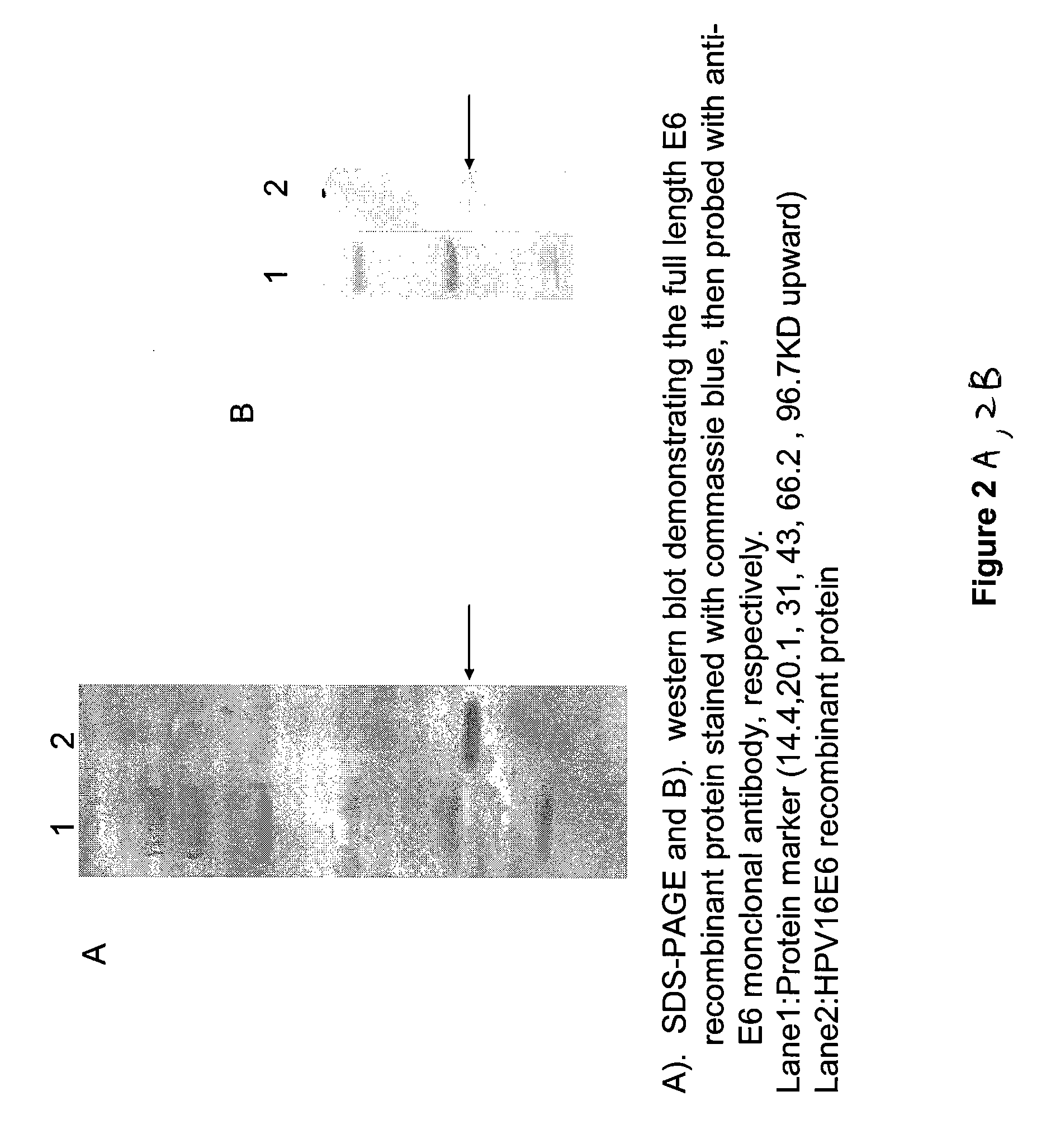
[0088] Cloning of an exemplary oncogenic E6 early gene from an exemplary HPV type, HPV-16, is described herein. A 474 base pair (b.p.) DNA fragment (SEQ ID NO. 1) containing the 157 amino acid coding region (SEQ ID NO. 2) of the HPV-16 E6 gene was obtained by polymerase chain reaction (PCR) amplification. Primers were used for cloning, for example, a pair of forward and reverse primers, 5′ cgcGGATCCcaccaaaagagaactgcaatgtttc 3′ (SEQ ID NO. 3) and 5′ cccAAGCTTttacagctgggtttctctacgtg 3′ (SEQ ID NO. 4), respectively. The DNA sequence of the isolated DNA fragment was confirmed by comparing with the sequence from Gene Bank database. All cloning procedures are carried out according to the protocols described in “Molecular Cloning”, A Laboratory Manual, eds. Sambrook, Fritsch and Maniatis, Cold Spring Harbor Laboratory Press, 1989. In addition, E6 DNA fragments from different strains of HPV-16 can also be...

example 2

Cloning and Production of Recombinant Proteins Encoded by HPV-16 Early E7 Gene

[0095] Cloning of an exemplary oncogenic E7 early gene from an exemplary HPV type, HPV-16, is described herein. A 294 base pair (b.p.) DNA fragment (SEQ ID NO. 7) containing the 99 amino acid coding region (SEQ ID NO. 8) of the HPV-16 E7 gene was obtained by polymerase chain reaction (PCR) amplification. Primers were used for cloning, for example, a pair of forward and reverse primers, 5′ cgcGGATCCcatggagatacacctacattgc 3′ (SEQ ID NO. 9) and 5′ ccgGAATTCttatggtttctgagaacagatgg 3′ (SEQ ID NO. 10), respectively. The DNA sequence of the isolated DNA fragment was confirmed by comparing with the sequence from Gene Bank database. In addition, E7 DNA fragments from different strains of HPV-16 can also be cloned from different clinical samples or sources.

[0096] The obtained 294 base pair (b.p.) DNA fragment was sub-cloned into a GST expression vector in order to express a recombinant HPV-16 E7 GST fusion protein...

example 3

Cloning and Production of Recombinant Proteins Encoded by HPV-16 Late L1 Gene

[0098] Cloning of an exemplary late gene from an exemplary HPV type, HPV-16, is described herein. A 1596 base pair (b.p.) DNA fragment (SEQ ID NO. 13) containing the 531 amino acid coding region (SEQ ID NO. 14) of the HPV-16 L1 gene was obtained by polymerase chain reaction (PCR) amplification. Primers were used for PCR cloning, for example, a pair of forward and reverse primers, 5′ ccgCTCGAGatgcaggtgacttttatttacatcc 3′ (SEQ ID NO. 15) and 5′ cccAAGCTTttacagcttacgttttttgcgttta 3′ (SEQ ID NO. 16), respectively. The DNA sequence of the isolated DNA fragment was confirmed by comparing with the sequence from Gene Bank database. In addition, L1 DNA fragments from different strains of HPV-16 can also be cloned from different clinical samples or sources.

[0099] The obtained 1596 base pair (b.p.) DNA fragment was sub-cloned into a baculovirus expression system in order to express a recombinant HPV-16 L1 protein. T...


PUM

PropertyMeasurementUnit
Fractionaaaaaaaaaa
Fractionaaaaaaaaaa
Fractionaaaaaaaaaa

Abstract

Embodiments of the invention provide methods, assays, and kits for detecting HPV infection and HPV associated epithelial cell abnormalities, most notably those associated with pre-malignant and malignant epithelial cell lesions. Detection of HPV DNAs, genomes, and / or oncoproteins by nucleic acid hybridization assays and immunological assays can be used in early clinical screening for HPV infection and diagnosis for cervical cancer. The polypeptides, recombinant proteins, antibodies, nucleic acids, and various detection methods thereof are particularly useful for diagnosing carcinomas of the uterine cervix and those at risk of developing cervical cancer.

Description

CROSS-REFERENCE TO RELATED APPLICATIONS [0001] This application claims benefit of U.S. provisional patent application Ser. No. 60 / 737,152, filed Nov. 15, 2005, which is herein incorporated by reference.BACKGROUND OF THE INVENTION [0002] Cancer of the uterine cervix or cervical cancer is the second most common cancer in women worldwide. Although screening programs to identify precursor lesions of cervical cancer have contributed to a reduction in mortality and morbidity, 500,000 new cases of invasive cervical cancer are diagnosed worldwide and 230,000 women die of cervical cancer annually. Early detection and diagnosis is critical for survival of cervical cancer. [0003] Infection by human papillomaviruses (HPV) at specific epithelium cells to induce epithelial proliferations plays an important role for cervical carcinogenesis. About 99 percent of confirmed cervical cancer cases are found to be associated with HPV infection with biopsy-confirmed squamous intraepithelial lesions (SIL) ...

Claims


Application Information

Patent Timeline
IPC IPC(8): C12Q1/70
CPCC12N2710/20051C12N2710/20022C07K14/005C12Q1/708
Inventor CHENG, SHU-LING
Owner HEER MEDICAL TECH DEV CO LTD
Learn More