Muscle Derived Cells For The Treatment Of Gastro-Esophageal Pathologies And Methods Of Making And Using The Same

a gastro-esophageal and muscle derived technology, applied in the field of muscle derived progenitor cells, to achieve the effect of improving at least one symptom

Active Publication Date: 2008-06-26
UNIVERSITY OF PITTSBURGH +1
View PDF14 Cites 12 Cited by
  • Summary
  • Abstract
  • Description
  • Claims
  • Application Information

AI Technical Summary

Benefits of technology

[0019]In one embodiment, when the MDCs and compositions thereof are used to treat gastroesophageal reflux they are injected directly into the esophagus. Preferably, they may be injected into the lower esophageal sphincter. In another embodiment, MDCs and co...

Problems solved by technology

These methods result in the enrichment of MDCs that have long-term sur...

Method used


Image

  • Muscle Derived Cells For The Treatment Of Gastro-Esophageal Pathologies And Methods Of Making And Using The Same
  • Muscle Derived Cells For The Treatment Of Gastro-Esophageal Pathologies And Methods Of Making And Using The Same
  • Muscle Derived Cells For The Treatment Of Gastro-Esophageal Pathologies And Methods Of Making And Using The Same

Examples


example 1

MDC Enrichment, Isolation and Analysis According to the Pre-Plating Method

[0053]Enrichment and isolation of MDCs: MDCs were prepared as described (U.S. Pat. No. 6,866,842 of Chancellor et al.). Muscle explants were obtained from the hind limbs of a number of sources, namely from 3-week-old mdx (dystrophic) mice (C57BL / 10ScSn mdx / mdx, Jackson Laboratories), 4 6 week-old normal female SD (Sprague Dawley) rats, or SCID (severe combined immunodeficiency) mice. The muscle tissue from each of the animal sources was dissected to remove any bones and minced into a slurry. The slurry was then digested by 1 hour serial incubations with 0.2% type XI collagenase, dispase (grade II, 240 unit), and 0.1% trypsin at 37° C. The resulting cell suspension was passed through 18, 20, and 22 gauge needles and centrifuged at 3000 rpm for 5 minutes. Subsequently, cells were suspended in growth medium (DMEM supplemented with 10% fetal bovine serum, 10% horse serum, 0.5% chick embryo extract, and 2% penicill...

example 2

MDC Enrichment, Isolation and Analysis According to the Single Plate Method

[0058]Populations of rapidly- and slowly-adhering MDCs were isolated from skeletal muscle of a mammalian subject. The subject may be a human, rat, dog or other mammal. Biopsy size ranged from 42 to 247 mg.

[0059]Skeletal muscle biopsy tissue is immediately placed in cold hypothermic medium (HypoThermosol (BioLife) supplemented with gentamicin sulfate (100 ng / ml, Roche)) and stored at 4° C. After 3 to 7 days, biopsy tissue is removed from storage and production is initiated. Any connective or non-muscle tissue is dissected from the biopsy sample. The remaining muscle tissue that is used for isolation is weighed. The tissue is minced in Hank's Balanced Salt Solution (HBSS), transferred to a conical tube, and centrifuged (2,500×g, 5 minutes). The pellet is then resuspended in a Digestion Enzyme solution (Liberase Blendzyme 4 (0.4-1.0 U / mL, Roche)). 2 mL of Digestion Enzyme solution is used per 100 mg of biopsy ti...

example 3

Soft Tissue Augmentation of the Gastro-esophageal Junction and Anal Sphincter

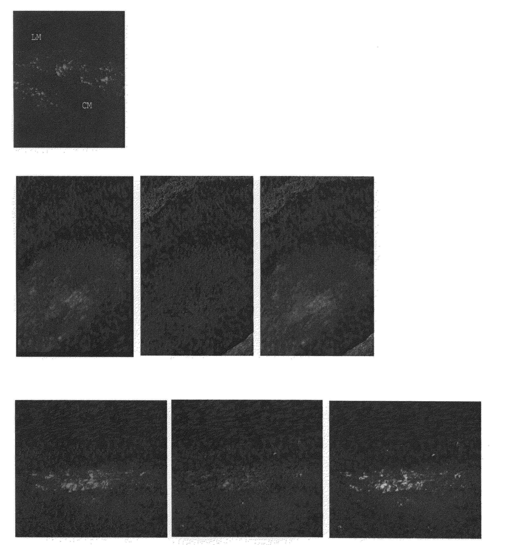
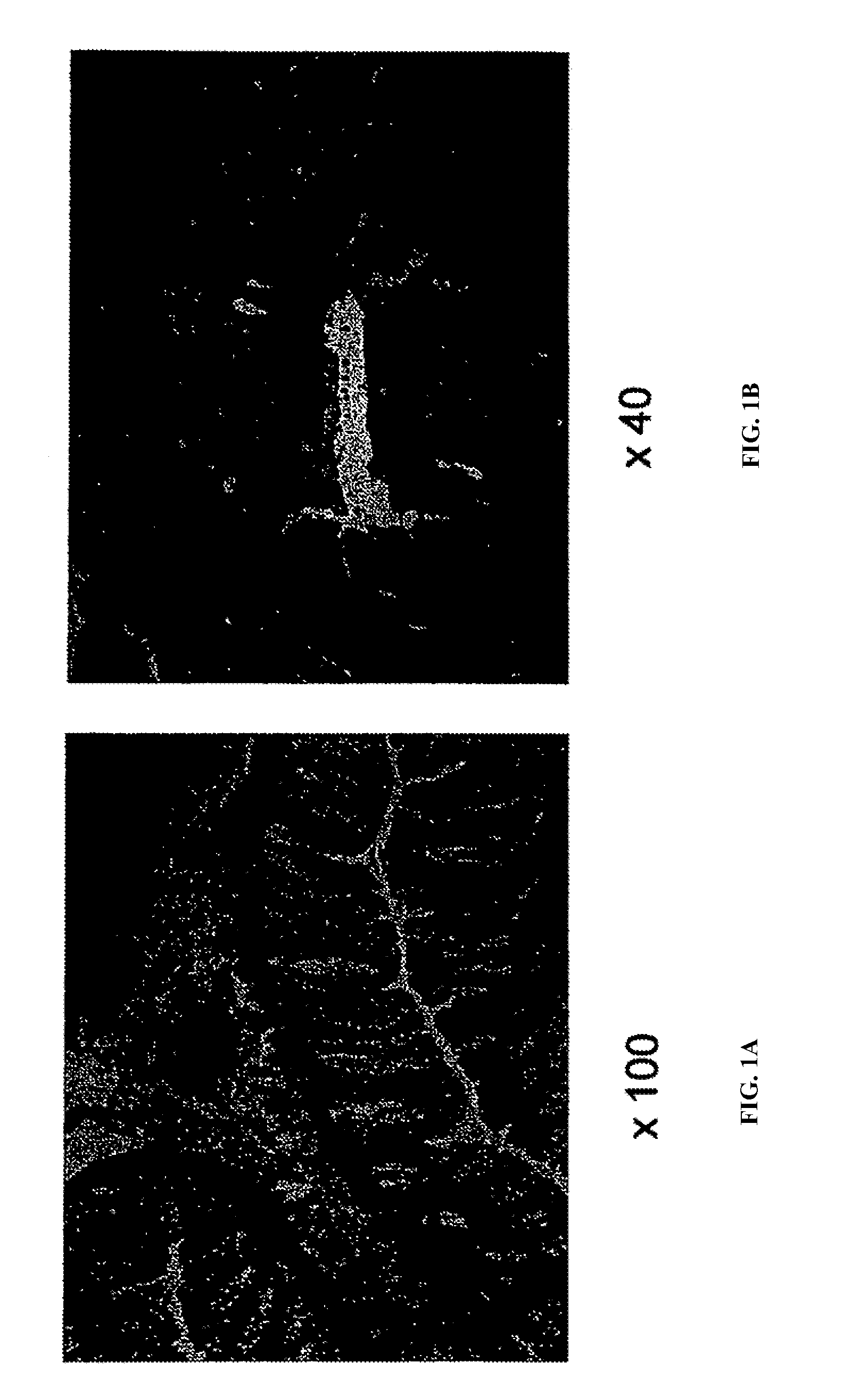
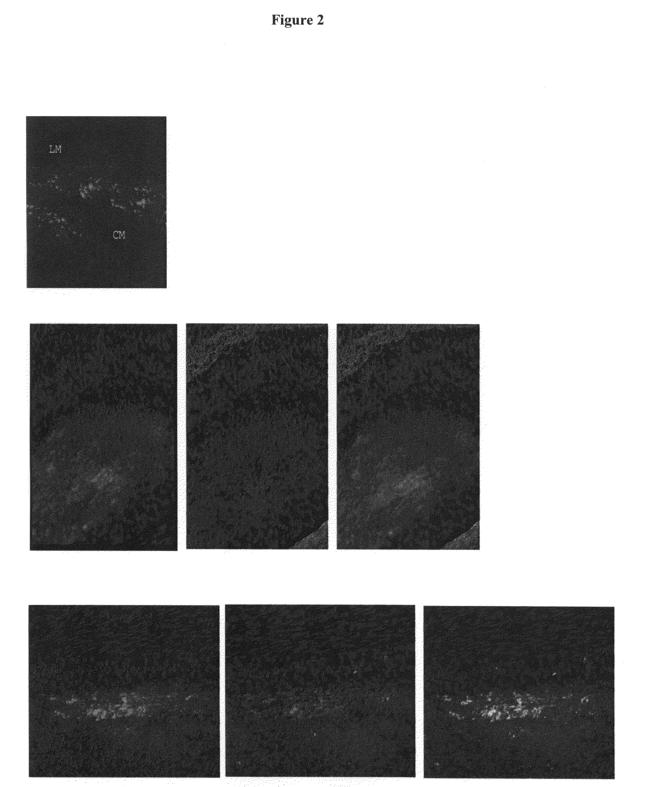
[0064]Sprague-Dawley (SD) rats were prepared for surgery by anesthetizing with halothane using standard methods, and washing the surgical site with Betadine® solution. A midline abdomen incision was made to expose the gastroesophageal junction and anal sphincter. The soft tissue was injected with 10 μl of a suspension of muscle-derived progenitor cells, prepared pursuant to the methods of Example 1, in HBSS (1-1.5×10⁶ cells) using a Hamilton microsyringe. At day 3 post-injection, the area surrounding each injection site was excised, prepared for histochemical analysis, stained for β-galactosidase to determine the location and viability of the cells carrying the LacZ marker, examined microscopically, and photographed. Results of these experiments demonstrate that MDC compositions can be used as esophageal and anal sphincter bulking materials (FIGS. 1A and 1B) for the treatment of gastroesophageal reflux or f...


PUM


Abstract

The present invention provides muscle-derived progenitor cells that show long-term survival following transplantation into body tissues and which can augment soft tissue following introduction (e.g. via injection, transplantation, or implantation) into a site of soft tissue. Also provided are methods of isolating muscle-derived progenitor cells, and methods of genetically modifying the cells for gene transfer therapy. The invention further provides methods of using compositions comprising muscle-derived progenitor cells for the augmentation and bulking of mammalian, including human, soft tissues in the treatment of various cosmetic or functional conditions, including malformation, injury, weakness, disease, or dysfunction. In particular, the present invention provides treatments and amelioration of symptoms for gastro-esophageal pathologies like gastro-esophageal reflux.

Description

FIELD OF THE INVENTION[0001]The present invention relates to muscle-derived progenitor cells (MDC) and compositions of MDCs and their use in the augmentation of body tissues, particularly soft tissue like gastric and esophageal tissue. In particular, the present invention relates to muscle-derived progenitor cells that show long-term survival following introduction into soft tissues, methods of isolating MDCs, and methods of using MDC-containing compositions for the augmentation of human or animal soft tissues, including gastric and esophageal tissue. The invention also relates to novel uses of muscle-derived progenitor cells for the treatment of functional conditions, such as gastro-esophageal reflux disease.BACKGROUND OF THE INVENTION[0002]Augmentation of soft tissue using synthetic materials such as silicone or polytetrafluoroethylene (PTFE) is well known in the art. U.S. Pat. No. 5,876,447 to Arnett discloses the use of silicone implants for facial plastic surgery. However, such...

Claims


Application Information

Patent Timeline
IPC IPC(8): A61K35/00C12N5/06A61P1/00A61K35/12A61K35/34C12N5/077
CPCA61K35/12A61K35/34C12N2533/54C12N2509/00C12N5/0658A61P1/00A61P1/04A61P1/06A61P1/14A61P11/00A61P11/06A61P11/14A61P29/00A61P43/00
Inventor CHANCELLOR, MICHAEL B.PASRICHA, JAYJANKOWSKI, RONALDPRUCHNIC, RYAN
Owner UNIVERSITY OF PITTSBURGH
Why Eureka
Try Eureka