Method of obtaining viable small tissue particles and use for tissue repair

a small, tissue technology, applied in the field of tissue particles, can solve the problems of limited durability limited healing ability of articular cartilage damage, and structurally weak tissue defects, etc., to improve the healing effect of tissue defects, facilitate tissue particle retention at the defect site, and enhance the effect of tissue particle retention

Active Publication Date: 2008-06-26
ZIMMER ORTHOBIOLOGICS
View PDF46 Cites 47 Cited by
  • Summary
  • Abstract
  • Description
  • Claims
  • Application Information

AI Technical Summary

Benefits of technology

[0011]Another embodiment is a method using the described compositions in ameliorating a tissue defect. In one embodiment, the defective tissue may be cartilage, bone, ligament, meniscus, tendon, muscle, nucleus pulposus, gingiva, annulus fibrosus, periosteum, perichondrium, fascia, and / or perineurium. In one embodiment, defects within articular cartilage are subjected to the method. In general, the method includes placing the isolated sized tissue particles into a tissue defect site. Retention of the tissue particles in the defect site is facilitated by the small particle size. In certain embodiments, retention of the tissue particles at the defect site may be enhanced by techniques such as microfracture and use of adhesives.

Problems solved by technology

The composition and highly complicated interaction of these components make regeneration and replacement techniques challenging.
For example, the lack of a direct blood supply and few cells distributed widely among a dense extracellular matrix leads to a limited healing ability of damaged articular cartilage.
Fibrocartilage is a relatively disorganized lattice of collagen fibers, as opposed to the natural hyaline cartilage, and thus partially fills the defect with structurally weak tissue that also exhibits limited durability.
The success of these procedures generally diminishes over time, possibly due to formation of fibrocartilage, inadequate development of repair tissue, poor cell differentiation, and / or poor bonding to the surrounding articular cartilage borders.
Intact full thickness grafts, such as osteochondral autografts and allografts, also may suffer from mismatched sizes, immunologic rejection, and poor adhesion of cartilage to bony surfaces.
Enzymatic digestion, which is often used to generate cells for autogenous chondrocyte cell transplantation, results in poor cell viability following initial isolation.

Method used


Image

  • Method of obtaining viable small tissue particles and use for tissue repair
  • Method of obtaining viable small tissue particles and use for tissue repair
  • Method of obtaining viable small tissue particles and use for tissue repair

Examples


example

[0045]Cartilage tissue particles were assessed for cell viability and evaluation in cartilage defect repair. All procedures were conducted in compliance with relevant regulations for the use of animal tissue. Porcine knee joints were obtained from a local abattoir. A knee joint was opened using a scalpel and the articular cartilage from the condyle load bearing area was exposed. Using a 7.5 mm diameter coring reamer, an osteochondral plug was obtained.

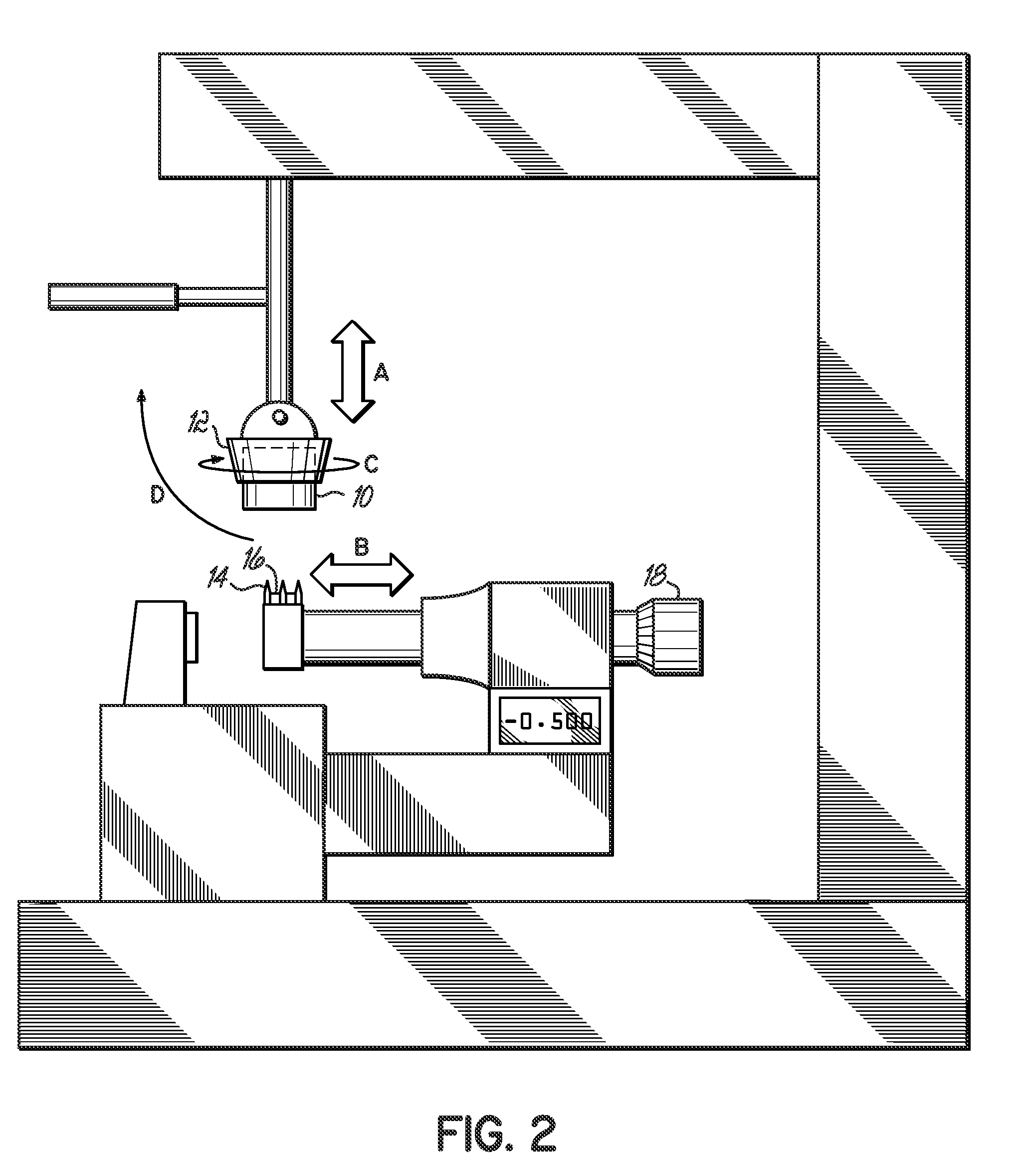
[0046]The osteochondral plug was mounted on the jaw of the cutting device (FIG. 2) and a series of cuts were made using three multiple blades to obtain viable small tissue particles.

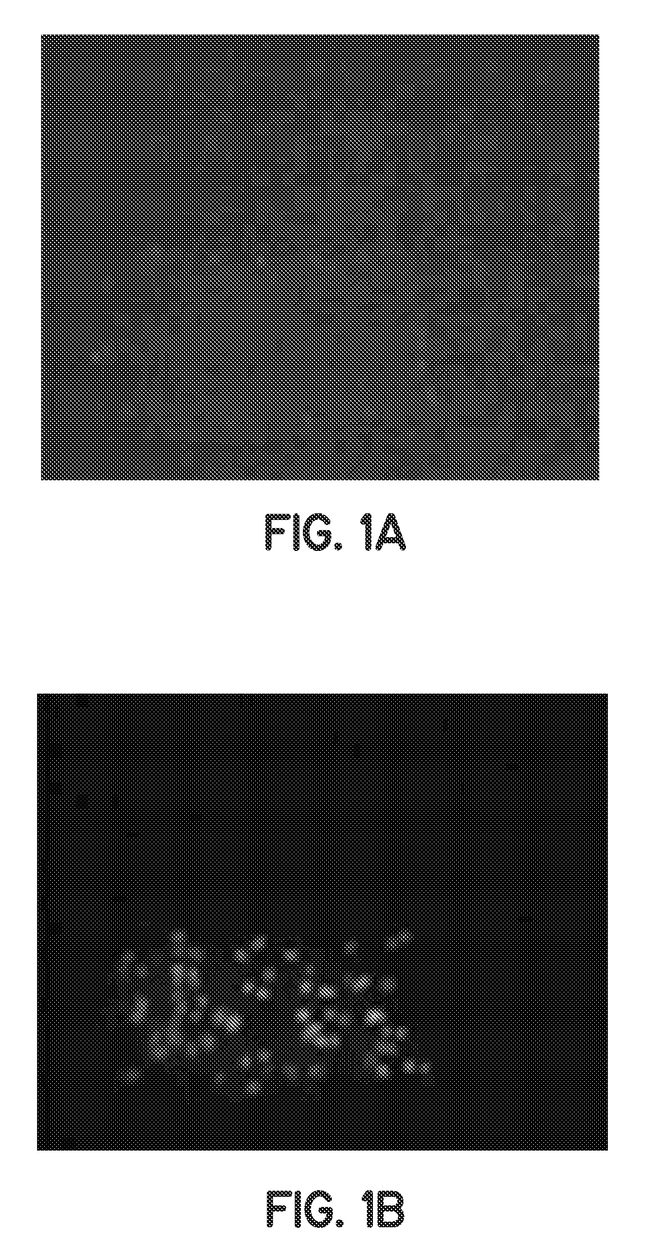
[0047]Tissue particles were stained using LIVE / DEAD® stain to determine cell viability. The LIVE / DEAD® stain uses a membrane-permeant CALCEIN AM that is cleaved by endogenous esterases in the live cells to yield cytoplasmic green fluorescence, and the membrane-impermeant ethidium homodimer-1 labels nucleic acids of membrane-compromised cells, e.g. dead cell...


PUM

PropertyMeasurementUnit
sizeaaaaaaaaaa
volumeaaaaaaaaaa
widthaaaaaaaaaa

Abstract

The invention provides a composition including isolated small living tissue particles, a method of making the tissue particles, and a method of using the composition to ameliorate a tissue defect. The tissue particles are composed of cells and their associated extracellular molecules and are sized, in certain embodiments, to be smaller than about 1 mm. Another aspect of the inventive tissue particles is the large percentage of viable cells. In certain embodiments, the tissue particles are made from cartilage and the composition may also contain additives such as adhesives, solutions, and bioactive agents.

Description

TECHNICAL FIELD OF THE INVENTION[0001]Preparation and use of tissue particles, sized from various sources, to repair tissue defects such as orthopedic tissue defects.BACKGROUND[0002]Articular cartilage is a thin, smooth, low friction, gliding surface composed of hyaline cartilage with resiliency to compressive forces. While only a few millimeters thick, it has excellent wear characteristics. Its mechanical and structural capacity depends on the integrity of its extracellular matrix, in which chondrocytes are sparsely distributed throughout structural macromolecules including collagen, proteoglycans, and noncollagenous proteins. Although chondrocyte cells produce the extracellular matrix, they compose less than 5% of the wet weight of cartilage.[0003]The composition and highly complicated interaction of these components make regeneration and replacement techniques challenging. For example, the lack of a direct blood supply and few cells distributed widely among a dense extracellular ...

Claims


Application Information

Patent Timeline
Patent Type & Authority Applications(United States)
IPC IPC(8): C12N5/06A61K35/32
CPCA61K35/32A61K38/4833A61K9/14A61K2300/00A61P19/02A61P19/04
Inventor YAO, JIAN Q.ZAPOROJAN, VICTOR
Owner ZIMMER ORTHOBIOLOGICS
Why Eureka
Try Eureka