
Blood Separator and Method of Separating a Fluid Fraction from Whole Blood

Inactive Publication Date: 2008-09-04
AKERS BIOSCI
View PDF13 Cites 14 Cited by
  • Summary
  • Abstract
  • Description
  • Claims
  • Application Information

AI Technical Summary

Benefits of technology

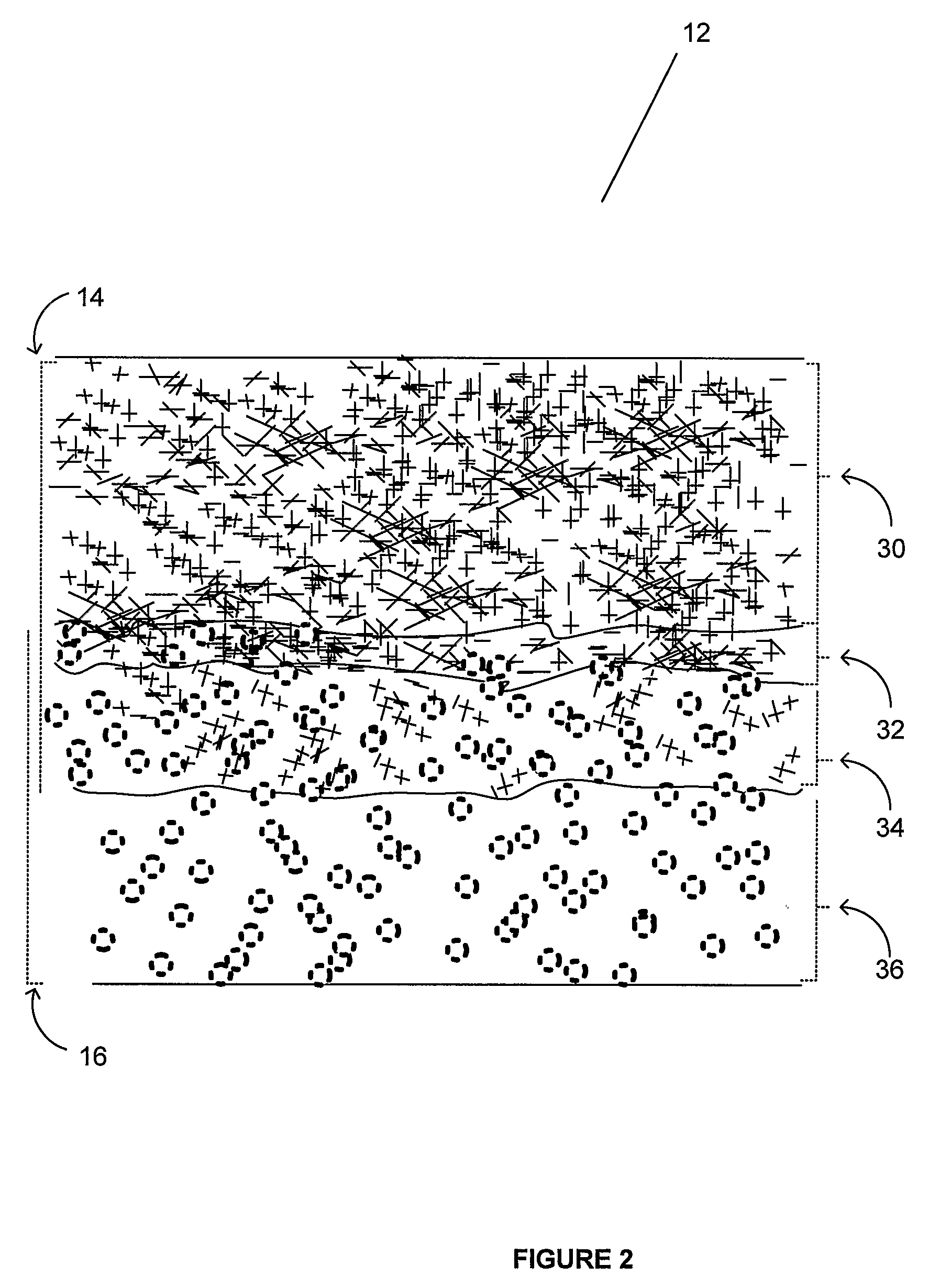
[0021]A novel composite having a layered structure is disclosed along with a device based on it for preparing a fluid fraction from whole blood. The novel composite comprises a fibrous separation medium impregnated into one side with a reinforcing material. The fibrous separation medium is anisotropic. Without intending to be bound by theory, it presents a first pore size in a first direction and a smaller pore size in a second different direction. The impregnating reinforcing material also reduces the pore size in a portion of the fibrous separation medium due to compaction in the impregnated part of the fibrous separation medium upon pressing in of the reinforcing material. The reinforcing material is preferably a hydrophilic polyester material that is bonded to the fibrous separation medium with a hot melt adhesive. In a preferred embodiment the hydrophilic polyester material is non-woven and the preferred hot melt adhesive is an ethylene vinyl acetate based web.
[0022]The composite may further comprise a semi-permeable or impermeable film that reinforces the composite and increases its stiffness. The thickness of the semi-permeable or impermeable film may be selected to achieve a particular degree of stiffness. In an preferred embodiment of the present invention, such a composite may comprise a film that is about 1.5 mils thick, less than about 5 mils thick, about 8 mils thick, about 10 mils thick, about 11 mils thick, about 12 mils thick, about 13 mils thick, about 14 mils thick, about 15 mils thick, and at least about 16 mils thick. Preferably the film include a hot melt adhesive coating that comprises ethylene vinyl acetate. Example films are supplied by TRANSILWRAP COMPANY, INC. of Chicago, Ill., USA. This novel composite comprising this secondary reinforcement is stiffer and easier to machine for high-speed manufacturing. The secondary reinforcement may be carried out in a single step with the reinforcement of the fibrous separation medium with the reinforcing material or, alternatively, in a separate step.

Problems solved by technology

Although whole blood is obtained relatively easily and in principle is capable of providing valuable information, its use is limited by problems in handling it for use in reliable rapid diagnostic assays.
For example, if a diagnostic assay is based on a colorimetric reaction, hemolysis of red blood cells introduces errors.
Even when the readings are not affected by colors contributed by hemolysis, the presence of cell lysates and its contribution variable recovery of the fluid fraction is a source of error.
The use of whole blood in the case of certain rapid diagnostic assays creates special problems.
For example, if the diagnostic assay is based on a calorimetric reaction, turbid or colored solutions, such as from red blood cells, disturb the readings.
In particular, it is desirable to separate cellular components from the fluid fraction of whole blood in the absence of hemolysis because hemolysis causes components of red blood cells to be released into the fluid fraction and potentially interfere with downstream applications.
However, higher molecular weight components of the blood, for example lipids or substrates bound to serum proteins, for example bilirubin, cannot be determined in this way because they are also not able to penetrate into the film or to pass through the semi-permeable membrane.
A disadvantage of certain filter systems is that blood penetrates through the membrane filter very slowly.
However, especially in the case of using small amounts of sample, such as 50 microliters or less, this gives rise to problems and the separation of supernatant and precipitated cellular components.
Further, conventional methods require more handling time by the doctor, nurse, technician, or tester.
Such additional handling is generally undesirable for efficiency as well as hygienic reasons.
Moreover, in some point of care situations, a centrifuge may not be available.
However, since the erythrocytes can pass through the filter along with the plasma, these filters are not suitable for obtaining plasma because the erythrocytes can interfere with the downstream application, such as a calorimetric test.
A potential drawback of these glass fiber-based separators is that the separation of blood cells from whole blood often does not occur without hemolysis.
Due to the hemolysis, certain components of erythrocytes contaminate the recovered fluid fraction, which may interfere with downstream applications.
None of the known methods teach demonstrably gentle methods for trapping red blood cells in a region where the blood sample is applied with little to no migration of red-blood cells while the fluid fraction is separated efficiently.
These separators also do not readily release the fluid fraction.
In another aspect, polysulfone based materials also require multiple steps during manufacturing and are expensive compared to glass fiber papers.
The methods also rely upon speedy removal of any recovered fluid fraction since red blood cells eventually reach and increasingly contaminate the recovered fluid fraction, both via direct contamination and due to hemolysis over time.
Significantly, the known methods and devices are not amenable to automation since, the most effective separation medium used therein, glass fibers, are brittle and difficult to handle.
Thus cutting the material into specific and accurate shapes and packaging thereof, often requires significant amount of manual labor rather than automated handling.

Method used


Image

  • Blood Separator and Method of Separating a Fluid Fraction from Whole Blood
  • Blood Separator and Method of Separating a Fluid Fraction from Whole Blood
  • Blood Separator and Method of Separating a Fluid Fraction from Whole Blood

Examples


Embodiment Construction

5.1 Composite:

[0061]In one aspect, the invention provides a novel composite comprising a fibrous separation medium impregnated with a reinforcing material. The reinforcing material is a hydrophilic polyester material. In a preferred embodiment the hydrophilic polyester material is non-woven.

[0062]According to one embodiment of the invention, the composite comprises a fibrous material with a multimodal distribution of fibers having a plurality of clusters having substantially aligned short large-diameter fibers and a plurality of clusters having substantially aligned long small-diameter fibers, wherein there are fewer of the short large-diameter fibers than the long small-diameter fibers; and a reinforcing material impregnating a bottom side of the fibrous material. Surprisingly, the present inventors have found that a family of products used as storage battery separators, e.g., those manufactured by the Whatman Company, including but not limited to Whatman glass fiber papers identif...


PUM

PropertyMeasurementUnit
Lengthaaaaaaaaaa
Lengthaaaaaaaaaa
Lengthaaaaaaaaaa

Abstract

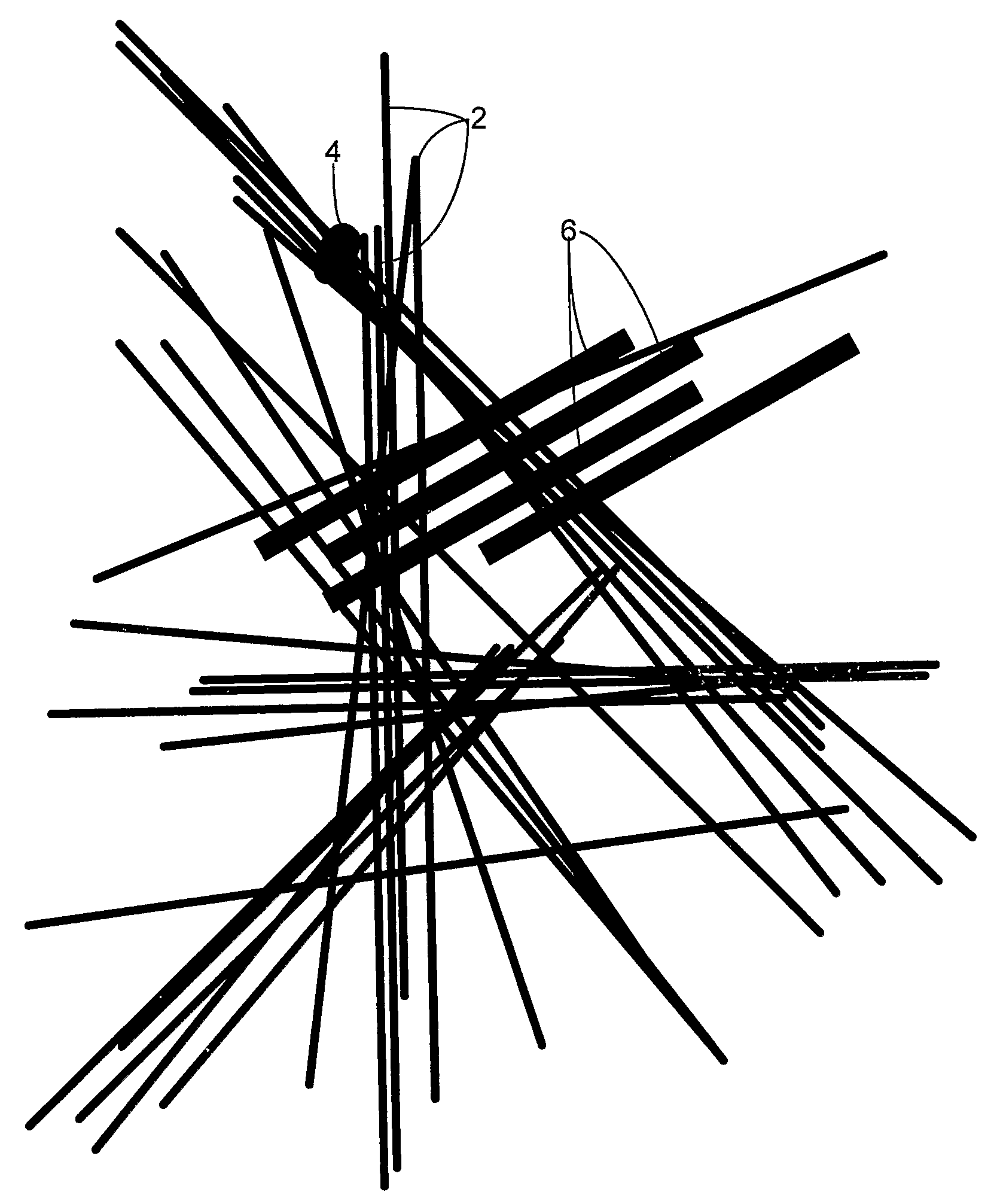
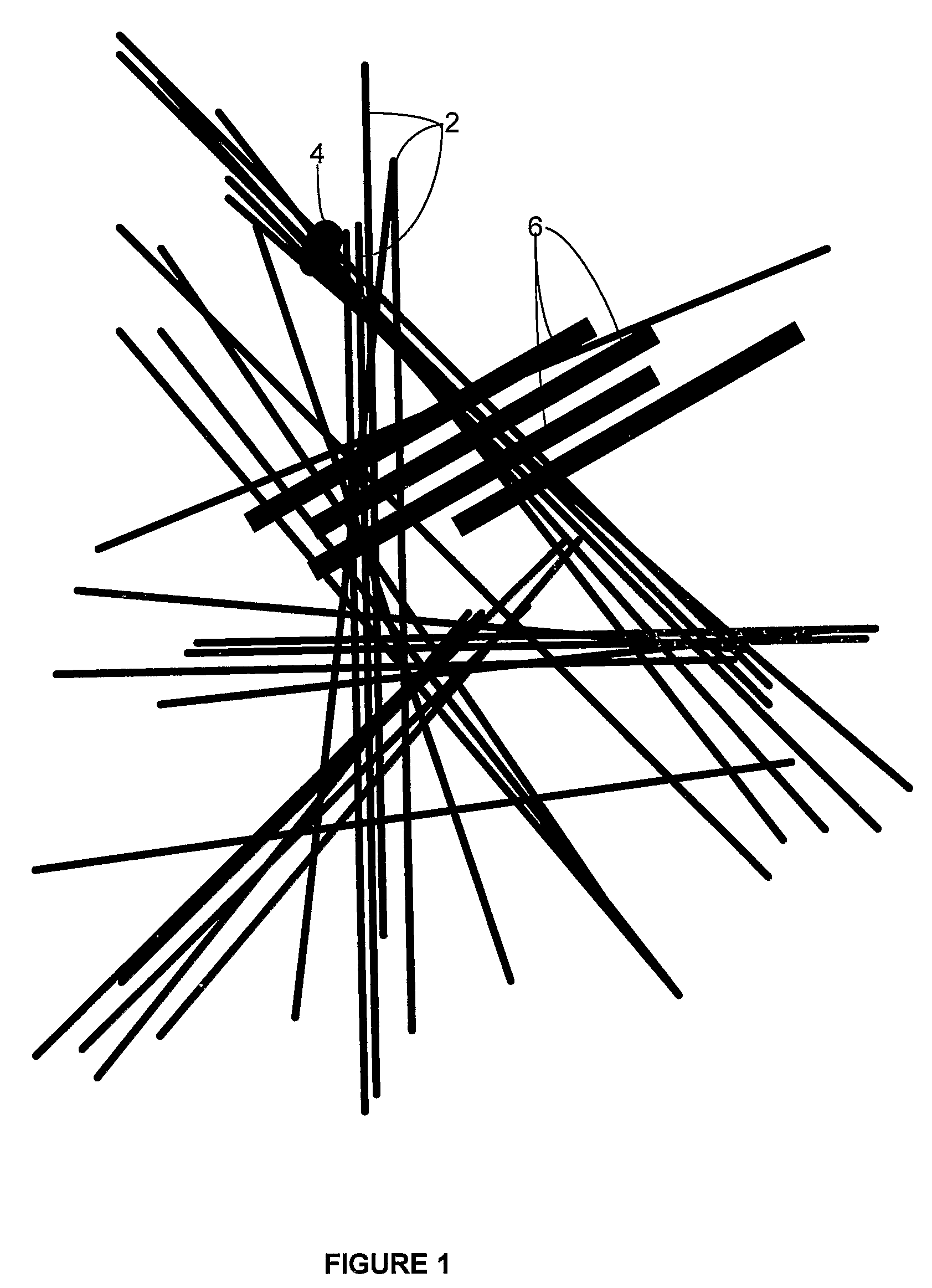
A novel composite of glass fiber paper and reinforcing material that is useful for high speed manufacturing of devices comprising it is disclosed. The composite is formed by pressing into a glass fiber paper, having multimodal (at least bi-modal) composition, a hydrophilic reinforcing material. The composite has anisotropic separation properties that are particularly useful for preparing a fluid fraction from whole blood. The preferred glass fiber paper comprises glass fibers such that there are a plurality of short fibers (6) having large diameters and long fibers (2) with smaller diameters. The preferred glass fiber paper also exhibits partial ordering. There are many more of the long fibers (2) with smaller diameters than of the short fibers (6) having large diameters. Also disclosed are several blood separator device designs for efficiently separating the fluid fraction of blood from whole blood using the composite.

Description

1 FIELD OF THE INVENTION[0001]The present invention relates to a novel composition of matter which is a composite material comprising partially ordered glass fiber paper impregnated, on one side with a hydrophilic polyester material. Preferably, the hydrophilic material is non-woven. The present invention also relates to a blood separator for separating blood fluids, whether serum or plasma, from blood cells in a sample, such as a sample of blood. More particularly, the present invention relates to an improved blood separator that allows for rapid separation of the fluid fraction from cellular components in even very small volumes of blood samples.2 BACKGROUND OF THE INVENTION[0002]The separation of the fluid fraction, whether serum or plasma, from blood is of great importance in the clinic. Blood is made up of two fractions: blood cells, and the liquid in which they are suspended, blood plasma (“plasma”), a proteinaceous fluid. The important cell types encountered in blood are the ...

Claims


Application Information

Patent Timeline
IPC IPC(8): B01D39/16B32B5/08B32B38/08D04H1/559
CPCB01D39/2017Y10T156/109G01N33/491D21H13/40Y10T442/2008
Inventor MILUNIC, DAVIDRUSSELL, DONALD
Owner AKERS BIOSCI
Learn More