
Methods, Reagents and Instrumentation for Preparing Impregnated Tissue Samples Suitable for Histopathological and Molecular Studies

a tissue sample and histopathological technology, applied in the field of histopathological and molecular studies, can solve the problems of affecting the morphology of frozen tissue samples, affecting the morphology of paraffin-embedded tissue samples, and severely compromising gene expression data, and achieve the effect of low melting point paraffin

Inactive Publication Date: 2008-11-06
HISTOGENEX NV
View PDF22 Cites 17 Cited by
  • Summary
  • Abstract
  • Description
  • Claims
  • Application Information

AI Technical Summary

Benefits of technology

The present invention provides a method for preparing a tissue sample for morphological and molecular analysis, which involves fixing the sample with a fixative solution, dehydrating and clearing it using a dehydration-clearing solution, and infiltrating it with an inert specimen matrix. The method includes the use of a cross-linker and an excess of organic solvent in the fixative solution, as well as a pH modifying substance. The resulting sample can be used for both morphological and immunohistochemical analysis. The method also provides a kit for preparing the sample, including the necessary solutions and an instrument for automation. The invention also provides a paraffin-embedded sample that can be used for molecular analysis. The technical effects of the invention include improved tissue preservation and reduced damage to the sample, making it easier to prepare for analysis.

Problems solved by technology

Paraffin-embedded tissue samples generally show good morphology but gene expression data are severely compromised.
Conversely, frozen tissue samples remain the gold standard for obtaining high quality gene expression information, but tissue morphology from frozen material is inferior compared to the morphology of paraffin-embedded tissue samples.
Given the empirical foundation of microscopic analysis for current methods of tissue diagnosis and prognosis, the transfer of molecular methods into pathology practice has been greatly hampered.
However, as known by those persons skilled in the art, the freezing process results in suboptimal microscopical detail of tissue architecture and cell morphology.
Moreover transport of frozen tissues requires specialist shipment and is more costly and risky (chance of defrosting) than transporting paraffin-embedded material.
Sometimes, international sample carriage is not possible.
Another problem in the art is the limited tissue biopsy availability.
A single small-sized biopsy (e.g. needle biopsy) may not provide sufficient tissue for both classical histopathological analyses and molecular pathology assays.
Consequently, it may be required to collect two tissue samples which causes additional distress and injury to the patient.
However, prolonged exposure of tissues to the fixative formaldehyde results in irreversible cross-linking of proteins and nucleic acids, causing the maximum PCR amplicon size to be limited (Finkelstein S D, Sayegh R, Christensen S, Swalsky P A: Genotypic classification of colorectal adenocarcinoma.
In addition extensive fixation in formaldehyde leads to nucleic acid scission, further diminishing the efficiency of PCR-based analysis and amplicon size.
Although DNA survives fixation and embedding reasonably well, RNA content is seriously decreased due to the combination of the presence of RNase activity in virtually all tissues and the use of excessive heating during the infiltration and embedding procedures of the tissue processing.
Indeed, it is more difficult to obtain high molecular weight RNA from (archival) paraffin-embedded material.
However, such RNA fragment sizes severely limit the suitability of the RNA for certain molecular profiling applications such as the construction of full length cDNA libraries.
Even if the average RNA fragment size would be sufficiently large to allow paraffin-embedded tissues or cells to be used for RT-PCR, nucleic acid amplification procedures and microarray analyses, the reliability and reproducibility of quantitative gene expression studies are questionable in the presence of degraded and chemically modified RNA, especially since the different mRNA species from the mRNA pool are most likely not affected to the same degree / extent.
However, the resulting tissue blocks must be sectioned and processed differently from routine paraffin blocks which may complicate the work-flow in a routine pathology lab.

Method used


Image

  • Methods, Reagents and Instrumentation for Preparing Impregnated Tissue Samples Suitable for Histopathological and Molecular Studies
  • Methods, Reagents and Instrumentation for Preparing Impregnated Tissue Samples Suitable for Histopathological and Molecular Studies
  • Methods, Reagents and Instrumentation for Preparing Impregnated Tissue Samples Suitable for Histopathological and Molecular Studies

Examples


example 1

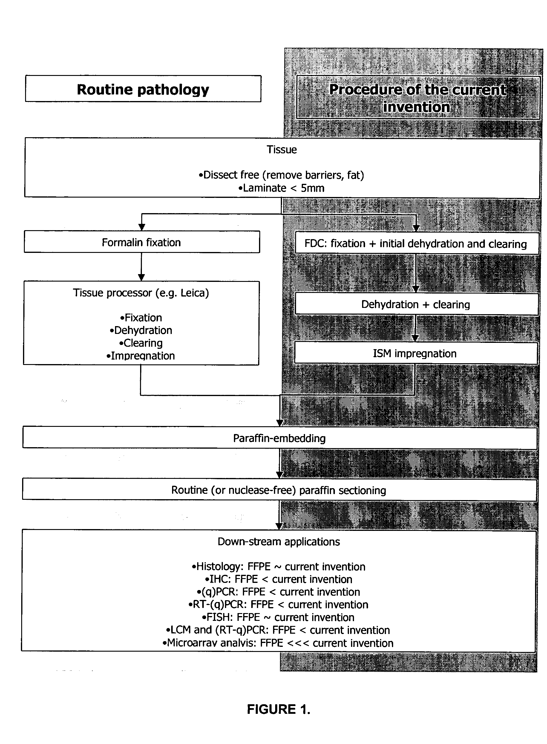
Exemplary General Manner of Tissue Processing

[0214]Tissue samples to be processed according to the current invention should be processed as quickly as possible after excision from the source after the onset of ischemia or death, or after removal from the soil or any other substrate solution or medium. In the case the samples posses a protective barrier that would interfere with FDC diffusion (e.g. a renal capsule or the waxy coating of plant material) this barrier should be removed prior to proceeding with the processing as described in the current invention. Large samples should be dissected / laminated into smaller fragments to maximize FDC diffusion in the first step of the present method. A general rule of thumb states the tissue thickness should not exceed 5 mm in at least one spatial dimension to allow proper fixation (Kiernan J P: Histopathological and Histochemical Methods, Theory, and Practice, ed 3. Oxford, Butterworth-Heinemann, 1999). Samples that consist of cell cultures ...

example 2

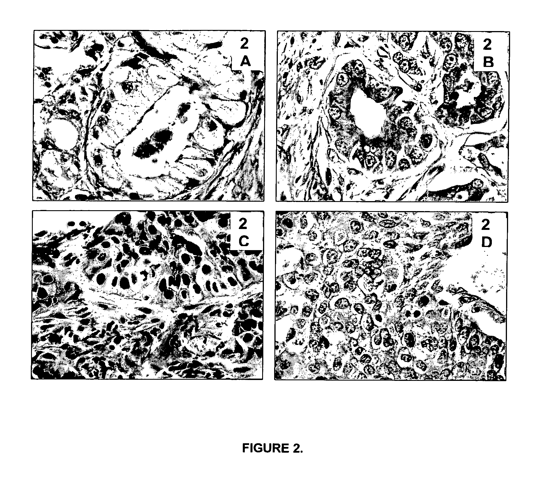
[0215]The tissue morphological detail and cytology of tissues processed according to the current invention is as good as or better than FFPE tissues (e.g. prostate tissue and bronchoscopic biopsy).

[0216]A prerequisite for the general acceptance and introduction of the current invention into routine pathology, research and clinical laboratories is that tissues processed according to the described invention have a morphology comparable or superior to the one of FFPE tissues.

[0217]Typical examples that demonstrate the importance of preservation of good morphological detail for histopathological diagnosis are prostatic and bronchoscopic biopsies. Prostatic (FIGS. 2A and 2B) and bronchoscopic biopsies (FIGS. 2C and 2D) are shown. FIGS. 2A and 2C show biopsies processed according to standard routine pathology procedures (4% formalin fixation for 16 hours at room temperature, followed by tissue processing in a Leica Tissue Processor). FIGS. 2B and 2D show biopsies processed according to th...

example 3

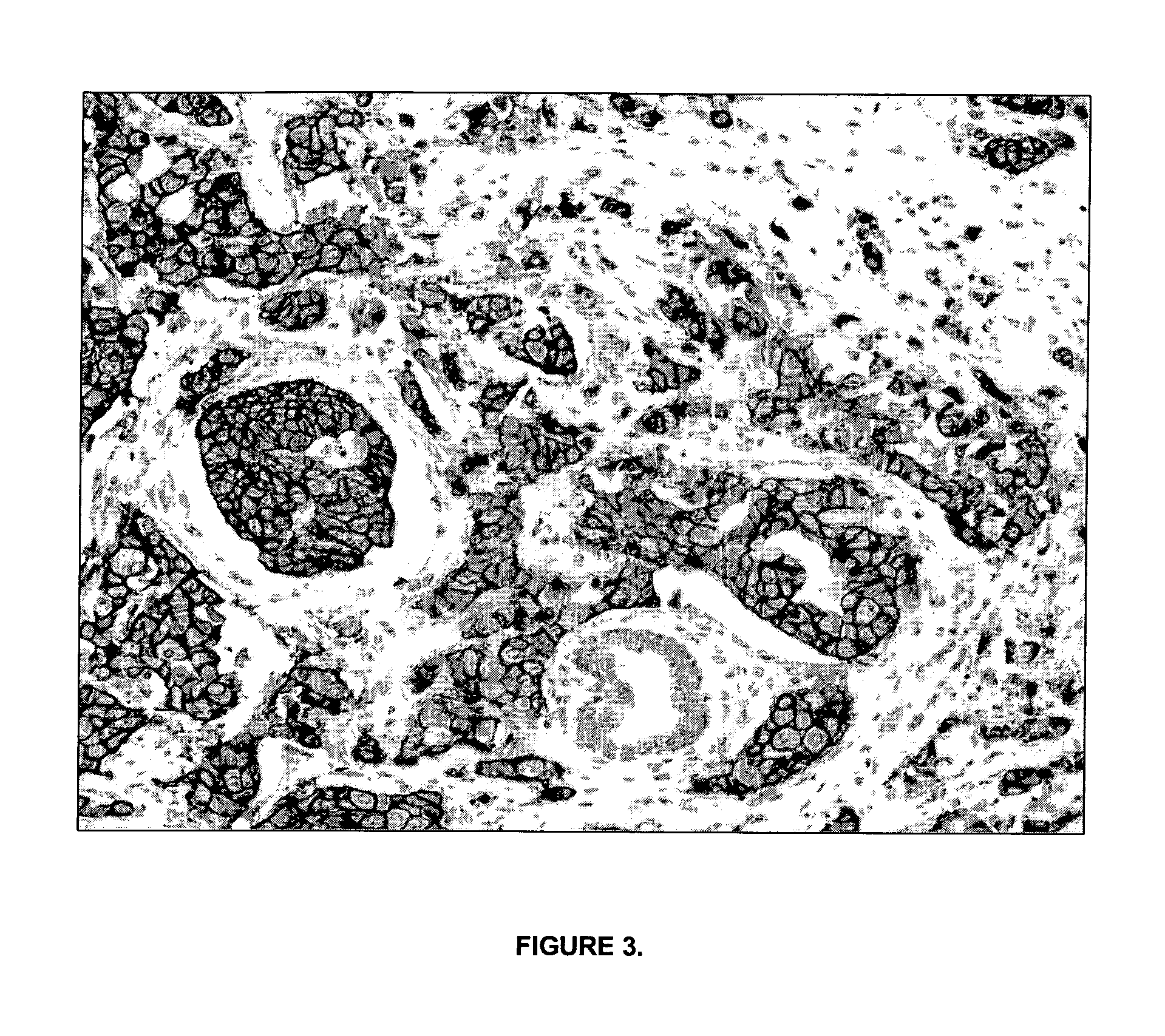
[0219]Immunohistochemical (IHC) stainings performed on tissue processed according to the described invention and embedded in paraffin.

[0220]The antigenic profile of the samples processed according to the current invention should be comparable to the one of FFPE tissues. In addition, the antigen retrieval procedures that are applied to FFPE tissues should be applicable without any (or only minor) adaptations on material processed according to the described invention. Table 1 lists 85 antibodies (both monoclonal and polyclonal) that have been applied successfully on human surplus tissues processed according to the described invention. In total, 4239 sections were stained immunohistochemically and evaluated by pathologists, who found no differences between standard FFPE sections and sections from tissues processed according to the current invention. Data is shown in Table 1.

Number of IHCAntibody targetstainings performedType of antibody□-1-antitrypsin10P□-actin, sarcomeric6M□-smooth mu...


PUM

PropertyMeasurementUnit
v/vaaaaaaaaaa
v/vaaaaaaaaaa
v/vaaaaaaaaaa

Abstract

A process and kit for the production of paraffin sections of biological tissue, especially for molecular pathology studies, is described. In the process, the tissue sample is simultaneously fixed, dehydrated and cleared in a first step, subsequently dehydrated and cleared in a second step and infiltrated with an inert specimen matrix in a third step. The specimen can then be further embedded in a casting supporting matrix according to the standard procedures followed by any local pathology or research laboratory. The method is uncomplicated, requires little hands-on time, is reproducible and is standardized thereby limiting cross-centre pre-analytical variance. A bio-indicator system for measuring the degree of crosslinking is also disclosed.

Description

BACKGROUND OF THE INVENTION[0001]Many researchers experience a trade-off between maintaining cell morphology and preserving gene expression information in the same tissue sample. Paraffin-embedded tissue samples generally show good morphology but gene expression data are severely compromised. Conversely, frozen tissue samples remain the gold standard for obtaining high quality gene expression information, but tissue morphology from frozen material is inferior compared to the morphology of paraffin-embedded tissue samples. Given the empirical foundation of microscopic analysis for current methods of tissue diagnosis and prognosis, the transfer of molecular methods into pathology practice has been greatly hampered. Indeed, the development of improved methods for tissue processing for transcript profiling of pathological samples are still necessary (Perlmutter M A, Best C J, Gillespie J W, Gathright Y, Gonzalez S, Velsaco A, Linehand W M, Emmert-Buck M R, Chuaqui R F: Comparison of sna...

Claims


Application Information

Patent Timeline
Patent Type & Authority Applications(United States)
IPC IPC(8): G01N1/28C12N5/06
CPCG01N1/31B01L2200/16G01N2001/315G01N1/30B01L3/563B01L3/508Y10T436/2575G01N2035/00782B01L2400/0487B01L2300/0816B01L2400/0683G01N1/36B01L2300/046B01L2300/049B01L2300/0609B01L3/523
Inventor DUYMELINCK, CARLAKOCKX, MARC
Owner HISTOGENEX NV
Learn More