Fluorescent Methods and Materials for Directed Biomarker Signal Amplification

a biomarker and signal amplification technology, applied in the direction of material analysis, instruments, solid-state devices, etc., can solve the problem of no such simple method for amplifying targeted materials

Active Publication Date: 2008-11-27
SIRIGEN
View PDF37 Cites 114 Cited by
  • Summary
  • Abstract
  • Description
  • Claims
  • Application Information

AI Technical Summary

Benefits of technology

[0032]In a further embodiment 1, and 2 can include one or more a-g linking groups having the structure:

Problems solved by technology

Comparatively, in the field of protein recognition, there are no such simple methods for amplifying the targeted materials.

Method used


Image

  • Fluorescent Methods and Materials for Directed Biomarker Signal Amplification
  • Fluorescent Methods and Materials for Directed Biomarker Signal Amplification
  • Fluorescent Methods and Materials for Directed Biomarker Signal Amplification

Examples


example 1

[0251]General protocol for the sandwich ELISA method with polymer-dye conjugated antibody:

[0252]1. Bind the unlabeled antibody to the bottom of each well by adding approximately 50 μL of antibody solution to each well (20 μg / m in PBS) in a 96 wells polyvinylchloride (PVC) microtiter plate. PVC will bind approximately 100 ng / well (300 ng / cm2). The amount of antibody used will depend on the individual assay.

[0253]2. Incubate the plate overnight at 4° C. to allow complete binding.

[0254]3. Wash the wells twice with PBS.

[0255]4. The remaining sites for protein binding on the microtiter plate must be saturated by incubating with blocking buffer. Fill the wells to the top with 3% BSA / PBS with 0.02% sodium azide. Incubate for 2 hrs. to overnight in a humid atmosphere at room temperature.

[0256]5. Wash wells twice with PBS.

[0257]6. Add 50 μL of the antigen (or sample) solution to the wells (the antigen solution should be titrated). All dilutions should be done in the blocking buffer (3% BSA / P...

example 2

General Protocol for Microarray Labeling with Polymer-Dye Conjugated Antibody

[0266]1. Prepare total RNA or mRNA.

[0267]2. Use T7-oligo(dT) primer to perform one-cycle or two-cycle cDNA synthesis.

[0268]3. Cleanup of double stranded cDNA.

[0269]4. Use IVT (in vitro transcription) amplification kit to incorporate biotin-labeled ribonucleotide into cRNA.

[0270]5. Fragmentation of cRNA.

[0271]6. Hybridize cRNA fragments on chip.

[0272]7. Wash off residual cRNA and stain the chip with streptavidin-polymer-dye conjugate (Example B or D)

[0273]8. Wash off residual reagents on chip.

[0274]9. Scan microarray.

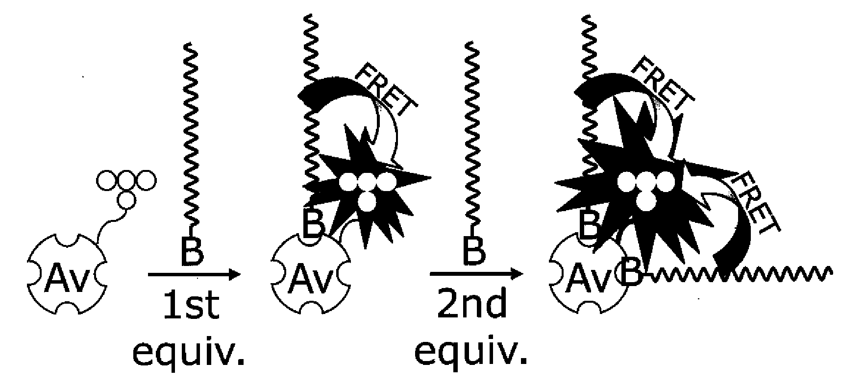
[0275]In the regular practice of microarray methodology, an integration of biotin-labeled nucleotides into the cRNA sequences is the means of sequestering the streptavidin phycoerythrin conjugate and biotinylated anti-streptavidin antibody for amplified signal reporting. Due to the manufacture complexity of streptavidin phycoerythrin conjugate, the batch-to-batch variation is significant. Theref...

example 3

Synthesis of Cationic Conjugated Polymer with an Amine Functional Group, CA001

[0277]

[0278]1-(4′-Phthalimidobutoxy)-3,5-dibromobenzene: 3,5-dibromophenol (970 mg, 3.85 mmol) was recrystallized from hexanes. After removal of solvent, N-(4-bromobutyl)phthalimide (1.38 g, 4.89 mmol), K2CO3 (1.88 g, 13.6 mmol), 18-crown-6 (53 mg, 0.20 mmol), and acetone (20 mL) were added. This was refluxed for 1 hour, and then poured into 100 mL of water. The aqueous layer was extracted with dichloromethane (4×30 mL). The organic layers were combined, washed with water, saturated NaHCO3, and brine, then dried over MgSO4 and filtered. Removal of solvent yielded a white solid, which was purified by column chromatography (4:1 hexanes:CH2Cl2) followed by recrystallization in hexanes to yield colorless needles (650 mg, 87%). 1H NMR (CDCl3): 7.860 (m, 2H); 7.733 (m, 2H); 7.220 (t, J=1.6 Hz, 1H); 6.964 (d, J=2.0 Hz, 2H); 3.962 (t, J=6.0 Hz, 2H); 3.770 (t, J=6.6 Hz, 2H); 1.846 (m, 4H).

[0279]Poly[(2,7-{9,9-bis(6...


PUM

PropertyMeasurementUnit
temperaturesaaaaaaaaaa

Abstract

Methods and compositions are provided that include a multichromophore and/or multichromophore complex for identifying a target biomolecule. A sensor biomolecule, for example, an antibody can be covalently linked to the multichromophore. Additionally, a signaling chromophore can be covalently linked to the multichromophore. The arrangement is such that the signaling chromophore is capable of receiving energy from the multichromophore upon excitation of the multichromophore. Since the sensor biomolecule is capable of interacting with the target biomolecule, the multichromophore and/or multichromophore complex can provide enhanced detection signals for a target biomolecule.

Description

CROSS-REFERENCE[0001]This application claims the benefit of U.S. Provisional Application No. 60 / 825,615, filed Oct. 6, 2006, which application is incorporated herein by reference.BACKGROUND OF THE INVENTION[0002]Fluorescent hybridization probes have developed into an important tool in the sequence-specific detection of DNA and RNA. The signals generated by the appended fluorescent labels (or dyes) can be monitored in real time and provide simple, rapid, and robust methods for the detection of biological targets and events. Utility has been seen in applications ranging from microarrays and real time PCR to fluorescence in situ hybridization (FISH).[0003]Recent work in the area of multichromophores, particularly regarding conjugated polymers (CPs) has highlighted the potential these materials have in significantly improving the detection sensitivity of such methods (Liu and Bazan, Chem. Mater., 2004). The light harvesting structures of these materials can be made water soluble and ada...

Claims


Application Information

Patent Timeline
Patent Type & Authority Applications(United States)
IPC IPC(8): G01N33/536C08G63/48
CPCG01N33/542G01N33/582Y10T436/13C08G2261/3142H01L51/0039C08G61/02H10K85/115C08G61/12C08G2261/3221C08G2261/3241C08G2261/3246C08G2261/522C08G2261/78C08G2261/94
Inventor GAYLORD, BRENT S.HONG, JANICE W.FU, TSU-JUSUN, CHENG-JUNBALDOCCHI, RUSSELL
Owner SIRIGEN
Why Eureka
Try Eureka