Apparatus and methods for mapping retinal function

a retinal function and retinal potential technology, applied in the field of electrophysiological neuroimaging, can solve the problems of difficult to administer psychophysical tests to patients with very low vision, inability to administer psychophysical tests to animals, and inability to spatially uniformize the potentials on the eye surface resulting from bioelectric activity of the retina

Active Publication Date: 2008-11-27
THE BOARD OF TRUSTEES OF THE UNIV OF ILLINOIS
View PDF15 Cites 54 Cited by
  • Summary
  • Abstract
  • Description
  • Claims
  • Application Information

AI Technical Summary

Benefits of technology

[0033]The present invention provides an electrode array device for simultaneously detecting electrical potentials at five or more locations on the anterior surface of an eye. The electrode array device comprises a dielectric contact lens substrate, e.g., a suitable polymeric material such as poly(methyl methacrylate), having a concave inner surface conforming to the anterior surface of the eye, and at least five (5) recording electrodes positioned in relation to the inner surface of the lens substrate so as to make electrical connection with the anterior surface of the eye when the lens substrate is contacted with or placed on the anterior surface of eye. Each recording electrode is in electrically conductive communication with a corresponding conductive contact pad, there being one conductive contact pad (or “contact”) for each active electrode. Each conductive contact is electrically insulated from the anterior surface of the eye, and is adapted for operable connection to a signal processor that preferably includes an amplifier and is capable of detecting and amplifying an electrical potential signal from each electrode. Preferably, the lens substrate includes 5 to 500 recording electrodes. The electrodes and contact pads can be made from a metal, such as a noble metal (i.e., a corrosion resistant metal of groups VIIb, VIII, and Ib of the second and third transition series of the periodic table, preferably gold, platinum or iridium), as well as from a conductive polymer, or a semiconductor material (e.g., a doped silicon). If desired, the lens can include an integral vacuum port or line so that a mild negative pressure can be applied to the undersurface of the lens, to help anchor the lens to the surface of the eye, and prevent movement during measurement of electrical potentials on the eye surface.

Problems solved by technology

However, this psychophysical test has several drawbacks including difficulty in administering the test to young patients or patients with very low vision, and the fact that it measures quality of visual perceptions, and does not directly reflect function at the retina.
Further, such psychophysical tests cannot be administered to animals.
It has long been known that the potentials on the eye surface resulting from bioelectric activity of the retina are not spatially uniform.
This is seen as a complication to ERG recording, and is usually mitigated by using an active electrode that contacts the eye surface over an extended area.
However, for any non-zero difference in eye surface potential along one axis, there are an infinite number of possible distributions of underlying retinal activity that could result in that difference.
Therefore, the size and location of a retinal lesion cannot be determined using the method proposed by Cringle.
Theoretically, it is not even possible to obtain such location and size information from the output of the system described by Cringle, because the underlying computational problem is under-constrained.
There simply is not enough information to make such calculations from the type of data obtained by the method and apparatus of Cringle, as explained below.
The simple difference measurements described by Cringle only determine the amount of asymmetry in the distribution of retinal activity, and cannot be used to determine the size and location of a retinal lesion.
These methods also have significant drawbacks.
The mfERG takes several minutes to record, during which the subject must fixate on a small target, making it difficult to record from very young patients or patients with low central vision.
Further, the mfERG signal is not a true bioelectric signal, and physiological interpretation of the signal remains a challenge.
As noted above, eye diseases often result in localized dysfunction of the retina.

Method used


Image

  • Apparatus and methods for mapping retinal function
  • Apparatus and methods for mapping retinal function
  • Apparatus and methods for mapping retinal function

Examples


second embodiment

[0089]A second embodiment is similar to that described above, but where the contact lens extends past the corneal margin (FIGS. 6, 7, 8, 14) onto the sclera. In this embodiment, a spherical section of lens material with radius of curvature appropriate to fit the subject's sclera extends the lens past the corneal margin. In this embodiment, this scleral extension contains a plurality of recording electrodes. The contact pads associated with each electrode are present on the convex surface of the scleral extension, and preferably are connected to each electrode by way of an insulated conductive trace as described above.

third embodiment

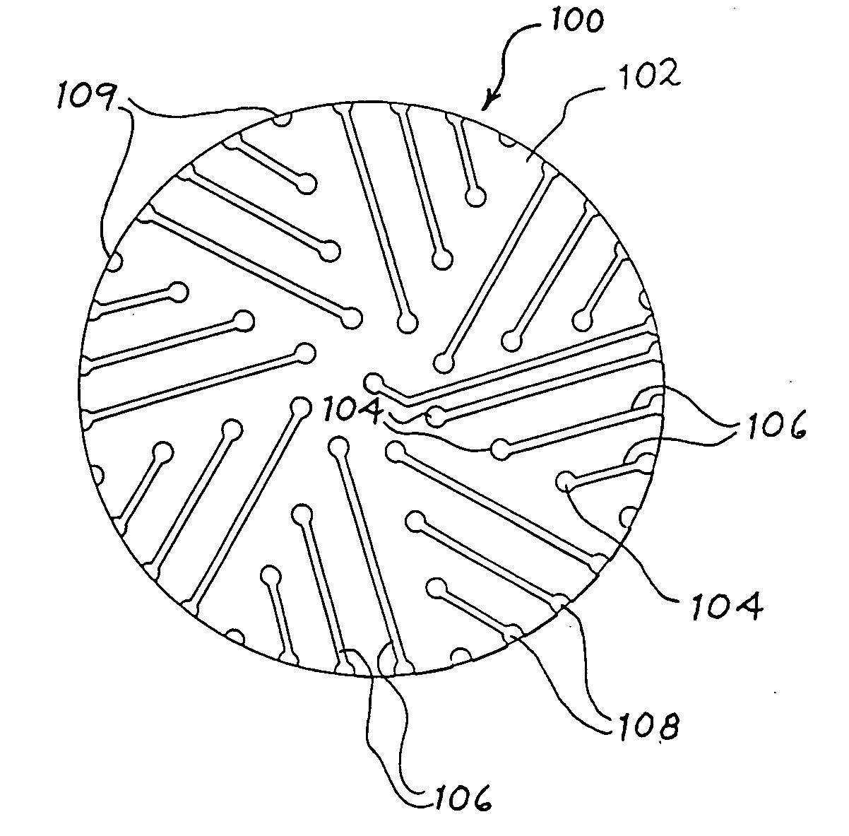
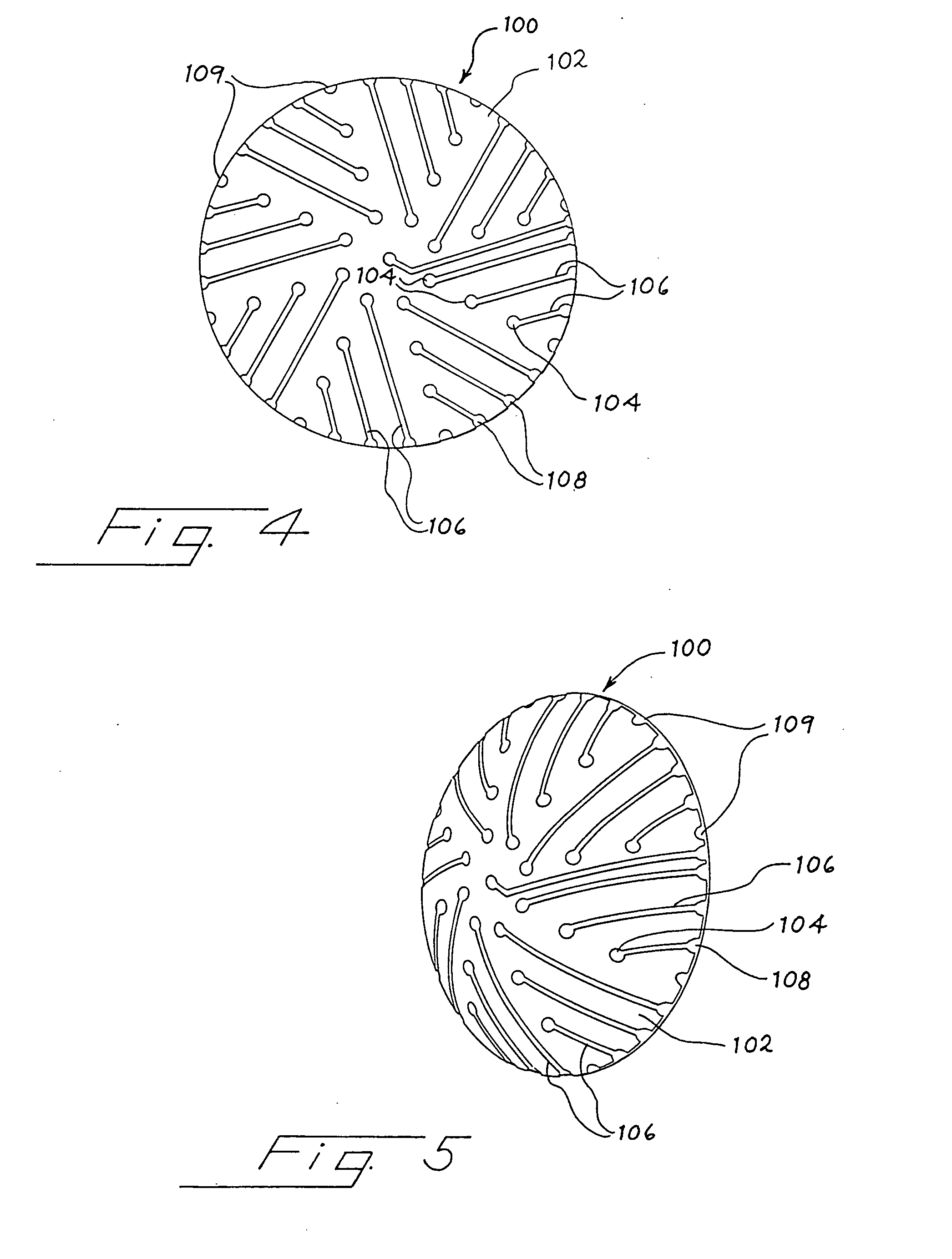
[0090]A third embodiment consists of a contact lens appropriate to fit the subject's cornea (FIGS. 12, 13), or the cornea and anterior sclera (FIG. 14). FIG. 12 shows two views of a portion of a contact lens 500 adapted to include a plurality of recording electrodes. Panel A shows a top plan view and Panel B shows a side cross-section. Through-holes 502 connect the corneal surface 503 of lens 500 with the distal side 505 of the lens 500 at each recording electrode location. In this embodiment, the recording electrode position comprises a through-hole 502, into which a conductive wire, a conductive hydrogel, or a conductive liquid can be placed as the electrode. Through-holes 502 can be lined by a conductive material, which can be in contact with a conductive trace attached to a contact pad.

[0091]FIG. 13 shows a contact lens of FIG. 12 in side cross-section, showing connections of the recording electrodes 504 to amplifiers 506 via contacts 512 and wires 511. Each amplifier 506 includ...


PUM


Abstract

The present invention provides an electrode array device for simultaneously detecting electrical potentials at five or more locations on the anterior surface of an eye. The device comprises a dielectric lens substrate having a concave inner surface conforming to the anterior surface of the eye, and at least five recording electrodes positioned in relation to the inner surface of the lens substrate so as to make electrical connection with the anterior surface of the eye when the lens substrate is placed on the anterior surface of eye. Each recording electrode is in electrically conductive communication with a corresponding conductive contact, there being one conductive contact for each recording electrode. Each conductive contact is adapted for operable connection to signal processor, and each conductive contact is electrically insulated from the anterior surface of the eye. A computational method for analyzing electrophysiological potentials recorded at five or more locations on the anterior surface of the eye, which reflect the spatial distribution of activity of the retina, is also provided.

Description

CROSS-REFERENCE TO RELATED APPLICATIONS[0001]This application is a continuation-in-part of U.S. Ser. No. 11 / 707,783, filed on Feb. 15, 2007, now U.S. Pat. No. 7,384,145, which claims the benefit of U.S. Provisional Application for Patent Ser. No. 60 / 774,097, filed on Feb. 16, 2006, each of which is incorporated herein by reference in its entirety.FIELD OF THE INVENTION[0002]The present invention relates to electrophysiological neuroimaging, also referred to as surface potential mapping, specifically as applied to the eye. More specifically, this invention relates to apparatus and methods for simultaneously determining electrical potentials at multiple locations on the surface of the eye, and for using the so-determined measurements of electrical potentials to calculate the magnitude of neural activity at specific locations on the retina. The resulting data can be used to map retinal activity, and can be used to study, diagnose, or monitor the progression of ocular diseases. The appa...

Claims


Application Information

Patent Timeline
Patent Type & Authority Applications(United States)
IPC IPC(8): A61B5/05
CPCA61B5/0496A61B5/398
Inventor HETLING, JOHN R.BAN, TAMASRAHMANI, SAFA
Owner THE BOARD OF TRUSTEES OF THE UNIV OF ILLINOIS
Why Eureka
Try Eureka