Method for the Treatment of Retinopathy of Prematurity and Related Retinopathic Diseases

a retinopathy and prematurity technology, applied in the field of retinopathy treatment methods, can solve the problems of ineffective prevention of visual loss, significant loss of visual function, and no treatment is currently available to specifically treat ocular vascular diseas

Inactive Publication Date: 2008-12-25
THE SCRIPPS RES INST
View PDF5 Cites 13 Cited by
  • Summary
  • Abstract
  • Description
  • Claims
  • Application Information

AI Technical Summary

Benefits of technology

[0023]A particular advantage of ocular treatments with the methods of the present invention is a vasculotrophic and neurotrophic rescue effect observed in eyes intravitreally treated with cells from the lineage negative he...

Problems solved by technology

For patients with choroidal neovascularization due to ARMD or inflammatory eye disease such as ocular histoplasmosis, photocoagulation, with few exceptions, is ineffective in preventing visual loss.
Since the retina consists of well-defined layers of neuronal, glial, and vascular elements, relatively small disturbances such as those seen in vascular proliferation or edema can lead to significant loss of visual function.
While significant progress has been made in identifying factors that promote and inhibit angiogenesis, no treatment is currently available to specifically treat ocular vascular disease.
Despite these observations, there are still no effective treatments to slow or reverse the progression of these retinal degenerative diseases.
In the normal adult, angiogenesis is tightly regulated, and is limited to wound healing, pregnancy and uterine cycling.
In these diseases, tissue damage can stimulate release of an...

Method used


Image

  • Method for the Treatment of Retinopathy of Prematurity and Related Retinopathic Diseases
  • Method for the Treatment of Retinopathy of Prematurity and Related Retinopathic Diseases
  • Method for the Treatment of Retinopathy of Prematurity and Related Retinopathic Diseases

Examples


example 1

Cell Isolation and Enrichment; Preparation of Murine Lin− HSC Populations A and B

[0128]General Procedure. All in vivo evaluations were performed in accordance with the NIH Guide for the Care and Use of Laboratory Animals, and all evaluation procedures were approved by The Scripps Research Institute (TSRI, La Jolla, Calif.) Animal Care and Use Committee. Bone marrow cells were extracted from B6.129S7-Gtrosa26, Tie-2GFP, ACTbEGFP, FVB / NJ (rd / rd mice) or Balb / cBYJ adult mice (The Jackson Laboratory, ME).

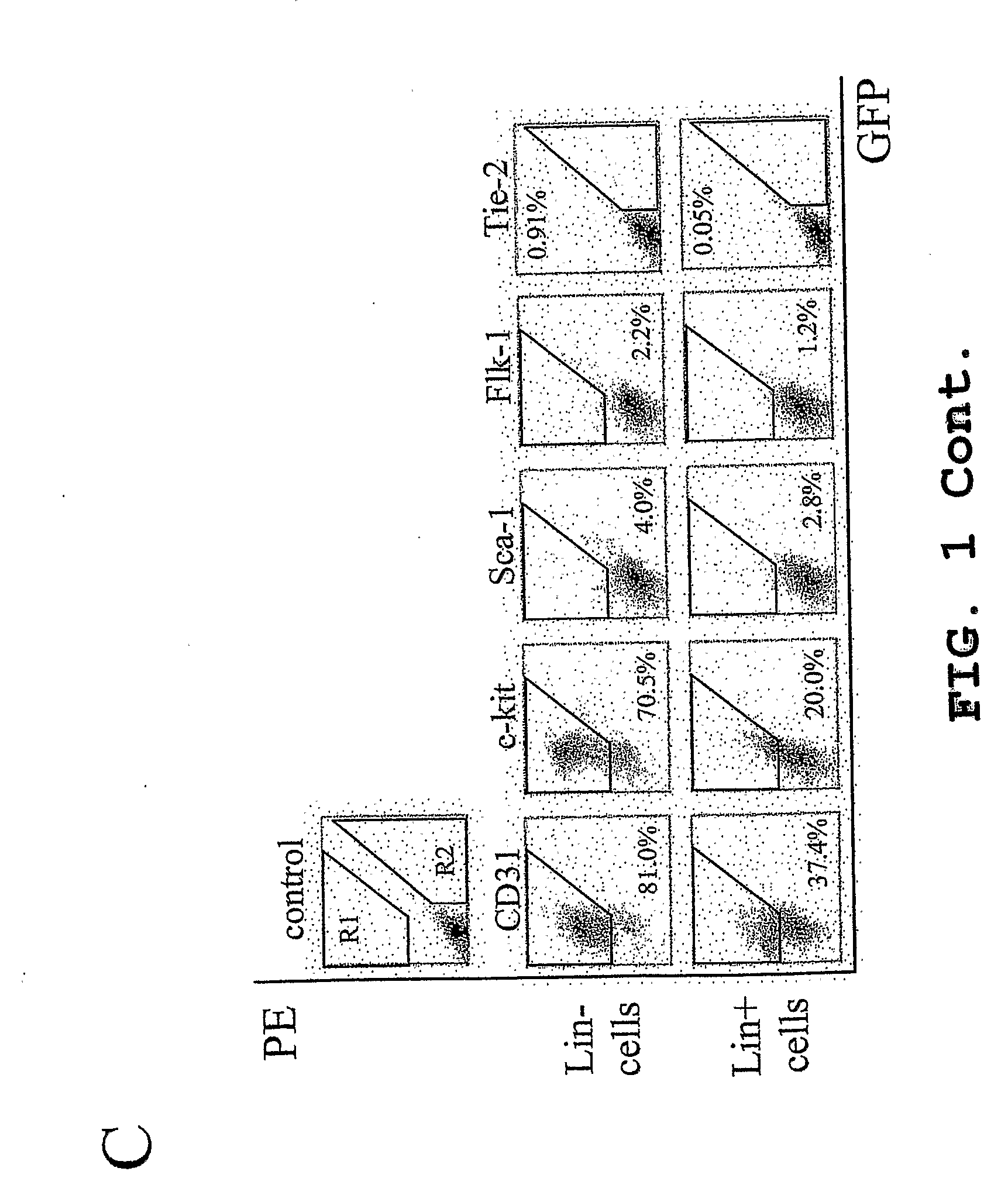
[0129]Monocytes were then separated by density gradient separation using HISTOPAQUE® polysucrose gradient (Sigma, St. Louis, Mo.) and labeled with biotin conjugated lineage panel antibodies (CD45, CD3, Ly-6G, CD11, TER-119, Pharmingen, San Diego, Calif.) for Lin− selection in mice. Lineage positive (Lin+) cells were separated and removed from Lin− HSC using a magnetic separation device (AUTOMACS™ sorter, Miltenyi Biotech, Auburn, Calif.). The resulting Lin− HSC population, containing en...

example 2

Intravitreal Administration of Cells in a Murine Model

[0133]An eyelid fissure was created in a mouse eyelid with a fine blade to expose the P2 to P6 eyeball. Lineage negative HSC Population A of the present invention (approximately 105 cells in about 0.5 μl to about 1 μl of cell culture medium) was then injected intravitreally using a 33-gauge (Hamilton, Reno, Nev.) needled-syringe.

example 3

EPC Transfection

[0134]Murine Lin− HSC (Population A) were transfected with DNA (SEQ ID NO: 1, FIG. 7) encoding the T2 fragment of TrpRS (SEQ ID NO: 3) also enclosing a His6 tag using FuGENE™ 6 Transfection Reagent (Roche, Indianapolis, Ind.) according to manufacturer's protocol. Lin− HSC cells (about 106 cell per ml) were suspended in Opti-MEM® medium (Invitrogen, Carlsbad, Calif.) containing stem cell factor (PeproTech, Rocky Hill, N.J.). DNA (about 1 μg) and FuGENE reagent (about 3 μl) mixture was then added, and the mixtures were incubated at about 37° C. for about 18 hours. After incubation, cells were washed and collected. The transfection rate of this system was approximately 17% as confirmed by FACS analysis. T2-TrpRS production was confirmed by western blotting. The amino acid sequence of His6-tagged T2-TrpRS is shown as SEQ ID NO: 2, FIG. 8.


PUM


Abstract

The present invention provides a method for treating retinopathy of prematurity (ROP) and related retinopathic diseases. The method comprises administering to the retina of a mammal suffering from, or at risk of developing, retinopathy of prematurity or a related retinopathic disease an amount of cells from a vasculotrophic lineage negative hematopoietic stem cell population, effective to promote beneficial physiological revascularization of damaged areas of the retina and to ameliorate damage to the retina caused by the disease. Preferably, the mammal is a human patient. In one preferred embodiment, the lineage negative hematopoietic stem cell population is a lineage negative hematopoietic stem cell population comprising hematopoietic stem cells and endothelial progenitor cells (i.e., Lin− HSC). In another preferred embodiment, the lineage negative hematopoietic stem cell population is an isolated myeloid-like bone marrow (MLBM) cell population in which the majority of the cells are lineage negative and express CD44 antigen and CD11b antigen. As an alternative, for treatment of newborn infants, a lineage negative hematopoietic stem cell population can be isolated from umbilical cord vein blood.

Description

CROSS-REFERENCE TO RELATED APPLICATIONS[0001]This application claims the benefit of U.S. Provisional Patent Application Ser. No. 60 / 656,122, filed on Feb. 24, 2005, which is incorporated herein by reference.STATEMENT OF GOVERNMENT INTEREST[0002]A portion of the work described herein was supported by grants number EY11254 and EY12598 from the National Eye Institute of the National Institutes of Health. The United States Government has certain rights in this invention.FIELD OF THE INVENTION[0003]This invention relates to methods for treating retinopathic diseases. More particularly, this invention relates to methods for treating retinopathy of prematurity and related retinopathic diseases by administering lineage negative hematopoietic stems cells to the eye of a mammal suffering from or at risk of developing said diseases.BACKGROUND OF THE INVENTION[0004]Vascular diseases of the retina, including diabetic retinopathy, exudative age related macular degeneration (ARMD), retinopathy of ...

Claims


Application Information

Patent Timeline
IPC IPC(8): A61K35/48A61K35/12A61P27/02C12N5/0789
CPCA61K31/137A61K48/00C12N2510/02C12N5/0647C12N2510/00A61K2035/124A61P27/02A61P9/00
Inventor FRIEDLANDER, MARTINBANIN, EYALAGUILAR, EDITH
Owner THE SCRIPPS RES INST
Why Eureka
Try Eureka