Coronary sinus access catheter with forward-imaging means

a catheter and coronary sinus technology, applied in the field of coronary sinus access catheters with forward-imaging means, can solve the problems of cellular death, discontinuation of rapid heart rate, and elusive opening, and achieve the effect of less rotation

Inactive Publication Date: 2009-03-12
OLYMPUS CORP
View PDF5 Cites 19 Cited by
  • Summary
  • Abstract
  • Description
  • Claims
  • Application Information

AI Technical Summary

Benefits of technology

[0027]The tight-radius deflection mechanism consists of one or two deflection wires pulling on a segment of the distal portion of the lead, creating deflections of about 60 degrees over the last centimeter of the catheter distal end. If two wires are used the deflection is bi-directional; one wire creates unidirectional deflections. If unidirectional deflection is used, the catheter can be torqued so that rotation on the proximal end results in a similar rotation on the distal end. The combination of rotating and deflecting permits the physician to navigate in 360 degrees about the catheter axis. The bi-directional system has the advantage of requiring less rotation to orient the catheter; the unidirectional deflection mechanism allows in a smaller catheter since only one wire is needed in the catheter. The deflection wire(s) is connected to a handle on the proximal end, which when manipulated, deflects the tip of the catheter.

Problems solved by technology

Alcohol contact results in cellular death and the discontinuation of the rapid heart rate.
1. Finding the opening (ostium) of the coronary sinus. Patients in CHF (congestive heart failure) have hypertrophied hearts, which alters the location and size of the coronary sinus. Physicians routinely place leads in the coronary sinus during routine EP (electrophysiologic) studies, however, they are dealing with normal-sized hearts with electrical conduction defects. With CHF patient candidates, finding the opening is much more elusive. Sometimes it is located significantly off-center from the normal location since the heart has hypertrophied. Other times, flaps of tissue prevent entry into the coronary sinus.
2. Advancing the lead through the coronary sinus to a branch in close proximity to the left ventricle so it can be chronically paced. Pacemaker implants are performed on the right side of the heart since implants in the left heart could lead to thrombi heaving deleterious consequences such as a stroke or heart attack. The coronary sinus is the only area of the heart anatomy by which a lead can be inserted from the right heart into close proximity to the left ventricle. In fact, the tip of the pacing lead needs to be within several millimeters of ventricular muscle to successfully pace the ventricle. The coronary sinus branches into segments, five of which traverse the left ventricle. Locating the proper left-ventricular branch (where the left ventricle can be chronically paced) has been difficult in biventricular pacing clinical studies. Hypertrophied hearts also alter the location and length of these branches. Finding the correct branch in these highly variable hearts has been the other major challenge in biventricular pacing.
3. Preventing the lead from dislodging in the first few months following implantation. Since the coronary sinus lead is not anchored in the coronary sinus and it is undergoing significant motion from the left ventricle beating vigorously, these leads have a high dislodgement rate of 10-20%. Dislodgement incidence is reduced if the lead is wedged far enough into a lateral branch of the coronary vein.
Using the coronary sinus lead to access the proper branch was difficult due to the size of the lead and the inability to make sharp-angled bends required to access a suitable coronary sinus branch.
The complexity in the curve geometries and stiffness characteristics of the above disclosures is due to the physician relying on “touch and feel” at the proximal end of the catheter.
The difficulty with pre-curved catheters is the extreme variability of coronary sinus location and geometry in hypertrophic hearts.
As a consequence of these limitations, implantation of a coronary sinus lead significantly increases the time of pacemaker implantation.
Biventricular pacers require 3-6 hours implantation time, simply because of the difficulty in implanting the coronary sinus lead.
Furthermore, the implantation success rate is only 80-90%, with cases abandoned because of inability to implant the coronary sinus lead.
Moreover, following the implant, coronary sinus leads are much more prone to lead dislodgement.
Coronary sinus leads dislodge because anchoring means such as tines or screws, commonly used in the right atrium and ventricle, cannot be used in the coronary sinus.
Magnetic resonance imaging systems are currently too slow to view in real-time, although future improvements may eventually render it a real-time imaging modality.
These systems would not be useful in the coronary sinus vasculature, since the mapping catheter must first be in the vicinity of a structure to allow the system to render an image of the structure.
However, in that respect they are no different than other feedback systems; they provide a real-time image of the relation of the catheter tip to the coronary sinus os.
These systems have not been employed to place coronary sinus catheters because of the length of time it takes to map the right atrium.
Visual feedback radically alters the design considerations for guide catheters.

Method used


Image

  • Coronary sinus access catheter with forward-imaging means
  • Coronary sinus access catheter with forward-imaging means
  • Coronary sinus access catheter with forward-imaging means

Examples


Embodiment Construction

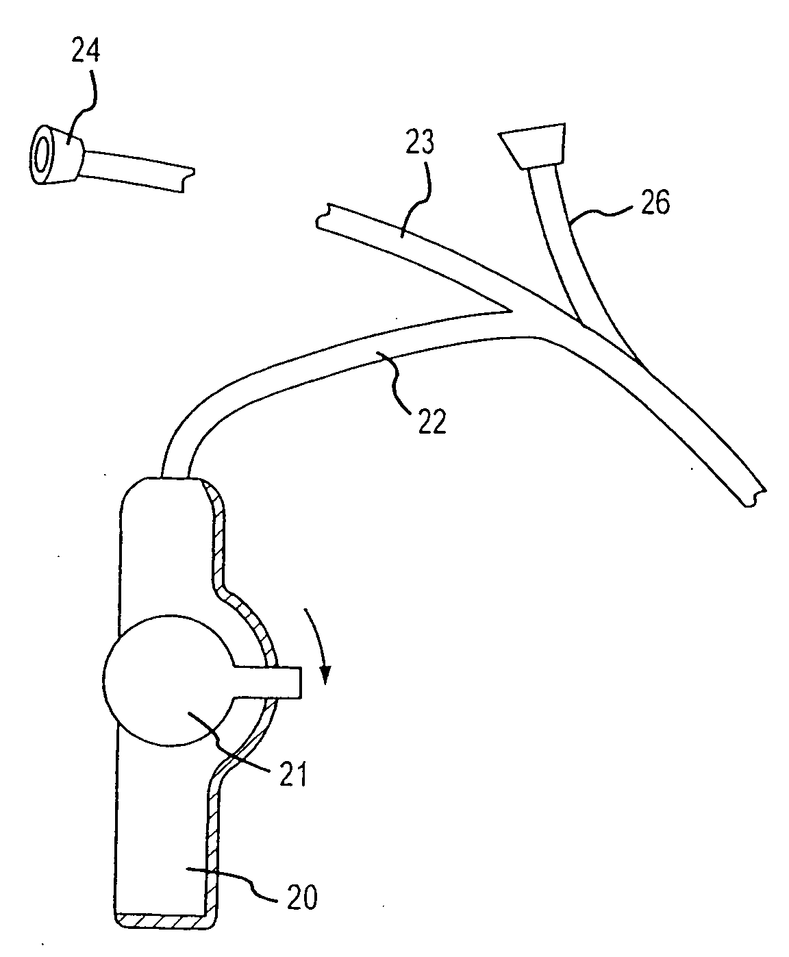
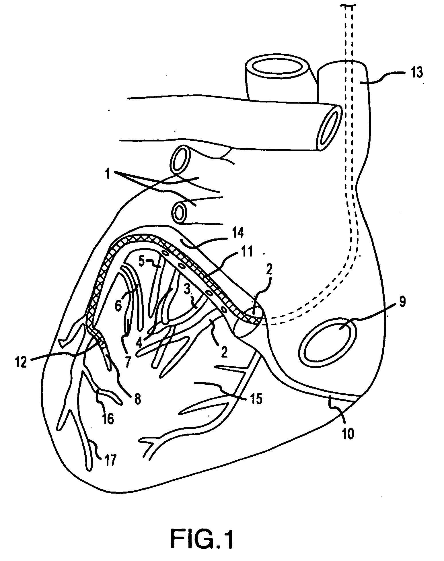
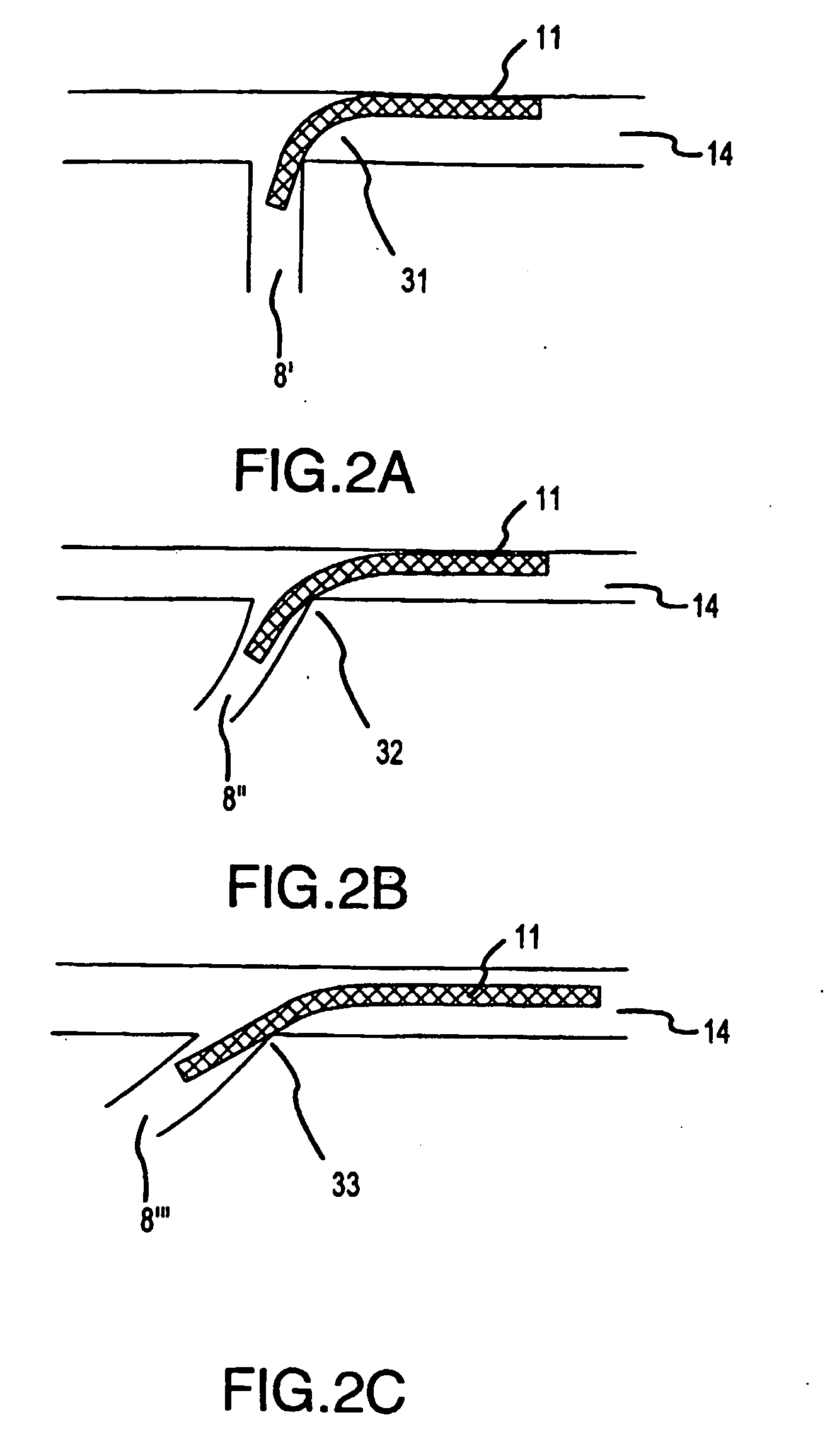
[0042]FIG. 1 shows the expected route of the catheter (11) as it is inserted onto the coronary sinus to a position in the anterior-lateral branch (8) of the coronary sinus vasculature. A sinus lead (12) is inserted through a puncture or cutdown technique into the subclavian vein where it eventually enters the superior vena cava (13). The lead is directed to the tricuspid valve plane in the lower right atrium where the os of the coronary sinus (1) is located. The os of the coronary sinus (1) is located near the tricuspid valve and the inferior vena cava (9). After entry into the coronary sinus os, the coronary sinus diverges into the great cardiac vein (14) and the right coronary vein (10). Directing the catheter (11) in the direction of the great cardiac vein (14) requires a tight radius deflection towards the left side of heart. As the catheter traverses the coronary sinus (1), several branch points called the posterior lateral coronary veins (2,3,4,5) run laterally along the left ...


PUM


Abstract

A coronary access catheter system simplifies the insertion of objects into distal branches of the coronary sinus. The system incorporates a real-time forward-imaging means to view the os and the branches of the coronary sinus. Preferably, the catheter uses near-infrared light as the forward-imaging means, but it could also include ultrasound or electromagnetic transducer. As the image is viewed, the catheter tip can be steered into the coronary sinus os and deflected in a tight radius bend on the distal end to navigate the short radius, right angle turns found in the coronary sinus branches. At that point, a flexible sheath can be placed over the guide catheter or objects such as guidewires can be inserted into channels of the guide catheter. The system consists of a catheter and image acquisition unit, which displays the forward image.

Description

[0001]This application claims the benefit of U.S. provisional patent application No. 60 / 332,654 filed on Nov. 9, 2002 and patent application Ser. No. 10 / 495,036 filed May 10, 2004.BACKGROUND OF THE INVENTION[0002]1. Field of the Invention[0003]This invention relates to cardiac catheters / introducers used to access the coronary sinus and navigate the sinus vasculature using a deflecting distal end with feedback provided by a forward-imaging means.[0004]2. Related Art[0005]The following references provide useful information in the filed of the present invention, and are incorporated by reference herein:Lurie 6 / 19955,423,772Adams 7 / 19955  433,729Jaraczewski 8 / 19955,445,148Toner 2 / 19965,488,960Avitall 7 / 19975,642,736Swoyer11 / 19975,683,445Randolph 7 / 19985,775,327Wang 3 / 20006,041,248Tockman10 / 20006,129,750Amundson 1 / 20016,178,346Lin 3 / 20016,200,269Williams 6 / 20026,408,214B1Zeylikovich 8 / 20026,437,867Ockuly10 / 20026,458,107B1[0006]Cardiac catheterizations are procedures in which a cardiologi...

Claims


Application Information

Patent Timeline
Patent Type & Authority Applications(United States)
IPC IPC(8): A61B8/14A61B1/04A61B5/00A61B8/12
CPCA61B5/0084A61B8/12A61B5/0086
Inventor AMUNDSON, DAVIDHANLIN, H. JOHNBLANKENSHIP, LARRY
Owner OLYMPUS CORP
Why Eureka
Try Eureka