
Compositions for the treatment of cancer, and methods for testing and using the same

a technology for cancer and compositions, applied in drug compositions, peptides, metabolic disorders, etc., can solve the problems of largely ineffective treatment, all too common devastating side effects, and inability to administer live bacteria to humans, so as to achieve rapid detection, rapid and effective determination, and rapid determination of activity

Active Publication Date: 2009-03-19
RUTGERS THE STATE UNIV
View PDF0 Cites 18 Cited by
  • Summary
  • Abstract
  • Description
  • Claims
  • Application Information

AI Technical Summary

Benefits of technology

[0015]Accordingly, in a second embodiment of the invention, a stable bioluminescent HL-60 cell line whose viability can be rapidly and effectively determined in the presence of whole blood and live animals has now been developed along with an assay that allows for the rapid determination of the activity of a given drug against a cell sample, such as leukemic cells, either taken from a patient or derived from a cell line. The assay is carried out in the presence of whole blood or serum. This quantitative assay can screen thousands of drugs at a time or multiple concentrations of a drug in a 96- or 384-well format.
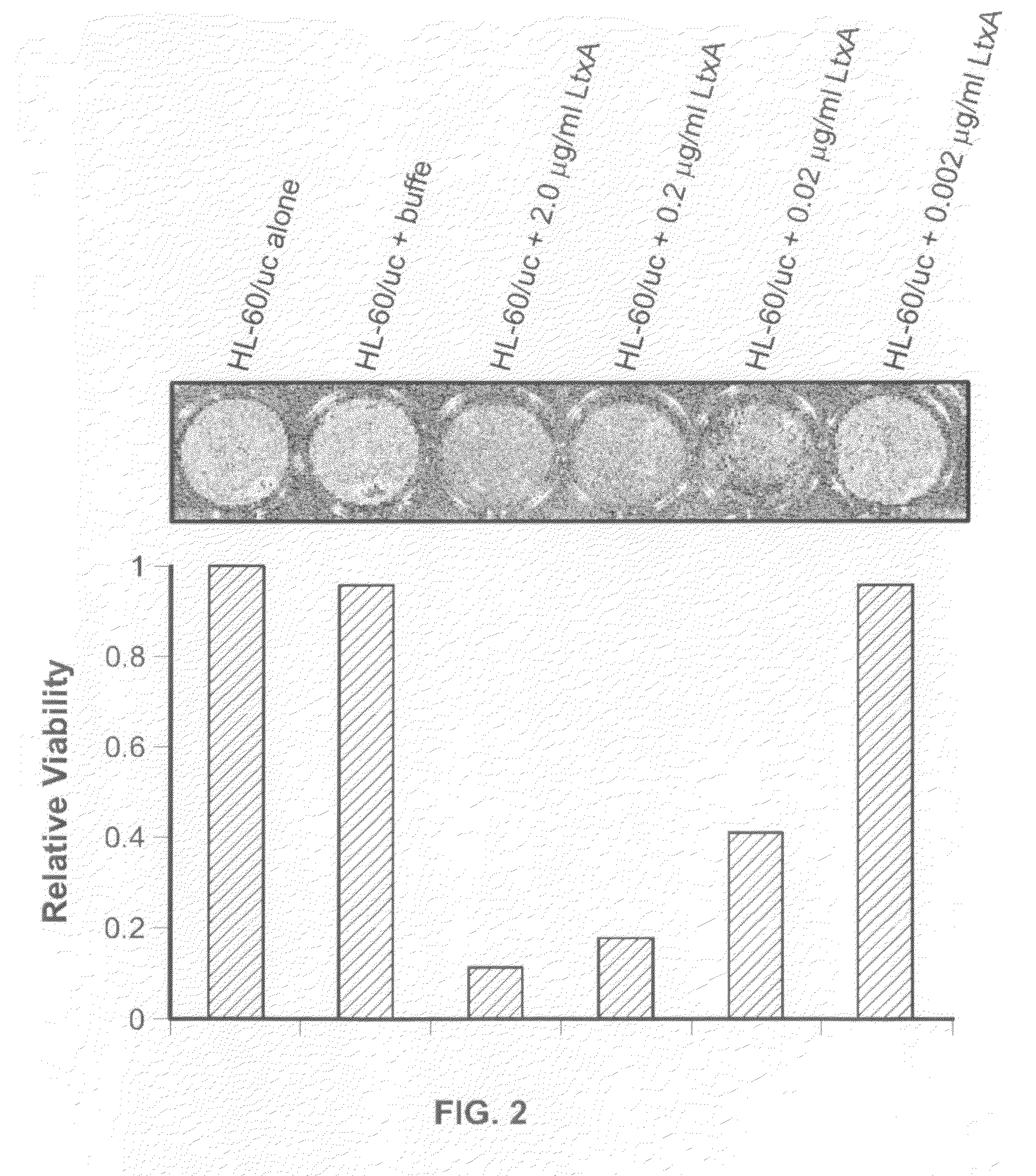
[0016]The present assay uses HL-60 cells that have been engineered that stably express firefly luciferase and produce light, whereby such bioluminescent HL-60luc cells may be rapidly detected in whole blood, eg. with a sensitivity of approximately 1000 viable cells. As demonstrated herein, treatment of HL-60luc cells with a bacterial leukocyte-specific toxin or the drug chlorambucil reveals that the bioluminescent viability assay is more sensitive than the trypan blue dye exclusion assay. HL-60luc cells administered intraperitoneally (i.p) or intravenously (i.v.) were visualized in living mice using an in vivo imaging system (IVIS). The rapidity and ease of detecting HL-60luc cells in biological fluid indicates that this cell line can be used in high throughput screens for the identification of drugs with anti-leukemia activity under physiological conditions.
[0019]It is a yet further principal object of the invention to provide a an assay and associated methodology, for the testing of candidate therapeutic agents for the treatment of cancers, that is rapid and efficient, and that can assess the activity of the candidate agents under physiological conditions.

Problems solved by technology

While their anti-tumor activities make many bacteria attractive therapeutic agents, there are inherent risks to administering live bacteria to humans.
As a result, devastating side effects are all too common.
Furthermore, a significant percentage of patients eventually show resistance to many of the drugs, thus rendering treatment largely ineffective.
While the drugs currently in use are toxic for cells, they are not highly specific.
While in vitro assays performed under cell culture conditions prove useful and necessary for preclinical testing of new therapeutics, extrapolation to the physiological conditions of a living organism is often difficult or impossible (27).
Testing the efficacy of anti-leukemia therapeutics against HL-60 cells in whole blood or other biological material is currently a challenge due to the inefficiency in differentiating the viability of HL-60 cells from other cells.

Method used


Image

  • Compositions for the treatment of cancer, and methods for testing and using the same
  • Compositions for the treatment of cancer, and methods for testing and using the same
  • Compositions for the treatment of cancer, and methods for testing and using the same

Examples


example 1

Purification of LtxA from the NJ4500 Strain of A. actinomycetemcomitans

[0093]The JP2 strain of A. actinomycetemcomitans produces abundant LtxA, but it does not represent a fresh clinical isolate. Here, LtxA was purified from the clinical isolate NJ4500 of A. actinomycetemcomitans . This strain also produces and secretes a large amount of LtxA, but the cells adhere to surfaces instead of growing planktonically. This type of adherent growth results in a relatively low number of cells per volume. The cell density of adherent cells was increased by increasing the surface area on which the cells can grow through the addition of spherical glass beads. Soda lime beads provided the greatest amount of LtxA when compared to Pyrex glass beads. The amount of LtxA that was purified from NJ4500 in the presence of soda lime beads was approximately twice that of JP2.

[0094]It is important to note that growth of A. actinomycetemcomitans in the presence of both types of glass beads was similar sugges...

example 2

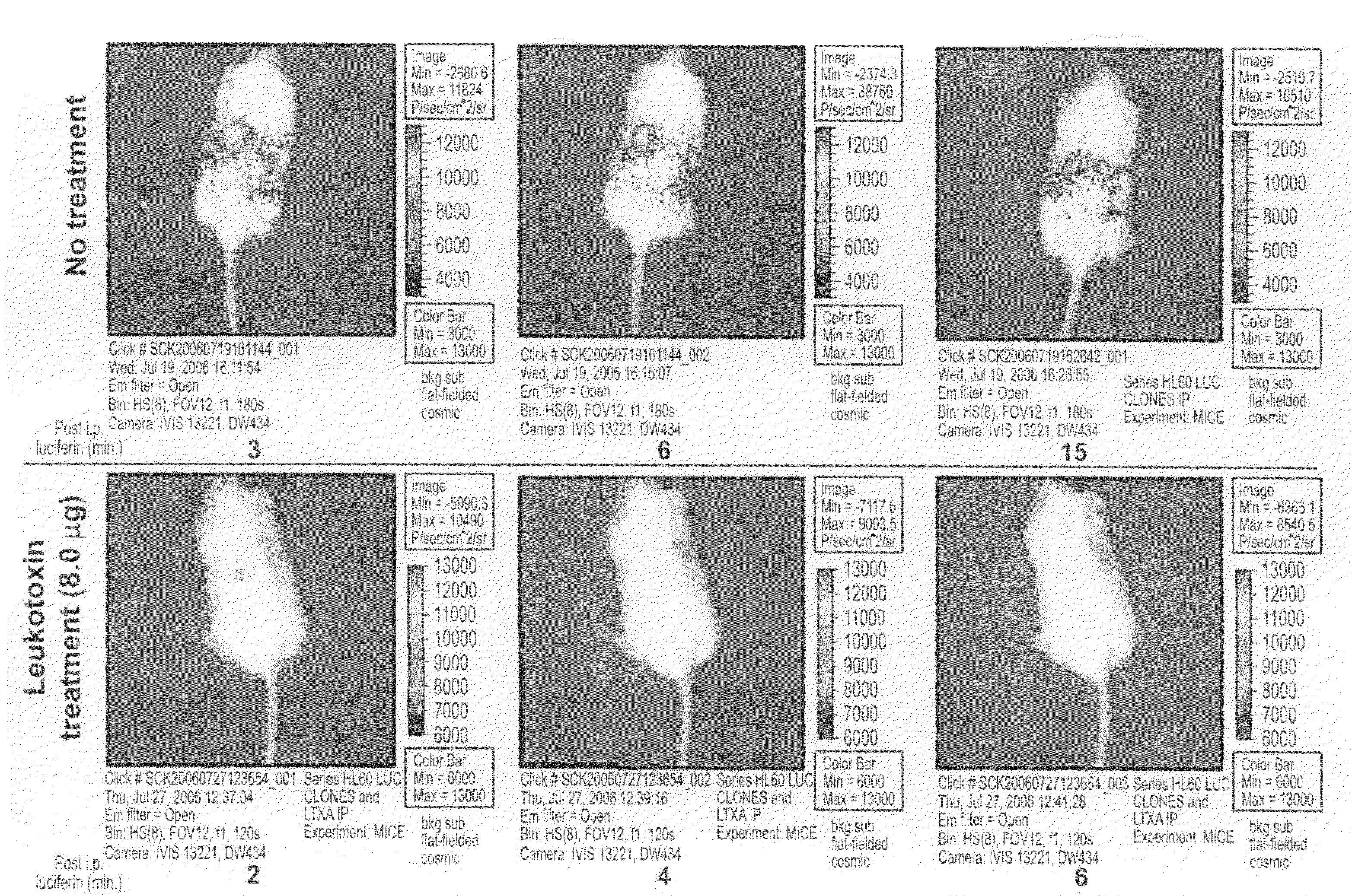
Imaging of Mice Injected with HL-60luc

[0099]The images of the mice shown in FIG. 3 were collected using in vivo bioluminescence imaging. The SCID mouse model has been used extensively for the study of hematologic malignancies, and the pattern of leukemia displayed in SCID mice closely resembles human clinical disease. In the model, leukemia cells are injected into SCID mice, usually intravenously. A commonly used leukemia cell line is HL-60 , originally isolated from a 36-year-old female patient with acute promyelocytic leukemia. Animal studies have shown that HL-60 cells can infiltrate bone marrow, the spleen, thymus, kidney, liver, lungs, and even the brain. It has been reported that the mean survival time for SCID mice that were injected with HL-60 cells was 42.5 days following injection; however, this time can vary depending on the passage state of the HL-60 cells being injected.

[0100]In vivo bioluminescence imaging (BLI) is a technology that allows visualization of live biolumi...

example 3

Prepareation of Assay System

Experimental

Cells and Growth Conditions.

[0103]HL-60 cells were obtained from American Type Culture Collection (ATCC) and maintained in RPMI +10% fetal bovine serum (FBS) (Invitrogen, Carlsbad, Calif.) at 37° C.+5% CO2. Escherichia coli was grown in LB medium at 37° C. A. actinomycetemcomitans strains were grown in AAGM at 37° C.+10% CO2 as previously described (12).

DNA Manipulations.

[0104]The luciferase-encoding plasmid for transfecting HL-60 cells was constructed by cloning luciferase gene from pGL3 (Promega, Madison, Wis.) into the geneticin resistance gene-containing plasmid pCI-neo (Promega, Madison, Wis.). Both plasmids were digested with BglII and XbaI and the Neo-containing fragment was then ligated to the pGL3 fragment that contained the luciferase gene. The mixture was transformed into E. Coli and the bacteria were selected on LB+carbenecillin (50 μg / ml). Plasmid from bacteria was prepared using the plasmid miniprep kit (Qiagen, Valencia, Calif.)...


PUM

PropertyMeasurementUnit
concentrationsaaaaaaaaaa

Abstract

A composition comprising leukotoxin proteins isolated from a bacterium is provided. In this composition, greater than 85% of the leukotoxin proteins are chemically modified at a basic amino acid residue, and the proteins induce cell death in myeloid leukocytes, while remaining substantially non-toxic to lymphoid leukocytes, lymphocytes, and red blood cells. Also provided is a method of selectively inducing cell death in myeloid leukocytes. The method comprises contacting the myeloid leukocytes with a composition comprising leukotoxin proteins. These leukotoxin proteins may be isolated from the NJ4500 strain of Actinobacillus actinomycetemcomitans. A method of purifying leukotoxin protein from the NJ4500 strain of Actinobacillus actinomycetemcomitans is also provided, as well as an assay that allows for the rapid determination of the activity of a given drug against leukemic cells either taken from a patient or derived from a cell line. The assay is performed in the presence of whole blood or serum.

Description

CROSS REFERENCE TO RELATED APPLICATIONS[0001]The present application is a Continuation In Part of PCT Application No. PCT / US2006 / 045258, filed Nov. 25, 2006, which, in turn, claims priority under 35 U.S.C. §119(e) from U.S. Provisional Application Ser. No. 60 / 739,537, filed Nov. 25, 2005; and co-pending U.S. Non-Provisional application Ser. No. 12 / 150,038, filed Apr. 23, 2008, which, in turn, claims priority under 35 U.S.C.§119(e) from U.S. Provisional Application Ser. No. 60 / 925,794, filed Apr. 25, 2007. The entire contents of all prior listed applications are incorporated by reference herein.RELATED FEDERALLY SPONSORED RESEARCH[0002]The work described in this application was sponsored at least in part, under Grant No. R01 DE16133, from the National Institute of Dental and Craniofacial Research, and under Grant No. NIH R01DE16133, from the National Institutes of Health. Accordingly, the Government has certain rights in the invention.FIELD OF THE INVENTION[0003]The present invention...

Claims


Application Information

Patent Timeline
Patent Type & Authority Applications(United States)
IPC IPC(8): A61K38/16C07K14/00C12N5/00C07K1/14C12Q1/02A61P35/00
CPCA61K38/00G01N2800/52G01N33/5011C07K14/285A61P35/00
Inventor KACHLANY, CHARLES S.
Owner RUTGERS THE STATE UNIV
Learn More