Antibody-induced apoptosis
a technology of apoptosis and antibodies, applied in the field of antiher2 antibodies, can solve the problems of increased relapse and low survival rate, poor clinical outcome, and no evidence of any of these factors
- Summary
- Abstract
- Description
- Claims
- Application Information
AI Technical Summary
Benefits of technology
Problems solved by technology
Method used
Image
Examples
example 1
Production of Her2, Her3 and Her4 Extracellular Domains
Cloning and Expression of Her2 Extracellular Domain (Soluble Her2)
[0032]A soluble Her2 receptor construct was made as follows. A cDNA clone of full-length Her2 in plasmid pLJ (pLJ is described in Korman et al. Proc. Natl. Acad. Sci. USA 84, 2150-2054 (1987) was digested with AatII which cuts once at position 2107 of the Her2 DNA sequence (numbering as in Coussens et al., supra). The linearized plasmid was cut with HindIII, which cuts 5′ of the initiating ATG, to release an approximately 2200 bp fragment. This fragment was cloned into pDSRα2 5′-HindIII to 3′SalI using an oligonucleotide linker (AatII-SalI) which contained an in-frame FLAG sequence and a translation termination codon. The resulting cDNA encodes for the Her2 extracellular ligand binding domain spanning amino acid residues 1-653 fused to the FLAG sequence (underlined):
[0033]Thr Ser Asp Tyr Lys Asp Asp Asp Asp Lys STOP
This construct was transfected into CHOd-cells. S...
example 2
Production of Anti-HER2 Antibodies
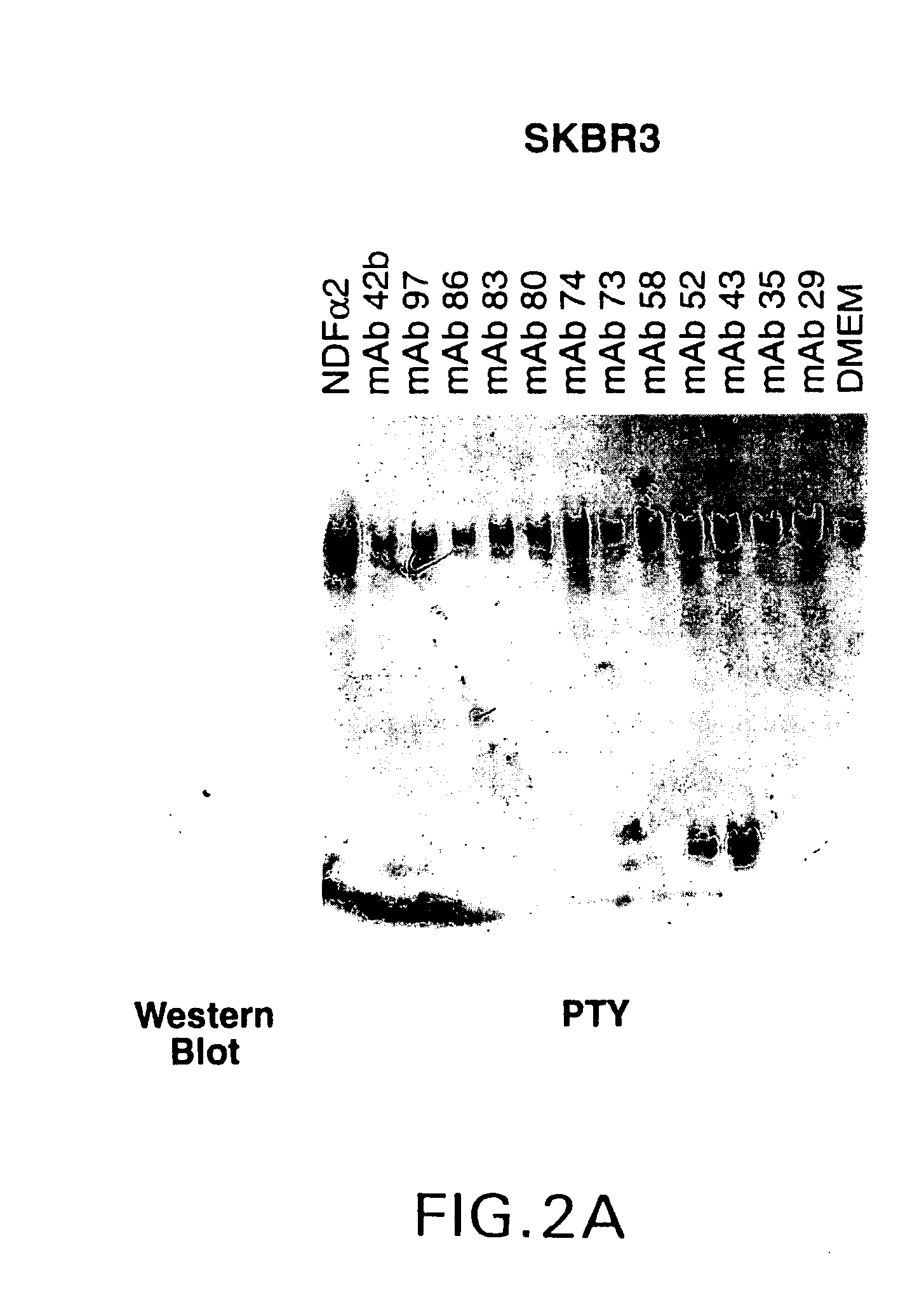
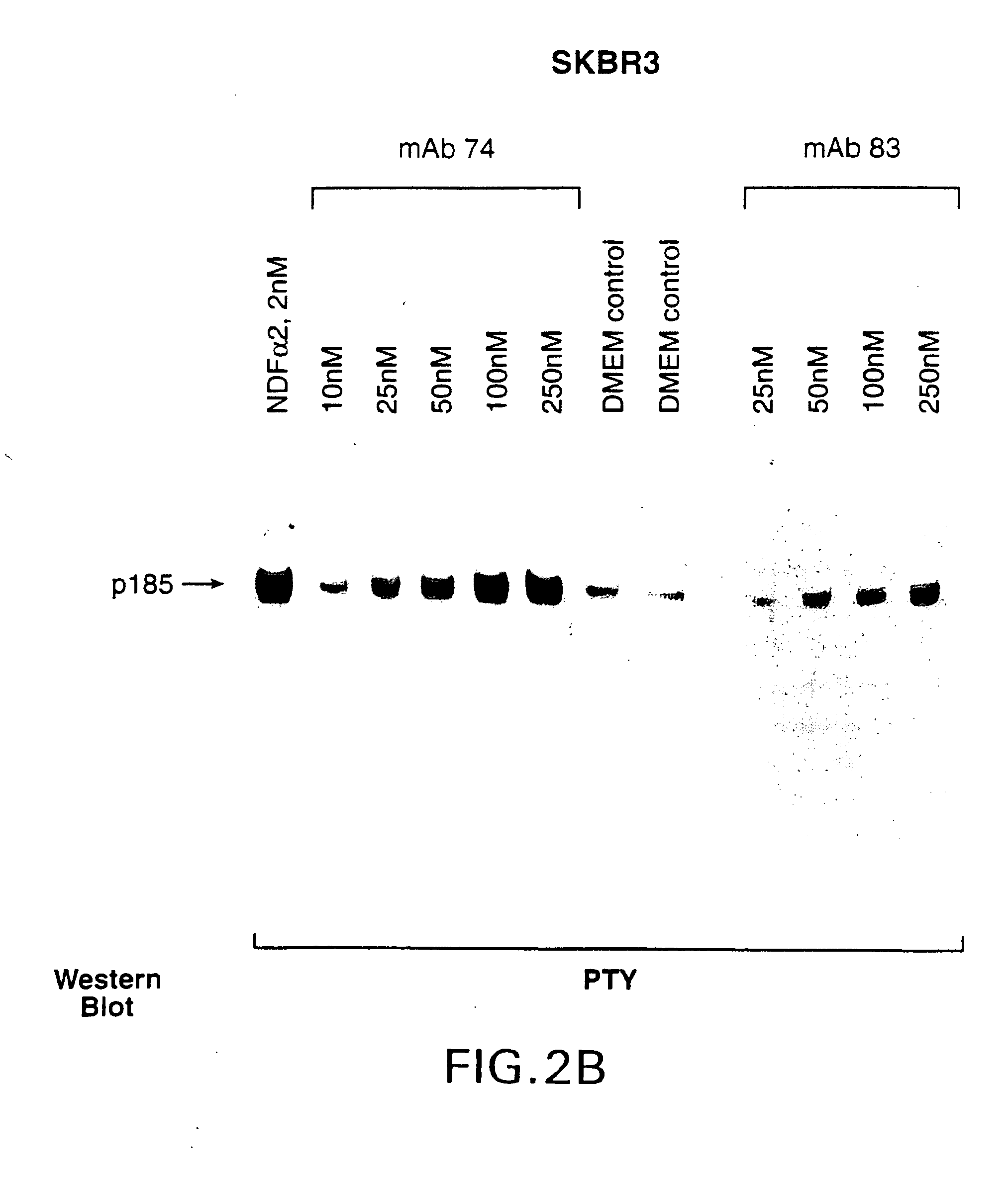
[0043]Procedures for immunizing animals, preparing fusions and screening hybridomas and purified antibodies were carried out generally as described in Harlow and Lane, Antibodies: A Laboratory Manual. Cold Spring Harbor Laboratory (1988).
Enzyme-Linked Immunosorbant Assay (EIA)
[0044]96-well plates were coated with 2 μg / ml sHer2, 2 μg / ml sHer3 or 2 μg / ml sHer4 in a carbonate-bicarbonate buffer. After blocking, hybridoma conditioned medium was added to the plate and incubated for 2 hours. The medium was aspirated and the plates were washed before addition of rabbit-anti-mouse IgG antibody conjugated with horseradish peroxidase (Boehringer Mannheim). After a one hour incubation, the plates were aspirated and washed five times. Bound antibody was detected with ABTS color reagent (Kirkegaard and Perry Labs., Inc.). The extent of antibody binding was determined by monitoring the increase in absorbance at 405 nm.
[0045]Cloning and IgG subtype determination. ...
example 3
Characterization of mAb74 Epitope on Her2
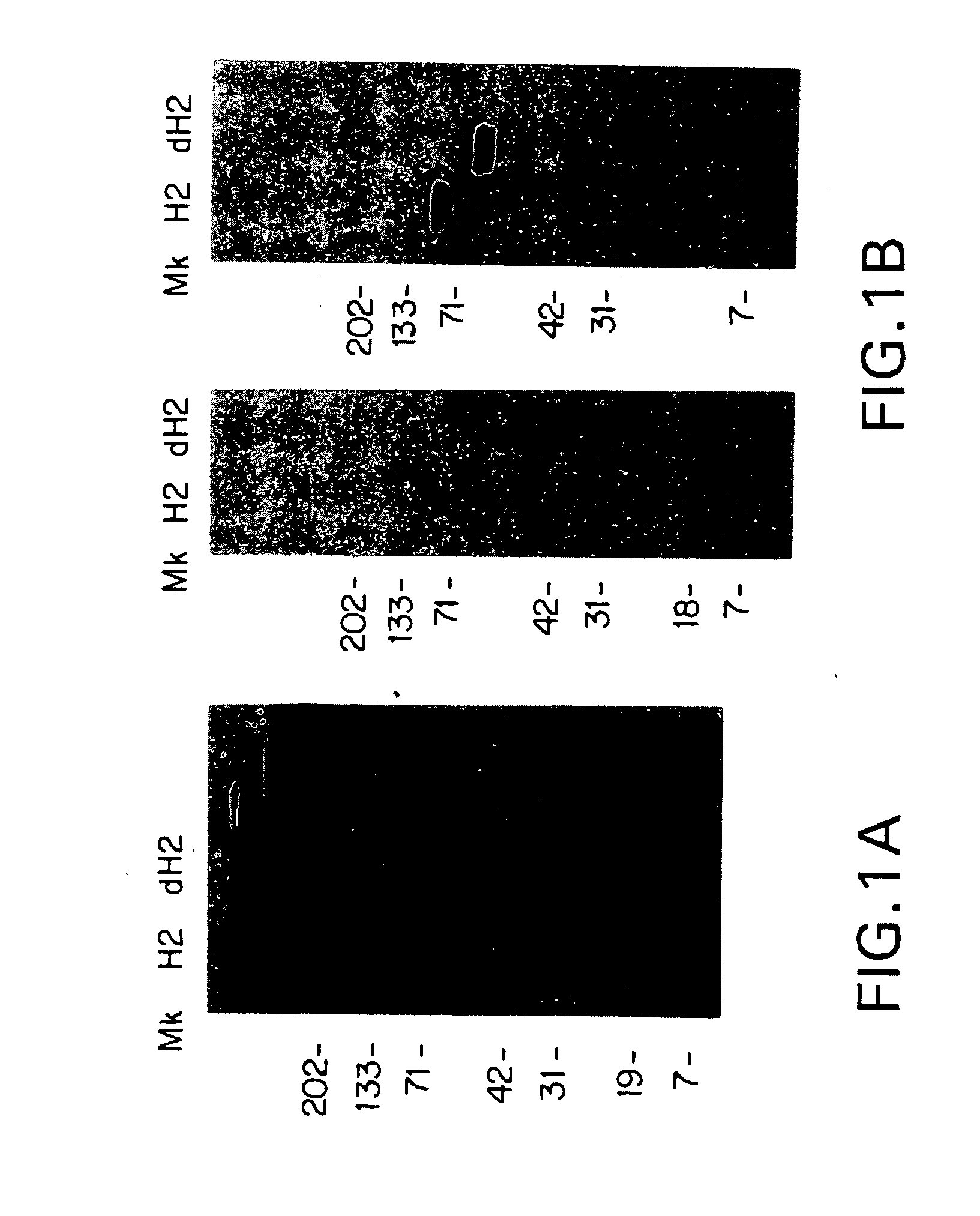
[0052]The effect of glycosylation on mAb74 interaction with sHer2 was determined as follows. Sixty μg of sHer2 in 20 mM BTP, 40 mM NaCl, pH 7.4, was denatured for five minutes in a boiling water bath in the presence of 0.4% SDS. After denaturation, NP-40 (Boehringer Mannheim) was added to 2% v / v, and the reaction diluted with an equal volume of DI H2O before adding 3 units of recombinant N-glycanase (Genzyme). The reaction was allowed to proceed with gentle shaking at 37° C. for 20 hrs.
[0053]An ECL glycoprotein detection system kit (Amersham Life Science) was used to determine the extent of deglycosylation. 0.25 μg each of sHer2 and deglycosylated sHer2 were run on a 4-20% gel (Novex) under nonreducing conditions and then blotted to nitrocellulose (Schleicher & Schuell) for 1 hour at 90 volts in a Bio-Rad mini PROTEAN II apparatus (BioRad) with cooling. After blotting, the membrane was treated with 10 mM sodium metaperiodate for 20 minutes, t...
PUM
| Property | Measurement | Unit |
|---|---|---|
| pH | aaaaa | aaaaa |
| pH | aaaaa | aaaaa |
| concentration | aaaaa | aaaaa |
Abstract
Description
Claims
Application Information
 - R&D
- Intellectual Property
- Life Sciences
- Materials
- Tech Scout
- Unparalleled Data Quality
- Higher Quality Content
- 60% Fewer Hallucinations
Browse by: Latest US Patents, China's latest patents, Technical Efficacy Thesaurus, Application Domain, Technology Topic, Popular Technical Reports.
© 2025 PatSnap. All rights reserved.Legal|Privacy policy|Modern Slavery Act Transparency Statement|Sitemap|About US| Contact US: help@patsnap.com


